What is 3D Atlas Of Neurological Surgery project?
MISSION
Our mission is to create a neuroanatomical 3D collection database facilitating learning of complex neurosurgical anatomy and approaches.
The project is nonprofit and is supported by the institutions involved, the European Association of Neurosurgical Societies (EANS) 2022 Research Fund and Sketchfab educational program.
The project is nonprofit and is supported by the institutions involved, the European Association of Neurosurgical Societies (EANS) 2022 Research Fund and Sketchfab educational program.
ACKNOWLEDGEMENTS
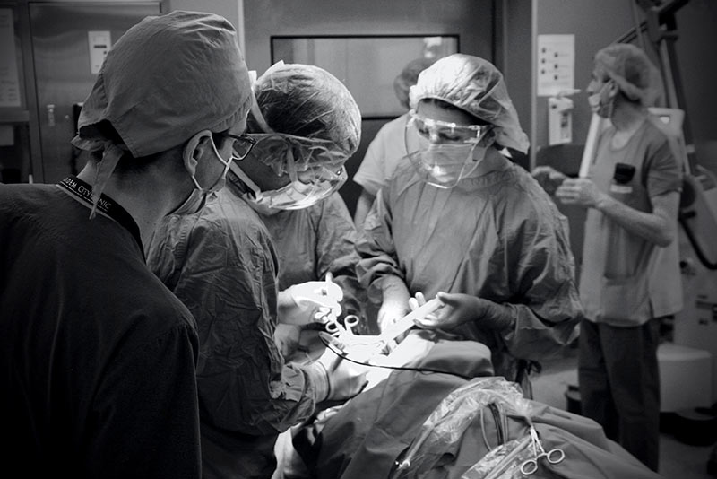
The anatomical dissections were done at the:
· Department of Anatomy and Histology, Pathology and Forensic medicine, University Hospital Lozenec, Medical Faculty, Sofia University, Sofia, Bulgaria,
· Department of Anatomy, Heinrich Heine University of Düsseldorf, Germany
· Department of Cellular and Molecular Medicine University of Copenhagen
· Laboratory of Neuroanatomy, Ebris institute of Salerno, Italy
The anatomical work at University of Düsseldorf was funded by the Department of Neurosurgery University Hospital of Düsseldorf and the Institute for Anatomy I, Medical Faculty & University Hospital Düsseldorf, Heinrich Heine University Düsseldorf, Germany.
The anatomical work at Copenhagen University Panum institute, was funded by the Department of Neurosurgery at Rigshospitalet, via the Copenhagen Neurosurgery Courses funding.
The specimens of individual skull bones were 3D scanned with permission at the Panum institute, Department of Cellular and Molecular Medicine and are part of the anatomical collection of University of Copenhagen.
We would like to thank Professor Jørgen Tranum Jensen for his great support as he is the reason we have had the opportunity to do all the anatomical work at the Panum institute, Copenhagen University.
The neuropathological photomicrographs in section Histology of the site are courtesy of the Department of Pathology and Neuropathology, Neuromed-Campus, Kepler-Universitätsklinikum, Linz, Austria.
The project is also supported by the European Association of Neurosurgical Societies (EANS) 2022 Research Fund and Sketchfab educational program.
AIM
The aim of the project is to create a photorealistic layered and annotated 3D anatomical models collection as well as DICOM CT and MRI based 3D models that illustrate relevant neurosurgical anatomy and approaches.
This atlas is a collaborative effort between different Medical Universities in several European countries and is continuously evolving.
This atlas is a collaborative effort between different Medical Universities in several European countries and is continuously evolving.
It comprises dissections of cranial, spinal and peripheral nerve anatomy as well as stepwise presentation of neurosurgical approaches captured by contemporary techniques of surface scanning allowing the creation of photorealistic 3D models.
All 3D models are processed using open source software - Blender .
The CT and MRI based 3D models that illustrate relevant surgical anatomy are processed also using open-source software - HorosTM and Slicer.
All 3D models are processed using open source software - Blender .
The CT and MRI based 3D models that illustrate relevant surgical anatomy are processed also using open-source software - HorosTM and Slicer.

Frequently Asked Questions
Can I use the information on this website for education purposes?
Yes, but you have to cite specifically that you use data from "3D atlas of neurological surgery" platform.
I am interested in one of the articles presented here. How can i contact the contributors?
I would like to use information from this website for a study.