NEWS
We are proud to present several new chapters of our platform:
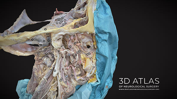
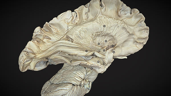
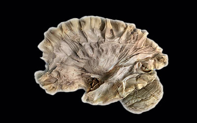
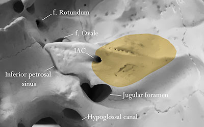
Completely updated and renovated section of Anatomy - with many new 3D models of brain anatomy, white matter dissections, brain ventricles, brainstem and posterior fossa, vascular anatomy, cavernous sinus, paranasal sinuses providing a 360° skull base anatomy.
The project was created with help of the Department of Neurosurgery, Medical Faculty & University Hospital Düsseldorf, Heinrich Heine University Düsseldorf and the support of the Institute for Anatomy I, Medical Faculty and University Hospital Düsseldorf, Heinrich-Heine-University, Germany. We would like to thank both departments and especially prof. Jan Frederick Cornelius MD, Kay M. Körner, Michael Wolf-Vollenbröker, MD, for their help in creating the material for this update and prof. Svenja Caspers for the institutional support.
The project was created with help of the Department of Neurosurgery, Medical Faculty & University Hospital Düsseldorf, Heinrich Heine University Düsseldorf and the support of the Institute for Anatomy I, Medical Faculty and University Hospital Düsseldorf, Heinrich-Heine-University, Germany. We would like to thank both departments and especially prof. Jan Frederick Cornelius MD, Kay M. Körner, Michael Wolf-Vollenbröker, MD, for their help in creating the material for this update and prof. Svenja Caspers for the institutional support.
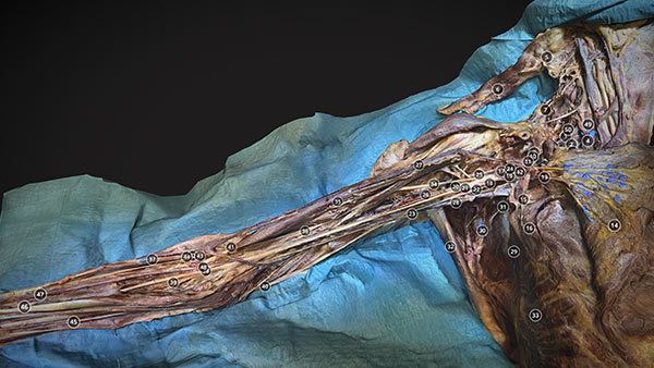
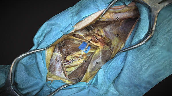
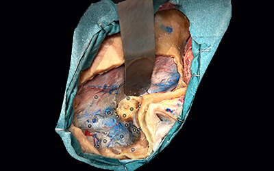
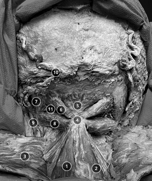
3D Photorealistic Atlas of Peripheral Nerve Approaches and Related Anatomy.
The project was created with the extensive help and collaboration of Kaare Fugleholm, MD PhD, Department of Neurosurgery, Rigshospitalet Copenhagen, University of Copenhagen, Denmark and Mr Mike Fox MD Consultant Surgeon in Peripheral Nerve Injury The Royal National Orthopaedic Hospital Stanmore UK & Cleveland Clinic London, UK. We would like to thank the Department of Cellular and Molecular Medicine University of Copenhagen, as well as Department of Neurosurgery, Rigshospitalet Copenhagen, University of Copenhagen for the support of this project and the 3D Atlas platform. The related content can be found in the menus "Approaches/Peripheral nerve and Anatomy/Peripheral nerve anatomy".
The project was created with the extensive help and collaboration of Kaare Fugleholm, MD PhD, Department of Neurosurgery, Rigshospitalet Copenhagen, University of Copenhagen, Denmark and Mr Mike Fox MD Consultant Surgeon in Peripheral Nerve Injury The Royal National Orthopaedic Hospital Stanmore UK & Cleveland Clinic London, UK. We would like to thank the Department of Cellular and Molecular Medicine University of Copenhagen, as well as Department of Neurosurgery, Rigshospitalet Copenhagen, University of Copenhagen for the support of this project and the 3D Atlas platform. The related content can be found in the menus "Approaches/Peripheral nerve and Anatomy/Peripheral nerve anatomy".
We would like to thank the - Italian Society of Neurosurgery namely the Head of the NeuroAnatomy Committee assistant prof. de Notaris for supporting a fellowship collaboration in the project „Skull base 360°" and Drs. Armocida and Carbone for their practical help.
We would also like to thank the European Association of Neurosurgical Societies (EANS) as well as all other contributors who support our platform and help maintain its open-access nature.
We would also like to thank the European Association of Neurosurgical Societies (EANS) as well as all other contributors who support our platform and help maintain its open-access nature.
Highlights
Each 3D model anatomical structure is annotated and the camera turns automatically to the region of interest when an annotation is selected.
A list of all annotation in each 3D model is present
3D models can be presented with or without annotations with the "Show/hide annotations" feature
Sections
ANATOMY
RESOURCES
NEUROSURGICAL APPROACHES
FOLLOW US ON SOCIAL MEDIA FOR NEWS AND UPDATES


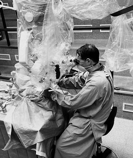
Dedicated
microsurgical
dissections
of complex neurosurgical anatomy
presented as comprehensive,
layered photorealistic 3D models

The project is one of the winners and supported by the European Association of Neurosurgical Societies 2022 Research Fund.

This project is supported by the Sketchfab educational program.
Anatomy is the foundation of everything what we do in the operating theater.
Built In Augmented reality
You can see every 3D model in Augmented Reality without the need of any additional app and regardless of the mobile phone type.
Powered by the "App free Augmented Reality" function of the Sketchfab platform.
Powered by the "App free Augmented Reality" function of the Sketchfab platform.
Virtual reality
3D models can be observed in an immersive VR environment using the Sketchfab VR features. The immersive VR experience can be seen with mobile phones and headset like Google Cardboard, with desktop VR headset, or with standalone headset.
Videos

Immersive 3D Operative Videos
