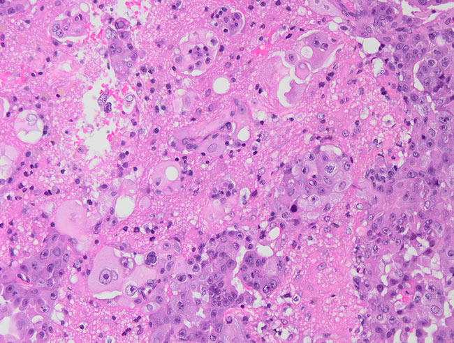
Pic 1. Groups of tumor cella with large polymorph nuclei with visibile nucleoli, sharply delineated from the Brain tissue; H&E stain; magnification x200.
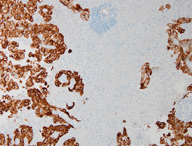
Pic 02. The tumor cells are pancytokeratin (AE1/AE3)-positive; x100.
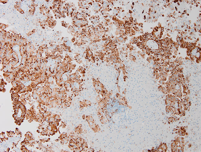
Pic 03. The tumor cells are Napsin A - positive and the tumor nuclei are TTF1-positive
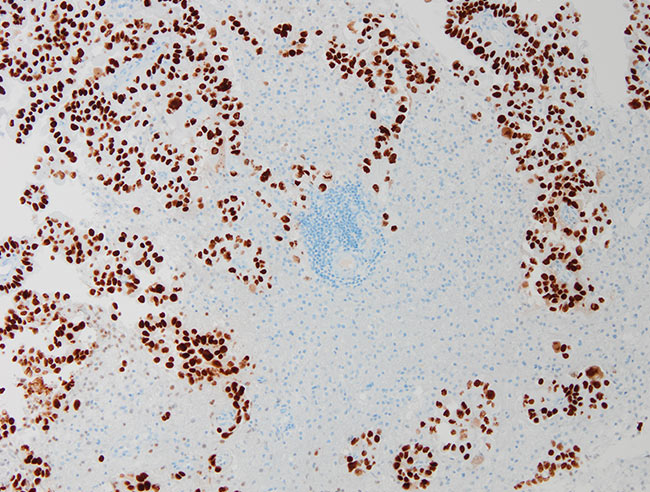
Pic 04. Both pictures - magn . x100 => lung adenocarcinoma metastasis.