Carotid body tumor (Glomus caroticum tumor)
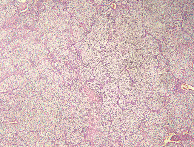
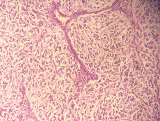
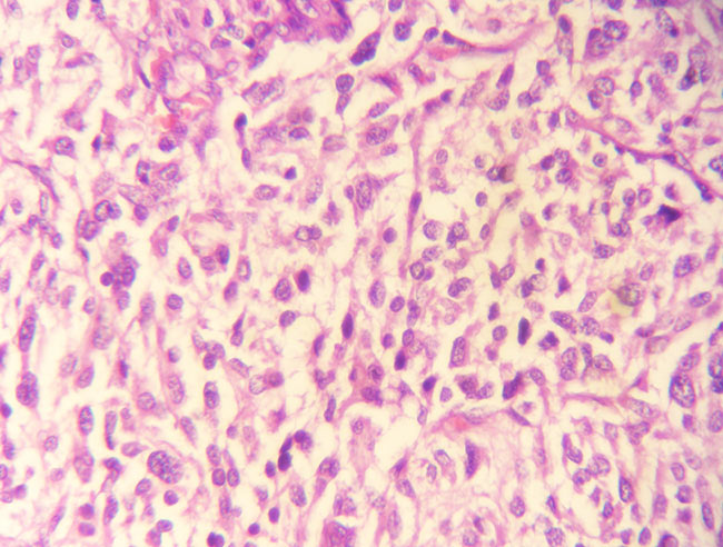
Micrograph of the pathological specimen with hematoxylin and eosin stain presenting typical arrangement of the tumor cells in cell balls (Zellballen), separated by fibrotic stroma and vessels. The cells are oval or polygonal with abundant granular eosinophilic cytoplasm and nuclear atypia.
Micrograph of the pathological specimen with hematoxylin and eosin stain presenting typical arrangement of the tumor cells in cell balls (Zellballen), separated by fibrotic stroma and vessels. The cells are oval or polygonal with abundant granular eosinophilic cytoplasm and nuclear atypia.


