А right sided supraclavicular skin incision is made from lateral border of the sternocleidomasoid m. and over its clavicular head to the lateral border of the trapezius m.
1. Superficial layers supraclavicular approach
The superficial layers of the approach and anatomical landmarks are exposed:
1) The lateral border of the trapezius m.
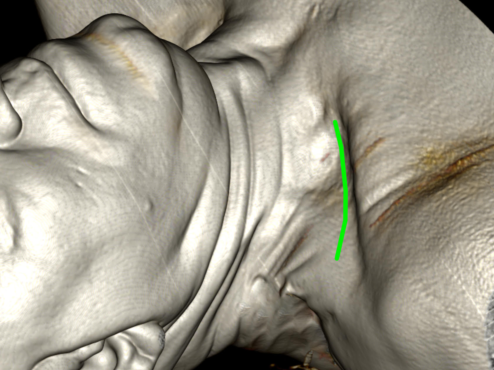
2) The lateral border of the sternocleidomastoid m.
The superficial layers of the approach and anatomical landmarks are exposed:
1) The lateral border of the trapezius m.
2) The lateral border of the sternocleidomastoid m.
2. Supraclavicular nerves
The supraclavicular nerves are dissected and mobilized. The accessory nerve supplying the trapezius muscle is identified
The supraclavicular nerves are dissected and mobilized. The accessory nerve supplying the trapezius muscle is identified
3. Superior and middle trunks of the brachial plexus
The supraclavicular deep fat pad is removed and the superior (C5, C6) and middle (C7) trunks of the brachial plexus are exposed.
The supraclavicular deep fat pad is removed and the superior (C5, C6) and middle (C7) trunks of the brachial plexus are exposed.
4. Final exposure
The omohyoid m. is divided. The brachial plexus trunks are further exposed revealing the superior (C5, C6), middle (C7) and inferior (C8, Th1) trunks between the middle and anterior scalene muscles. The associated nerves - suprascapular nerve, long thoracis nerve as well as the phrenic nerve are exposed. The transverse cervical artery passing posteriorly to supply the trapezius m. is identified between the middle and inferior trunks of the brachial plexus.
The omohyoid m. is divided. The brachial plexus trunks are further exposed revealing the superior (C5, C6), middle (C7) and inferior (C8, Th1) trunks between the middle and anterior scalene muscles. The associated nerves - suprascapular nerve, long thoracis nerve as well as the phrenic nerve are exposed. The transverse cervical artery passing posteriorly to supply the trapezius m. is identified between the middle and inferior trunks of the brachial plexus.