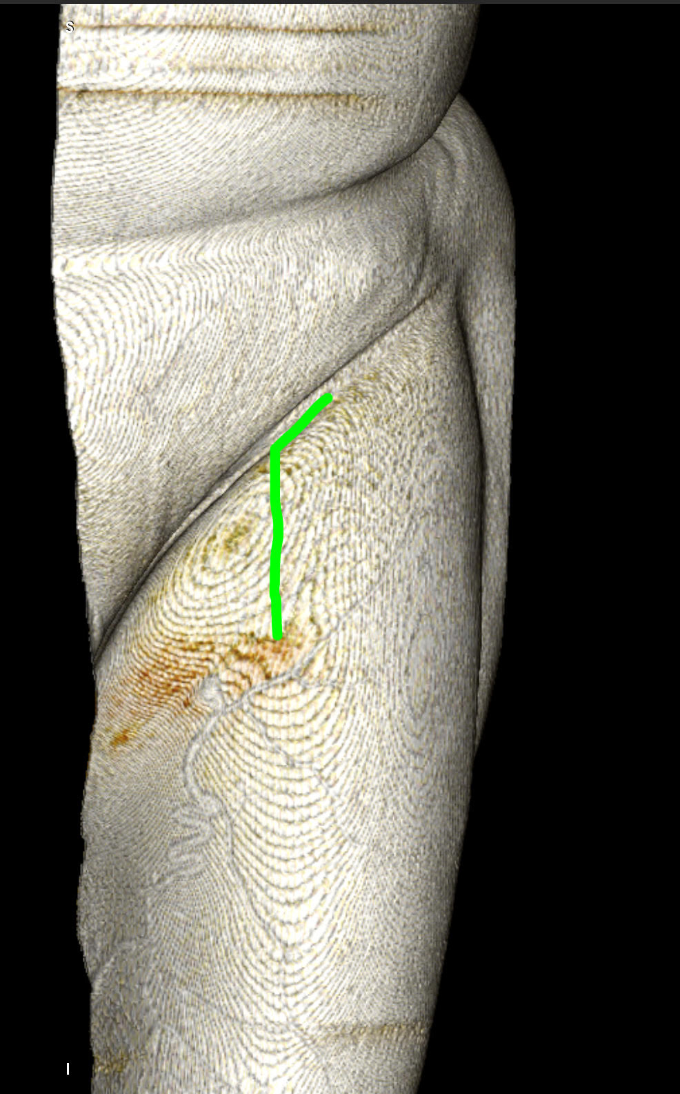
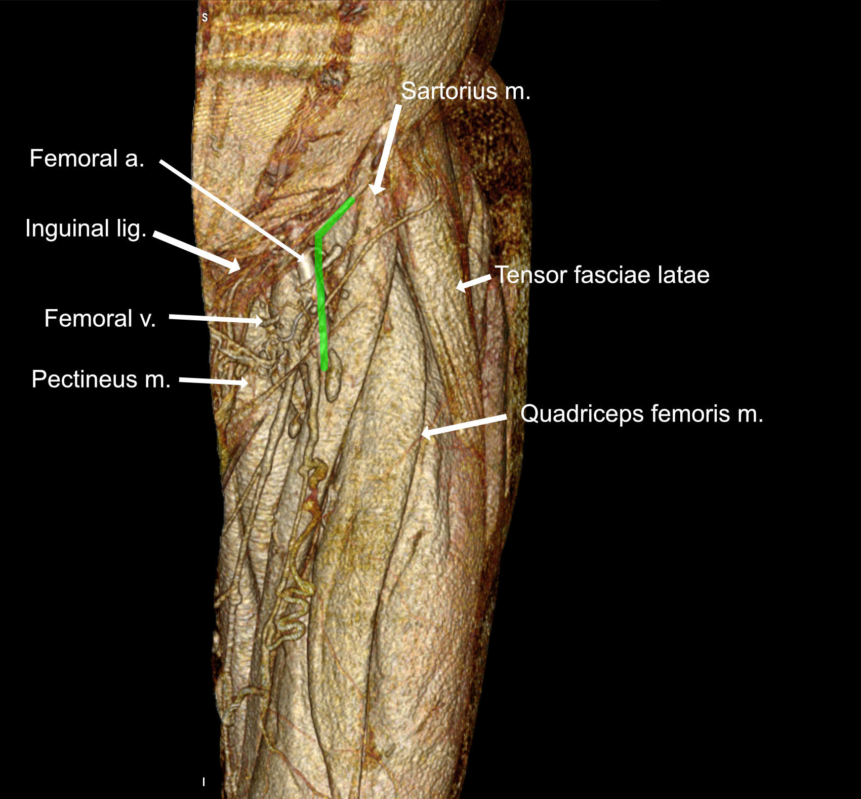
A slightly curved skin incision (left side marked with green) is made lateral to the palpable pulse of the femoral artery.


1. Superficial exposure
The femoral nerve is identified lateral to the femoral artery. The motor branches can be identified with the help of monopolar stimulation. The deep circumflex a. and v. can be identified in the upper border of the dissection.
The femoral nerve is identified lateral to the femoral artery. The motor branches can be identified with the help of monopolar stimulation. The deep circumflex a. and v. can be identified in the upper border of the dissection.
2. Femoral nerve branches
The femoral nerve branches are further identified (medially) going to the quadriceps femoris m. The sensory branches (saphenous n.) are identified more laterally. The femoral artery is presented.
The femoral nerve branches are further identified (medially) going to the quadriceps femoris m. The sensory branches (saphenous n.) are identified more laterally. The femoral artery is presented.
