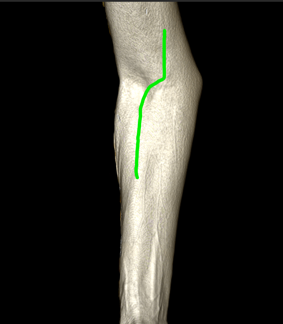

A curved skin incision is made over the cubital fossa (right side) extending along the forearm.
1. Superficial dissection
A curved skin incision is made over the cubital fossa. The brachioradialis m. is reflected laterally. The biceps tendon is visible as well as the pronator teres m. and flexor carpi radialis m.
A curved skin incision is made over the cubital fossa. The brachioradialis m. is reflected laterally. The biceps tendon is visible as well as the pronator teres m. and flexor carpi radialis m.
2. Median nerve exposure
The pronator teres m. is divided and the median nerve at the forearm is exposed just before entering the muscular canal of the flexor digitorum superficialis m.
The pronator teres m. is divided and the median nerve at the forearm is exposed just before entering the muscular canal of the flexor digitorum superficialis m.
3. Anterior interosseus nerve
The nerve branches to pronator teres m. are visualized. Flexor digitorum superficialis m. is partially sectioned exposing the anterior interosseus nerve which is presented lateral and posteriorly to the median n.
The nerve branches to pronator teres m. are visualized. Flexor digitorum superficialis m. is partially sectioned exposing the anterior interosseus nerve which is presented lateral and posteriorly to the median n.