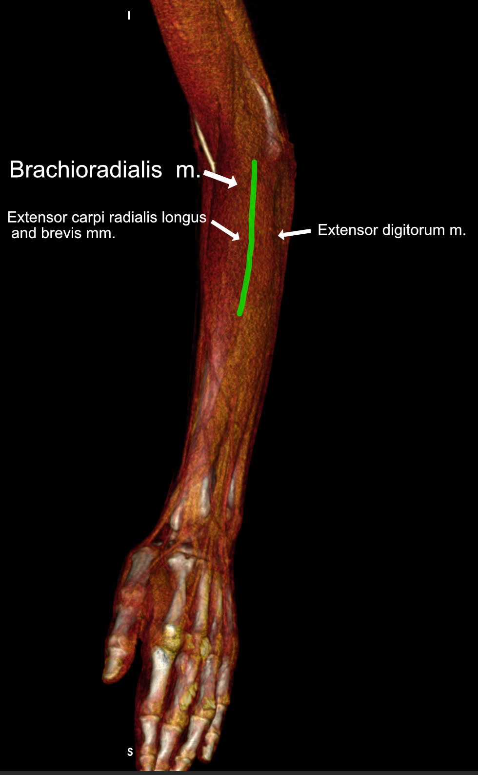
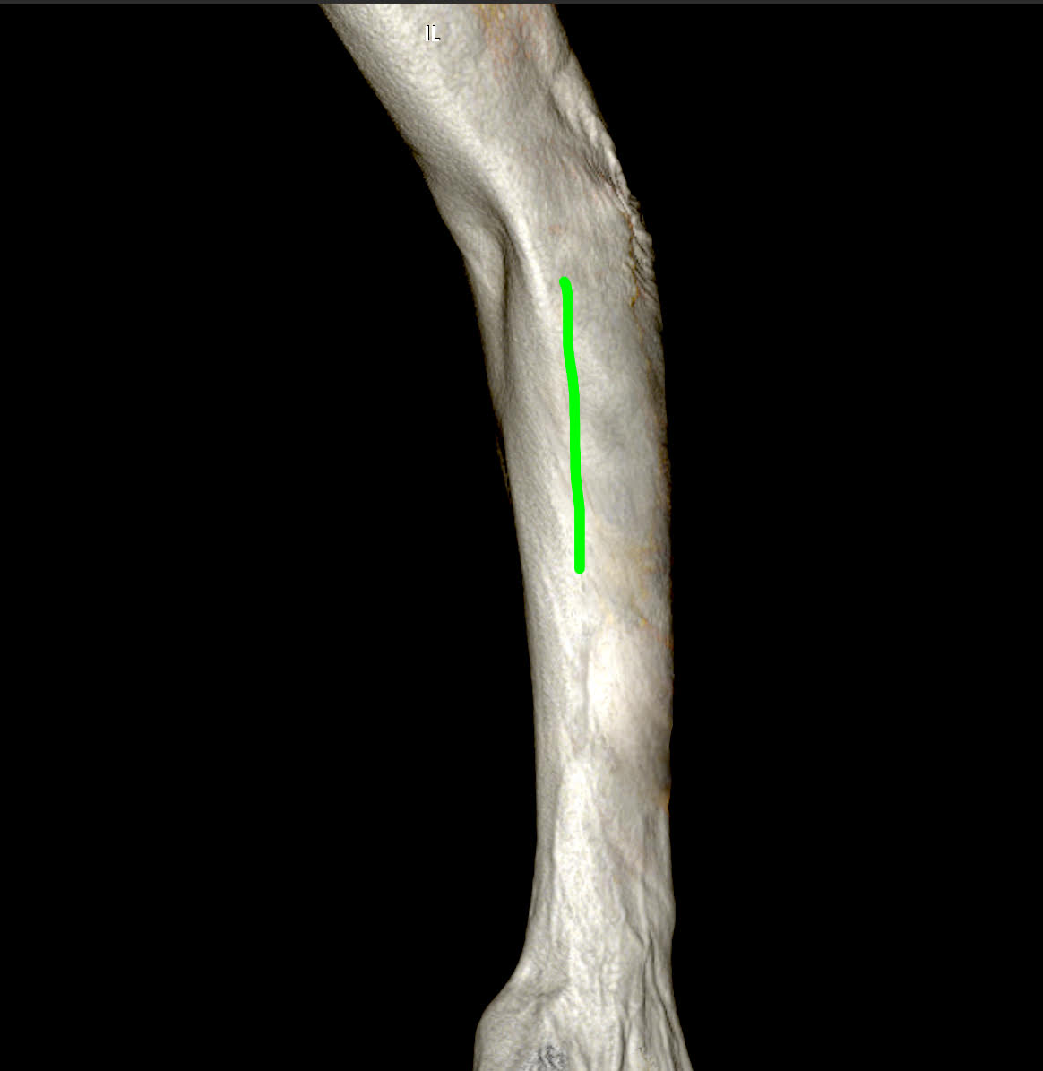
Left side: A linear skin incision on the forearm is made over the projection of the extensor carpi radialis longus/brevis mm. and extensor digitorum.
1. Supinator muscle exposure
A linear skin incision is made over the projection of the extensor carpi radialis longus/brevis mm. and extensor digitorum. A fascial plane is followed to the deeper muscular layer which is the supinator m.
A linear skin incision is made over the projection of the extensor carpi radialis longus/brevis mm. and extensor digitorum. A fascial plane is followed to the deeper muscular layer which is the supinator m.
2.Posterior interosseous nerve
The posterior interosseus n. (PIN) is exposed and a branch to the extensor digitorum m. is visualized. The fibro-muscular canal of the PIN is presented (4) within the supinator m.
The posterior interosseus n. (PIN) is exposed and a branch to the extensor digitorum m. is visualized. The fibro-muscular canal of the PIN is presented (4) within the supinator m.
3.Posterior interosseous nerve decompressed
The supinator m. is transacted and the whole course of the posterior interosseous nerve within the muscle is visualized.
The supinator m. is transacted and the whole course of the posterior interosseous nerve within the muscle is visualized.