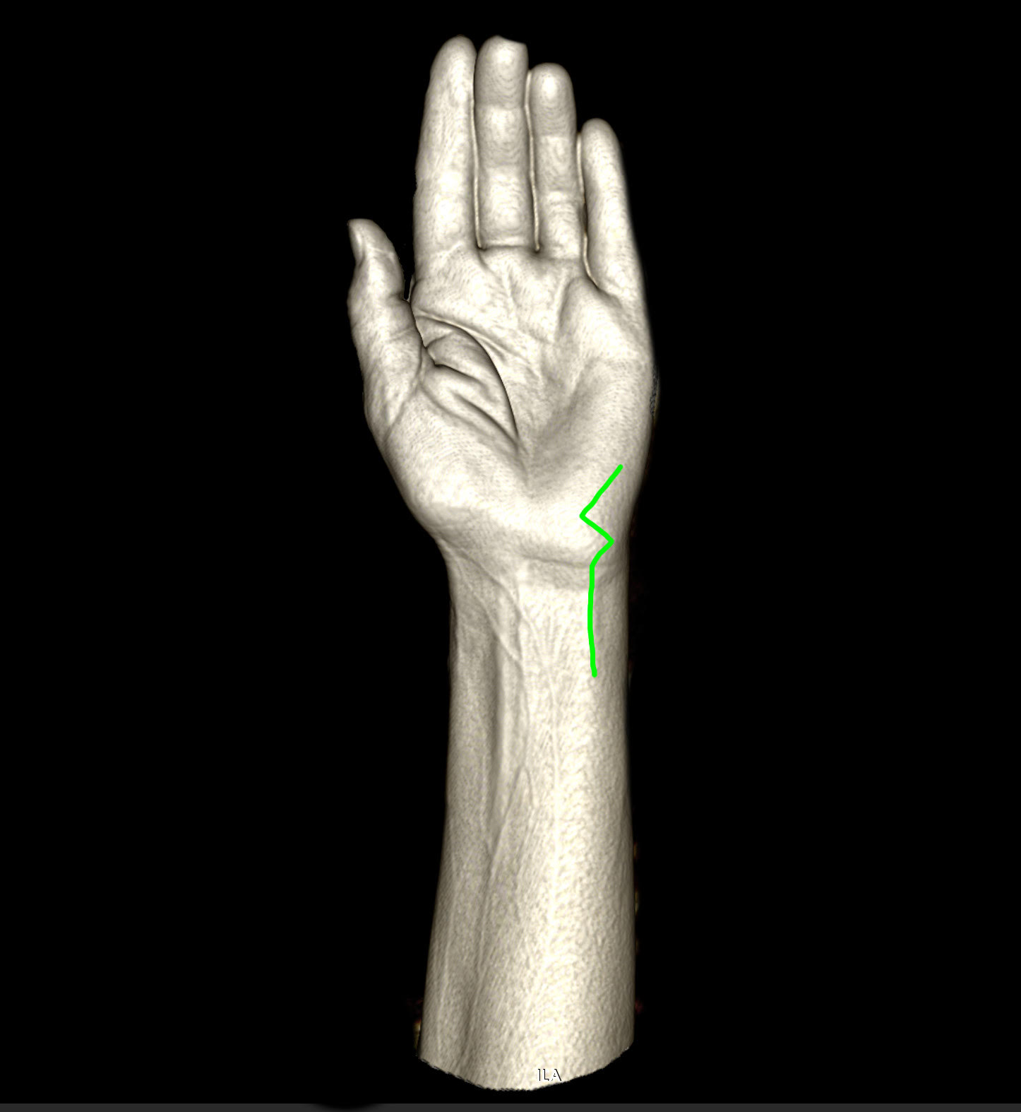
A curved skin incision (left side marked with green) is made over the projection of the Gyon's canal.
1. Ulnar nerve exposure at Gyon's canal
Initial exposure of the ulnar nerve before entering the Gyon’s canal. The Palmar carpal ligament is visible with a yellow rubber band placed through the Gyon’s canal for better presentation. The ulnar artery is situated medially to the ulnar nerve.
Initial exposure of the ulnar nerve before entering the Gyon’s canal. The Palmar carpal ligament is visible with a yellow rubber band placed through the Gyon’s canal for better presentation. The ulnar artery is situated medially to the ulnar nerve.
2. Ulnar nerve decompressed
The palmar carpal ligament is transacted and the ulnar nerve is decompressed. The ulnar artery is visible medial to the nerve, as well as the superficial branch of the nerve can be followed anteriorly.
The palmar carpal ligament is transacted and the ulnar nerve is decompressed. The ulnar artery is visible medial to the nerve, as well as the superficial branch of the nerve can be followed anteriorly.
