Legacy: "Atlast of Neurosurgery" / L.Karaguiosov, A. Ramadan, K.Karaguiosov / Kiwait/ 1998
PREV>15. Malformations of The Spine and Spinal Cord
NEXT<17. Infections of the Spine and Spinal Cord
16. ARTERIOVENOUS MALFORMATIONS OF THE SPINAL CORD
Arteriovenous malformations (AVM) of the spinal cord are rare, being one tenth as common as brain arteriovenous malformations. They appear more commonly in males (4:1) and generally occur at an older age than do those in the brain. According to the classification of M.Hamilton, J. Anson, and R. Spetzler, arteriovenous malformations of the spinal cord are of four types. Type 1 (or intradural type) is the most common, and is found predominantly in the lower thoracic region and in the conus medullaris. The lesion consists of a feeding artery that enters the dura at the area of the root sleeve, where a small cluster of vessels within or just beneath the dura makes up a fistula. Occasionally additional one or more arterial vessels are present at adjacent levels. These arterial vessels communicate with the fistula through small vessels coursing within or just beneath the dura. The feeding arteries originate from dural branches of the radiculo-medullary arteries and usually do not contribute any blood supply to the spinal cord. The venous outflow from the dural fistula drains intradu-rally into the surface of the spinal cord. Usually one draining vein as a single-coiled vessel carrying arterialized blood is located at the pial level of the cord, rarely penetrating the cord, and is usually dorsally situated.
The Type 2 spinal arteriovenous malformation has a true compact nidus within the parenchyma of the spinal cord. The feeding arteries are branches from the anterior spinal artery. The malformation drains into the coronal venous plexus that surrounds the spinal cord. This plexus becomes tortuous and distended for a variable distance from the lesion. The differentiation of the branches of the anterior spinal artery supplying the malformation from the branches supplying the spinal cord is difficult.
Type 3 is most commonly seen in adolescents and young adults as a more diffuse arteriovenous lesion, which often encircles the spinal cord, situated intra- and extramedullarily. It has feeding arteries, originating from multiple vertebral levels. The blood flow is fast and the blood supply of the malformation and the cord may be closely related.
Type 4 spinal arteriovenous malformations are intradural, extramedullary, or premedullary in location. They are fed by the anterior spinal artery and lie completely outside the spinal cord and its pia mater. The malformation drains directly into an enlarged venous outflow tract with no intervening network of small vessels. Type 4 spinal (AVMs) are located on the anterior surface of the spinal cord, but they may occur posteriorly when they are supplied by the posterior spinal artery.
The majority of cases with spinal cord AVMs have an insidious onset with progressive deterioration and half of them are severely disabled within 2 years of the onset of the disorder.
Subarachnoid haemorrhage with sudden onset or sudden onset of spinal cord symptomatology is unusual. A bruit over the spine is diagnostic and the presence of a cutaneous angioma helps diagnostically in localising the level of the lesion.
Most spinal cord AVMs are discovered at myelography, presenting as serpentine filling defects of the subarachnoid space. MRI has replaced myelography and provides a precise localisation of the arteriovenous malformation nidus in type 2 and type 3. Spinal angiography is always required in the preoperative evaluation of a patient with spinal AVM before treatment options can be discussed. Spinal angiography details the vascular anatomy and helps to categorise the type of malformation. This knowledge allows a rational treatment strategy to be selected. In addition, spinal angiography establishes indications for endovascular therapy.
Because prognosis is poor for patients with spinal cord AVMs that are untreated or simply decompressed, surgical exploration to assess resection is important. The satisfactory treatment of AVMs of the spinal cord is total removal. The role of embolization is limited and usually is not indicated for type 1 dural AVMs because surgery is safer and more efficacious. Embolization of type 2 can be helpful if enlarged feeding arteries exist that can be obliterated without excessive risk to the normal spinal supply. This embolization is usually applied as a prelude to surgical removal. Embolization of type 3 spinal AVMs is mandatory. These lesions are so complex and extensive that attempting surgical resection without the aid of embolization would be extremely risky. Embolization in type 4 spinal AVMs depend upon the size. Embolization is most useful and feasible in big fistulae, whereas embolization typically is impossible in small types.
SURGICAL TECHNIQUE
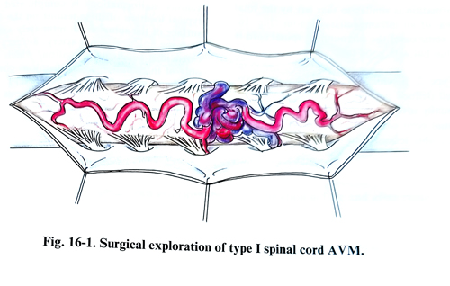
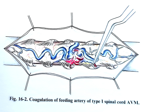
In type 1 AVM, the goal of the treatment is to eliminate the transmission of venous hypertension to the spinal cord. Simple interruption of the vein that carries blood from the dural AV fistula to the venous plexus helps in most patients by eliminating the venous hypertension of the spinal cord. After the laminectomy, the bone overlying the intervertebral foramina is removed and dissection exposes the glomus of the AV fistula in the dural covering of the nerve root. The spinal dura is then opened in the midline. After separating the arachnoid from the underlying vessels and laterally retracting the arachnoid, the site of intradural penetration of the arterialised vein draining the dural AV fistula is identified. This vessel is coagulated and divided. The dural AV fistula is either coagulated with bipolar coagulation or excised. In many cases, the dural artery that supplies the AV fistula, and the medullary artery, which supplies the spinal cord, are so close at the site of dural penetration that it is not possible to separate them. In such a case simple interruption of the vein that drains the fistula intrathecally is preferable. At this stage of the operation, the dorsal spinal cord veins should demonstrate decreased expansion and decreased blood flow within large vessels. If this has not happened, additional feeding arteries should be found and interrupted. The stripping of the large coiled veins from the dorsal surface of the spinal cord is not recommended because this may compromise the normal cord blood supply and cause ischaemic changes beyond the area of the malformation (Figs. 16-1; 16-2).
The type 2 spinal cord malformation can be removed on many occasions. The laminectomy can be moderately broad and the dura opened widely with preservation of the arachnoid until the extent of the lesion is visualised through the arachnoid. Microsurgical technique is essential. The arachnoid is then opened widely with small scissors or an arachnoid knife. If the nidus reaches the spinal cord surface, circumferential dissection is usually best started on the side where the margin of the nidus is most clear. If the nidus is intrinsic and does not reach the spinal cord surface at any point, a myelotomy is made in a longitudinal direction allowing visualisation of the anterior and posterior margins of the lesion. After the nidus has been exposed, resection is performed under high magnification, with low-power bipolar coagulation to shrink and obliterate the small penetrating vessels. The lesions are approached at the site of the arterial feeders. In some cases the arterial pedicle is closed to the source of major supply from the anterior spina! artery. Once the arterial side is interrupted the venous pedicle is easy to manage. Sharp dissection is always preferred in the removal instead of a blunt or tearing type of dissection technique. These AV malformations are frequently associated with a large venous aneurysmal dilatation, often partially thrombosed, with thin walls, predisposed to intraoperative bleeding. Careful use of a broad bipolar tip under irrigation may allow shrinkage of venous aneurysms, but care must be taken not to violate the aneurysm wall.
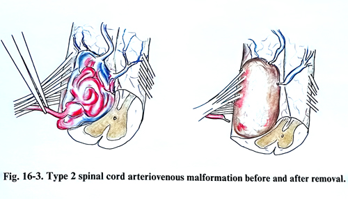
The residual cavity after removal of an AV malformation is similar to that left by the total removal of an intramedullary tumour (Fig. 16-3). In some cases the surrounding spinal cord is very thin but with good function.
Type 3, juvenile AV malformations have not well-defined margins permitting dissection, and usually involve the interior and exterior of the spinal cord over many segments. They are nonresectable lesions, but reduction of flow by embolization or by ligation of a feeding artery may diminish the further incidence of haemorrhage and reduce the degree of vascular steal and associated ischaemic lesion of the cord.
Surgical treatment of type 4 spinal arterio-venous malformations is complicated by the typical location of these lesions on the anterior surface of the spinal cord, intimately associated with the anterior spinal artery. Surgery is indicated in a small malformation, and it consists only of interruption of the fistulous connection between the anterior spinal artery and the dilated draining veins. The big lesions are best treated by endovascular techniques.