Legacy: "Atlast of Neurosurgery" / L.Karaguiosov, A. Ramadan, K.Karaguiosov / Kiwait/ 1998
PREV>19. General Principles of Peripheral Nerve Repair
20. CRANIAL NERVES
FACIAL NERVE
The most frequent injuries of the facial nerve, in which surgical intervention is necessary, are skull base fractures through the temporal bone pyramid, conditions following removal of tumours from the cerebellopontine angle and facial injuries. Not infrequent also are injuries of the nerve during mastoidectomy, removal of tumours of the parotid gland, and incisions of abscesses in the same area.
Surgical anatomy. From the point of view of the surgical anatomy of the facial nerve, three parts are distinguished: 1. Intracranial part, situated in the cerebellopontine cistern in the posterior fossa; 2. Middle part, which is found in bone canals of the temporal bone structure and 3. The peripheral part, situated outside the stylomastoid foramen.
The facial nerve arises from the brain stem in the lower area at the boundary between the pons and brachium pontis, medially from the intermedius and the statoacustic nerves. Together with these nerves it is directed laterally and anteriorly and enters the internal acoustic meatus. On the bottom of the internal acoustic meatus the facial nerve and intermedius nerve join and go together into the bone canals of the pyramid. In the initial part of the canal (the labyrinthic part) the facial nerve passes anteriorly and laterally, forming a small fold with its concavity forward. In this part of the canal above the nerve there is only a thin plate from the bone on the cerebral surface of the pyramid, below which lays the labyrinth. After that the canal of the facial nerve turns backward through an angle of about 60 - 70 degrees, where the nerve forms the so called genu or"knee". From the most convex part of the genu the major superficial petrosal nerve separates off. It contains preganglionic parasympathetic fibres, which reach the lacrimal gland. Further on, the canal goes obliquely posteriorly, laterally and inferiorly parallel to the axis of the pyramid. In this part of the canal, the facial nerve is in proximity with the tympanic cavity. After that, it changes direction almost in a right angle, goes vertically downward, and ends at the level of the stylomastoid foramen. In the last descending part of the canal, a branch separates from the facial nerve for the stapedius muscle and the chorda tympani, which conveys the gustative sensation from the anterior two thirds of the tongue and innervates the submandibular gland. After leaving the temporal bone, the facial nerve forms an arch under the external acoustic meatus and enters the parotid gland. At about 5 – 10 mm from its point of emergence from the bone canal the nerve divides into two branches, which innervate the facial muscles. Before its separation the posterior auricular nerve separates from it for the muscles behind the auricle, for the posterior belly of the digastric muscle and for the stylohyoid muscle.
FACIAL NERVE OPERATIONS
The surgical approaches and procedures to the facial nerve differ according to the nerve segment that should be reached. In a case of a facial nerve root lesion, end to end suturing can be performed during the removal of a cerebello-pontine neurinoma. In the same area microdissection of the facial root can be performed in cases of hemifacial spasm. For the middle part, the common task is revealing the nerve in the bony canal of the temporal pyramid for decompression or suturing. The other group of operations includes reconstructive procedures in injuries to the extracranial nerve part and its branches and anastomosis of the facial nerve with some neighbouring nerves: hypoglossal, accessory, or phrenic.
Reconstructive operations on the facial nerve should be performed in the first weeks after the injury, when there are no clinical and electromyographic data for regeneration. Operations done after 12 months cannot lead to good results. In old injuries when is impossible to expect regeneration after operation on the nerve, another kind of operations can be advised: transposition of the muscle parts from neighbouring areas, or operations aimed to fix the paralysed side of the face in a determined position to obtain facial symmetry.
FACIAL NERVE OPERATIONS IN THE TEMPORAL BONE.
These operations are applied to injuries of the facial nerve, provoked by fractures of the pyramid, to paralysis from otogenic origin or nerve tumours.
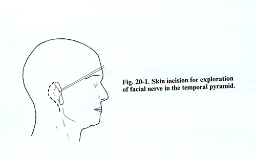
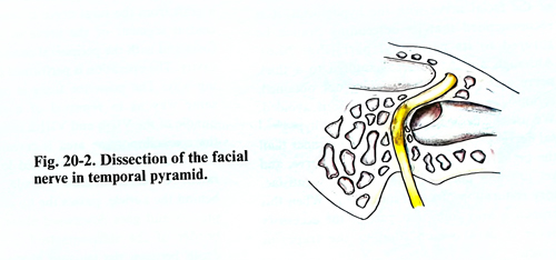
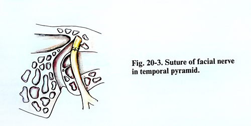
Operative technique. A curved incision of the soft tissues is made as in a mastoidectomy, continuing 3 cm below the inferior wall of the external acoustic meatus (Fig.20-1). First the facial nerve is revealed between the stylomastoid foramen and the place where the nerve penetrates into the parotid gland. After that an antrotomy is made to expose the facial nerve, which is prominent in the antrum, enclosed only under a thin bone plate, which is removed with a small bone curette. Having a clear view of the nerve in the tympanic cavity and its exit from the stylomastoid foramen, the entire canal can easily be opened. The bone is carefully excised with high speed drill until a thin plate remains on the nerve. This plate is subsequently removed with a fine curette (Fig. 20-2) When the nerve is intact along the entire bone canal, only changes in its colour and thickness can be observed. In such cases the operation consists of a longitudinal incision of the epineurium, so that additional decompression be obtained. If a nerve defect not longer than 20 mm is found, a suture of the nerve can be made, taking the nerve out of the canal in advance as that makes the approximation of the two ends possible (Fig. 20- 3).
FACIAL NERVE ANASTOMOSIS WITH NEIGHBOURING NERVES
Anastomosis of facial with the accessory or the hypoglossal nerves. The patient is placed in prone position on the operating table with the head slightly turned to the opposite side. A small cushion is put under the nuchal region.
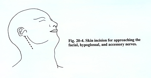
The skin incision begins from the apex of the mastoid process and goes along the anterior edge of the sternomastoid muscle with a length of 7 - 8 cm (Fig. 20-4). After the incision of the skin and the subcutaneous tissue, the great auricular nerve is revealed on the surface of the sternomastoid muscle, it is dissected and retracted laterally. The accessory nerve can be found on the internal surface of the sternomastoid muscle. If the hypoglossal nerve is suitable for anastomosis it can be found at the point where it crosses the internal carotid artery. The dissection of the facial nerve may sometimes be difficult, due to the changes provoked by its degeneration. It is of a slightly yellowish colour and has a diminished calibre, which sometimes makes it difficult to distinguish from the parotid gland tissue. Practically it is important and is recommendable that the parotid gland be retracted together with its capsule. The nerve is revealed by dissecting upward and following the styloid process, immediately laterally from its base. Its dissection must be made as high as possible. The nerve trunk must be cut at the stylomastoid foramen level. The small nerve branches, that separate from it immediately after its exit from the bone are cut, so that the nerve is mobilised distally about 2 cm within the parotid gland.
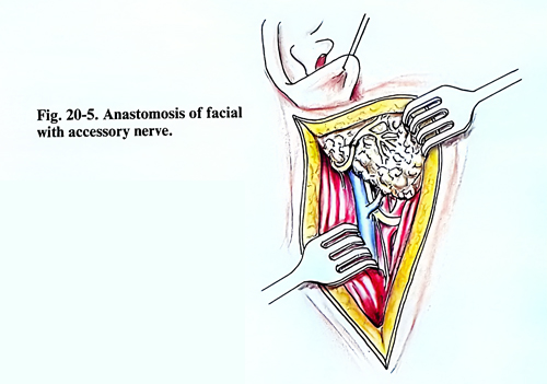
Next the accessory nerve is dissected proximal to the sternomastoid muscle, which permits approach to the facial nerve peripheral section (Fig. 20-5). Certain modifications of the operation are known, in which the entire trunk of the accessory nerve is not used. Some neurosurgeons use only that part of the nerve which innervates the sternomastoid muscle and avoid provoking paralysis and atrophy of the trapezius muscle. We use only the branch of the nerve, which perforates the sternomastoid muscle and innervates the trapezius muscle. If this part of the nerve is not sufficiently thick and the branch of the cervical plexus for the trapezius muscle is thicker, the incision is continued inferiorly and laterally, the trapezius muscle nerve is separated, and it is turned upward and sutured to the facial nerve.
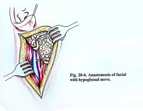
The hypoglossal nerve is transected in the vicinity of the hyoid bone and from there the facial nerve can be reached easily. In anastomosis of the facial with the hypoglossal nerve, it is recommended that its descending branch be sutured to its peripheral part (Fig. 20-6). Although a thin branch is sutured to a thicker nerve end and good regeneration is not obtained, it yet provides nonetheless a sufficiently satisfactory restoration of facial movement. When this branch is very thin, the trapezius branch of the accessory nerve is used. In that way serious hemiatrophy of the tongue is avoided.
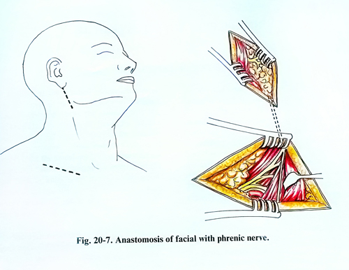
In anastomosis of the facial with phrenic nerves, the facial nerve is dissected after a vertical incision 5 - 6 cm long, beginning from the auricular base and passing along the anterior border of the sternomastoid muscle. The parotid gland is retracted forward to a depth of 2 - 4 cm where the facial nerve is revealed. It is divided as near as possible to its point of exit from the cranial cavity. A second incision 5 - 6 cm long is made parallel to the clavicle and 2 cm above its medial half, to reveal the phrenic nerve. It is exposed, retracting medially the lateral border of the sternomasoid muscle in the supraclavicular region on the anterior surface of the scalenus anterior muscle (Fig. 20-7). After that, with a thin blunt instrument, a tunnel is made under the sternomastoid muscle, connecting the two incisions. The proximal end of the phrenic nerve is turned upward and sutured with the facial nerve peripheral segment.
FACIAL NERVE GRAFTING
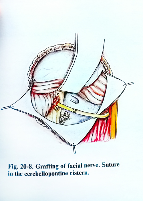
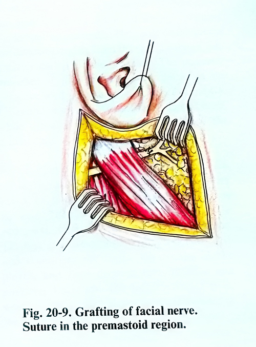
Direct method. After complete removal of a large cerebellopontine neurinoma, the facial nerve is frequently interrupted near to the middle of its cisternal part. The tumour usually elongates this part of the nerve and it is possible to approximate the two segments and to perform microsuturing. When this is impossible, N. Dot has introduced a surgical method, by which a graft from the sural nerve is interposed between the central segment of the nerve in the posterior fossa and the peripheral outside the cranial cavity. The operation is performed in the following way: The posterior fossa is opened in the same way as for the removal of the tumour and at the end of removal the bundle of the VIIth and VIIIth cranial nerves in the cerebellopontine area is revealed and only the facial nerve that is situated lower is separated. A second incision is made, which starts behind the auricle, passes the tip of the mastoid process and goes downward along the anterior border of the sternomastoid muscle. In the depth between the mastoid process and ramus mandibulae, the facial nerve is exposed and cut as near as possible to the cranial base. After that with a blunt instrument is made a tunnel, which connects the two incisions under the sternomastoid muscle. The graft is pushed through this tunnel and its superior end is sutured to the facial nerve in proximity with the brain stem, while the other end is sutured extracranially (Figs. 20-8; 20-9).
Cross grafting. The operation consists of grafting so that one of the ends of the graft of the sural nerve is sutured with the branch of the unaffected facial nerve, and the other end to the peripheral segment of the interrupted nerve. The graft is introduced subcutaneously through the tunnel, crossing the midline through the upper lip. The graft can be a little shorter if introduced through the retropharyngeal space. The advantages of this operation are that the reinnervation is from the same nerve fibres from the other side of the face. Hence, the patient recovers spontaneous movements in response to emotions and voluntary movements without any special training. Despite these possible advantages the recovery of these movements is minimal according to the aleady obtained observations.