
11. PRINCIPLES OF SPINAL CORD SURGERY
APPROACHING THE SPINAL CANAL
ANATOMICAL BASIS OF SURGICAL OPENING OF THE SPINAL CANAL
All operations on the spinal cord, its meninges and nerve roots are performed after operative opening of the spinal canal. The spinal canal is approached through different routes, which depend on the location of the lesion in the canal, its relation to the structure of the spine and spinal cord and the biomechanical consequences, which will result from the approach alone.
All surgical methods have two technical stages: penetration through the soft tissues on the way to the spine and the opening of the canal itself. The posterior wall of the spinal canal is situated relatively superficially in the posterior midline of the neck and the back; this makes it the most accessible to exposure. The lateral and anterior walls of the spinal canal are surrounded by more complex and vitally important structures at different levels of the spine corresponding to the neck, thorax and abdomen. Many lesions that require surgical treatment simultaneously affect both the spine and the spinal canal, and the approach is adjusted accordingly.
The same refers to the conditions of compromised spinal stability, either by the lesion or the proposed approach. In these last cases the surgery is concluded by an appropriate stabilizing technique.
The posterior approach to the spine remains the "classical", standard for reaching the neural structures. It will be presented in the introductory chapter of the spinal section. Several other important techniques, especially the microscopic ones such as the anterior approach to the cervical spine will be dealt with completely when dealing with the corresponding problems. Some methods related more to orthopedic spinal techniques, will be mentioned only in principles.
Paravertebral soft tissues. From the surgical anatomical point of view the paravertebral soft tissues can be divided in four regions: nuchal, thoracic, lumbar and sacrococcygeal (Fig.11-1).
Nuchal region. The topographical limits of the nuchal region are considered as follows: superiorly - the external occipital protuberance and the superior nuchal line, inferiorly - the horizontal line, passing through the apex of the spinous process of the VIIth cervical vertebra, which is easily palpated. The lateral border of the trapezius muscle serves as a lateral limit. The skin is compact and thick especially in the superior half and sometimes presents some difficulties in suturing.
The subcutaneous fatty tissue, as that of the scalp, is compact and contains fibrous bands that connect the skin and the underlying aponeurosis. Due to this, it is difficult during the operation to separate the external surface of the aponeurosis, which covers the trapezius muscle. Superiorly the aponeurosis is adherent to the external occipital protuberance and the superior nuchal line. Covering the trapezius muscle laterally, it continues into the superficial neck fascia. Along the midline the fascia is connected with the external edge of the nuchal septum, which is extended from the spinous process of the VIIth cervical vertebra to the external occipital protuberance. The nuchal septum is triangular in shape and is situated along the midline on the sagittal plane, connecting the external occipital crest with the spinous processes of the cervical vertebrae.
The muscles of the nuchal region are rather voluminous and can be divided into four layers in a transverse section. The trapezius muscle forms the first layer. Under it, as a second layer is revealed the splenius muscle. The third layer consists of muscles situated vertically: longitudinal major and minor muscles, longissimus capitis and cervicis, and multifidus muscles. The fourth layer, the deepest one, contains the following muscles: the two straight muscles, the two oblique muscles, transversospinal and inter-spinal muscles. These last muscles are short and serve for extension and rotation of the head.
These four layers of muscles are separated from one another in the occipital region by fine fascias.
The soft tissues in the nuchal region draw their blood supply from the occipital and deep cervical arteries. The occipital artery is a branch of the external carotid artery and enters the occipital region medially to the mastoid process. At its very beginning, it is situated deeper than the place of attachment of the sternocleidomastoid muscle. After that, its course changes upward and inward and 2,5 - 4 cm laterally from the external occipital protuberance, it penetrates into the scalp. The deep cervical artery is a branch of the subclavian artery, which goes to the neck upward between the iliocostal and transversospinal muscles, where it branches.
The posterior branches of the cervical nerves supply the nuchal region. The posterior branch of the second cervical nerve, which is the thickest one (the great occipital nerve) penetrates into the occipital region between the Ist and the 2nd cervical vertebrae. It envelopes the inferior edge of the oblique muscle, perforates the splenium and trapezius muscles and leaves the muscles 2 cm under and 2 - 4 cm lateral to the external occipital protuberance. Its branches innervate the soft tissues of the occipital region of the head. The nerve is situated medially and close to the occipital artery. The nuchal region
is rich in venous vessels, situated in the inter-muscular spaces. The veins flow into the vertebral veins, the posterior jugular vein and often have anastomoses with the sinuses of the dura.
Thoracic region. The thoracic region has a quadrangular form. The lines passing along the angles of the ribs serve as the lateral limits. The superior limit is the horizontal line, passing at the level of the spinous process of the VIIth cervical vertebra. The inferior limit follows the course of the XIIth ribs.
The skin in the thoracic region is thick and movable regarding the fascia, the spinous processes and the ribs.
The fascia covering the muscles is thin. The muscle can be divided as in the occipital region into four layers. The trapezius and latissimus dorsi muscles, which are flat, are the most superficial. The rhomboid muscles are situated under them as a second layer. The serrate muscles are situated as a third layer. Under them, in the deepest layer, are the iliocostal and longissimus muscles and the small muscles - transversospinal, interspinal and intertransversal muscles. The thoracolumbar fascias separate the third from the fourth layers.
Lumbar region. The paravertebral soft tissues in the lumbar region occupy a quadrangular area. Its limits are: superiorly - the line passing along the course of the XIIth rib on both sides, inferiorly - the arcuate line, which begins from the sacral bone and continues along the iliac crests to end laterally at the vertical line along the lateral border of sacrospinal muscles.
The skin and the subcutaneous fatty tissues have the same characteristics as they do in the thoracic region. The aponeurosis is more compact and is more easily separated than in the thoracic region. The muscles are of a larger volume and are also in four layers. The latissimus dorsi muscle is situated superficially with its aponeurosis. Under it in a second layer is located the inferior serratus muscle. The sacrospinal muscles are situated deeper as a third layer and in the last fourth layer, are the interspinal and intertransversal muscles.
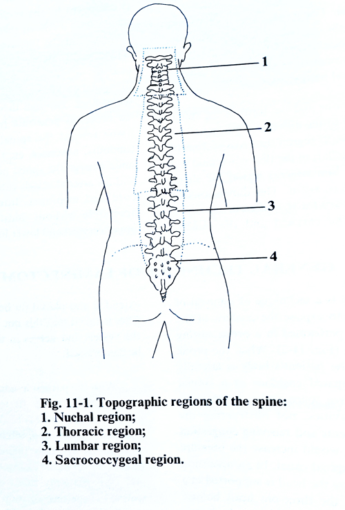
Sacro-coccigeal region. This region is triangular in shape and corresponds to the posterior surface of the sacrum and coccyx. The muscular layer is thinner than in the thoracic, as the fibrous beginnings of paravertebral muscles are attached on the posterior surface of these bones.
Spinal canal. The spinal canal occupies the internal part of the entire length of the spine. Its upper end communicates with the cranial cavity through the foramen magnum and its caudal end is in the sacral hiatus.
The dimensions and the form of the spinal canal are specific to the different segments of the spine. In the cervical region the cross-section of the canal is triangular, in the thoracic region it is oval, and in the lumbosacral region the shape becomes triangular again. The dimensions of the spinal canal vary for the different segments. It is broad in the cervical and the lumbar regions, while in the thoracic region, where the vertebrae are less mobile, it is narrower. The narrowest part is in the sacral region. These dimensions accommodate the corresponding changes of diameter in the spinal cord. The anterior wall of the spinal canal is formed by the posterior surface of the vertebral bodies and the intervertebral disks, and is covered by the posterior longitudinal ligament. The posterior wall is composed by the vertebral laminae and the ligamenta flava between them. The pedicles of the laminae and the intervertebral joints form the lateral walls. Between every adjacent pair of pedicles, are formed foramina through which the spinal canal communicates with the paravertebral area. Through these openings pathological processes penetrate from the paravertebral tissue toward the spinal canal or vice versa (tumours, abscesses, etc.). Through these foramina emerge the spinal nerves and spinal veins and the arterial vessels enter to supply the spinal cord and meninges. Interlaminar spaces are accessible without additional flexion in the upper cervical and lower lumbar areas.