GENERAL TECHNIQUE OF LAMINECTOMY
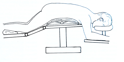
Laminectomy is a technique of removal of vertebral laminae to expose the content of the spinal canal. It is performed in a prone, sitting or lateral position (Fig. 11-2). When the prone position is used, the patient's body is laterally supported by elongated cushions or a special bridge, leaving a free space under the thoracic and abdominal cavities, not obstructing the respiratory movements and relieving congestion of the veins, which would increase the pressure in the veins of the spinal canal. In an operation on the cervical region the head is supported in a special headrest or the three-pin head holder.
The head and the upper part of the body are slightly elevated to decrease the venous pressure in the cervical region. For operations in the thoracic and lumbar region, with the patient in prone position, the head is turned to one side and the arms are extended and placed on both sides of the head. Care is taken for this not to be excessive and that the vessels and nerves in the axillar region are not compressed.
With the patient in the lateral position during laminectomy, many of the problems of the prone position are avoided. There is less compression on the abdominal veins resulting in minimal congestion inside the spinal canal, better control of the respiration, and blood and CSF naturally drain out of the operative field. The sitting position may also be used in operations on the cervical spine, and the conveniences of avoided venous bleeding and drained blood and CSF are obvious. This position presents some danger of brain ischaemia, air embolism, and quick drainage of CSF, which may lead to the accumulation of a subdural haematoma.
Anaesthesia. The opening of the spinal canal is almost always performed under general anaesthesia combined with infiltration of the soft tissues around the spine with local anaesthetic or saline mixed with adrenaline (1 mg to 400 ml).
The infiltration with local anaesthetic begins with an intradermal injection along the path of the incision. After that the subcutaneous fatty tissue and the paravertebral muscles are successively infiltrated over a larger area. This is accomplished by introducing the needle vertically until its tip reaches the resistance of the vertebral laminae. The injections are made at a distance of 2,5 - 3 cm on both sides of the spinous processes. The direction of the needle should be strictly vertical or with a slight downward inclination in order not to penetrate into the spinal canal. The use of a local anaesthetic considerably decreases the bleeding during the incision and helps the separation of soft tissues from the vertebrae.
Operative technique. In order to determine the level at which the operation is to be performed correctly, the following main reference points must be considered:
1. Spinous process of the VIIth cervical vertebra, which must be prominent and easy to palpate.
2. The horizontal line, which passes at the level of the inferior angle of the two scapulae, indicating the VIIth thoracic vertebra.
3. The line that passes through the iliac crests is situated at the level of the IVth lumbar vertebra.
4. X-ray film with a metallic marker in the presumed level of the pathology as a reference before incision.
5. X-ray film or fluoroscopic control during the surgery.
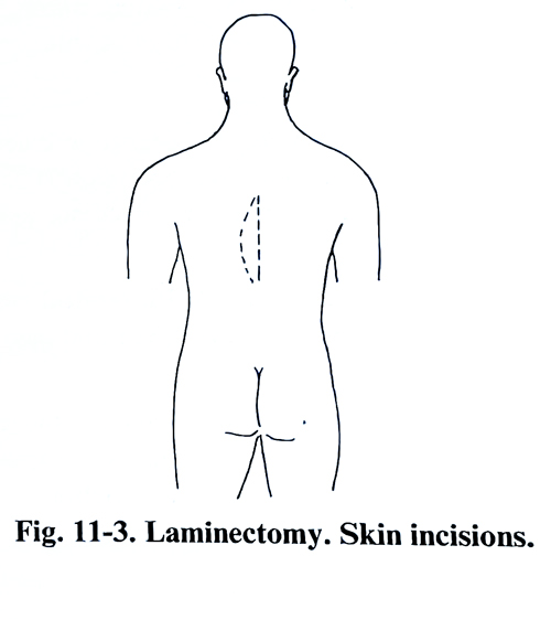
The skin incision is made along the midline over the spinous processes or in the shape of an arch, whose ends are on the midline; the convex part is separated 2 - 3 cm away from it (Fig. 11-3). The incision must be prolonged to expose one vertebra above and one below the place where the lesion is. With obese patients and those with well-developed muscles, the skin incision must be even longer. Together with the skin, the subcutaneous fatty tissue is also incised until the spinous processes are reached. The surface of the muscle fascia is dissected at a distance not more than 2 - 3 cm from the midline on both sides along the entire incision. The fascia and the tendinous attachments of the muscles are divided from the spinous processes as close as possible to the bone along the whole length of the skin incision. From this layer down diathermy can be used as a dividing tool and that reduces bleeding.
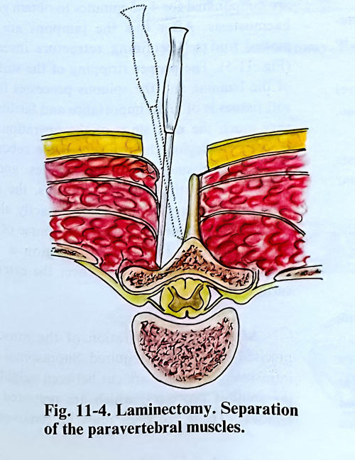
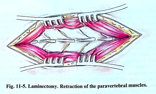
The next stage of the operation is separation of the paravertebral muscles from the spinous processes, the vertebral laminae and the ligaments situated between them. This separation is made with a periosteal elevator or plane chisel, in order to lift the muscle subperiostally (Fig. 11-4). Sometimes tendinous attachments of muscles, especially those in the lower part of the spinal column, are divided with a scalpel. The separation continues until the articular processes are reached. The separated muscles and the stripped surface of the laminae are packed for 4 - 5 minutes to obtain good haemostasis. After this the packs are removed and self-retaining retractors inserted (Fig. 11-5). The proper stripping of the surface of the laminae and the spinous processes from soft tissues is of great importance and facilitates very much the next stage of the operation. In the cervical region the incision of the subcutaneous fatty tissue exposes the muscles, and in order to reach the spinous processes, the surgeon must penetrate more deeply exactly along the midline and strictly follow the course of the nuchal septum. In the sacral region a few muscles and more ligaments cover the external surface of the sacrum. After a good separation of the muscles, precise haemostasis is required.
Supraspinal and intraspinal ligaments are cut between neighbouring spinous processes, which are prepared for removal. These processes must be removed as low as possible together with their bases, exposing the laminae and the flavum ligaments. Ligamentum flavum in the space between two laminae is opened until epidural fatty tissue appears. This opening is done at one of the ends of the exposed spine - above or below the lesion.
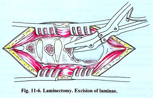
After that the opening is enlarged until it is possible to insert an angulated bone rongeur, and in small fragments remove the necessary number of laminae (Fig. 11-6). The resection of the laminae advances laterally until the joint processes are reached but without opening the joints. Always before removing the laminae the content of the canal (fat, dura, etc.) should be dissected away. When manipulating with the bone rongeur, the dural sac must always be protected from compression. In case of subdural tumours, when the dura is under pressure, the resection of the laminae is made on both sides of the lesion if possible. Last is removed the laminar part located over the compressed area of the spinal cord.
Bleeding from the resected bone is easily stopped with haemostatic wax. When the sacral canal is opened, the surgeon must begin from the lumbo-sacral interlaminar space or hiatus sacralis.
The epidural space is revealed under the laminae and ligamenta flava. It is occupied by friable fatty tissue with multiple venous vessels. This tissue is rather easily separated with any dissecting instrument and, if venous bleeding occurs, it is easily stopped by bipolar coagulation. In this way the dural surface is exposed along the entire length of the laminectomy. Before undertaking any further step, a thorough revision is made of the entire operative wound with a final haemostasis. The whole operative field, with the exception of the dura is covered with wet cottonoids, to prevent blood collection from structures external to the dura and to isolate it from the other tissues. The unchanged dura of the spinal canal is a brilliant grayish-blue layer with pulsations simultaneous with ventilator movements and the heart rate.
Before opening the dura the patient's head must be placed slightly lower than the level of the laminectomy in order to prevent the loss of large quantities of CSF. The revision of the epidural space can be completed by gentle palpation of the dural sac or with the ultrasonic probe.
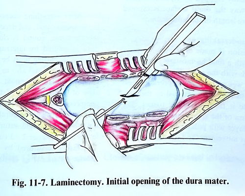
The dura is opened with a longitudinal incision on the midline, after elevating it with a fine sharp hook (Fig. 11-7). It is desirable that during dural opening, the arachnid, which has the tendency to protrude as a transparent bubble, be preserved. After a sufficiently large incision has been made a thin wet cottonoid is inserted under the dura in the direction of the continuing incision, serving as a protection from the scissors. The edges of the dura are lifted with traction sutures.
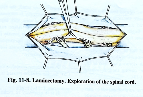
After that the spinal cord is inspected. The arachnoid is opened with a fine hook or with the tip of a scalpel and in normal conditions, CSF begins to leak immediately. The posterior surface of the spinal cord is inspected first, followed by the lateral, using a dissector for slight retraction. If this inspection uncovers a pathological process, situated anteriorly to the spinal cord, the exploration continues in greater detail. Two or three dentate ligaments are divided after lifting them with a hook. Slight elevation of the spinal cord by the free ends of the divided ligaments induces rotation of the spinal cord (Fig. 11-8). When there is a complete block of the subarachnoid space the amount of CSF that leaks is much less.
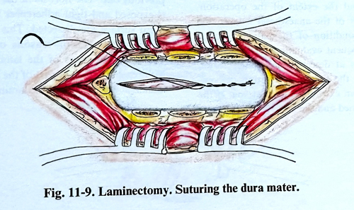
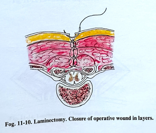
After everything necessary has been done in the spinal canal, and depending on the lesion. the operative wound is closed in layers. The dura is sutured by interrupted or continuous sutures. Its closing must be watertight to avoid leakage of CSF and also to prevent blood from entering the subarachnoid space (Fig. 11-9). An indication of good quality of the suture is when compression of the jugular veins does not evoke CSF leak between the dural edges. The muscles are closed by single sutures in two or three layers at a distance 2 - 3 cm one from another. The fascia is sutured by single sutures at a shorter distance (Fig. 11-10). The subcutaneous tissue and the skin are sutured last. As a cavity always remains between the dura and the deepest layer of sutured paravertebral muscles, haemostasis can be fragile, and vacuum drainage should be left there for 24 hours.
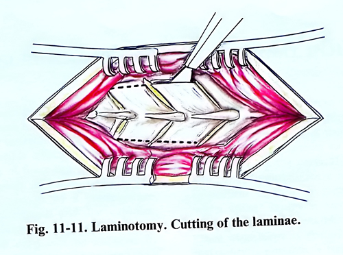
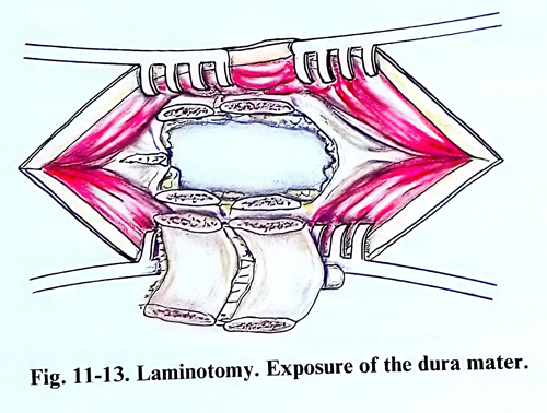
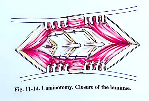
The spinal canal can be opened in an osteoplastic way (laminotomy), with cutting the laminae laterally with the the saw or a small drill, and at the end of the operation closing them by wiring (Figs. 11-11; 11-12; 11-13)
The postoperative care of patients, who have undergone a laminectomy varies according to the characteristics of the lesion, the localization and the extent of the operation. After the effect of the anaesthesia is over, the neurological condition of the patient is reassessed. Frequent evaluations in the following hours aims at detecting any deterioration and detecting the first manifestation of compression from an extradural haematoma. If the dura is not opened and the resection of the bone is comparatively small, the biomechanical effect of the laminectomy is minimal and the patient can get up and walk with support even on the day after surgery. After extended laminectomies, patients must remain in bed for several days to allow the paravertebral muscles to initiate the healing process before being loaded with normal activity.
Postoperative complications are rare. The CSF fistulae must be treated in the same way as after cranial operations.
In case of a second operation on the same site, the incision passes along the scar of the previous one. The fibrosis in the epidural space is exposed and tight adhesions to the external dural surface are dissected. This dissection can be difficult and, for reasons of safety, it is started at the edges of the laminectomy over unaffected dura, even with removal of small part of the intact adjacent lamina at the borders of the laminectomy.