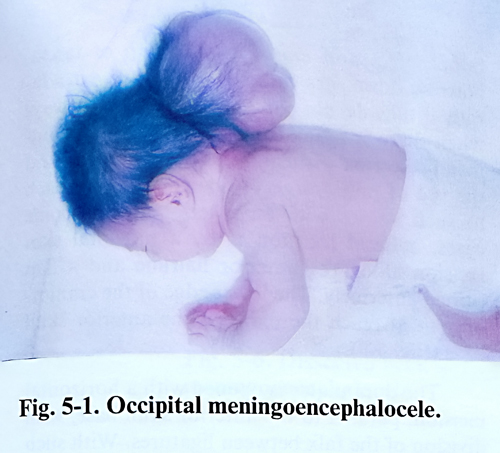
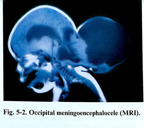
Cranium bifidum represents a congenital defect in the closure of the mesodermal layer covering the neural tube through which herniation of meninges and cerebrospinal fluid (meningocele) or meninges and brain tissue (encephalopcele) takes place. This is always at or near the middline (Figs. 5-1; 5-2).
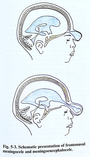
Referring mainly to their location, these lesions are subdivided into basal, sincipital, convexital, and atretic. Defects in the basal type are found in different areas, named after the structures affected. Basal encephaloceles can be sphenophayngeal, intranasal, sphenoorbital and sphenomaxillary. All basal types lesions are internal. Sincipital encephaloceles can be found in a similar way as nasofrontal, frontoethmoidal or nasoorbital. They are all located in the anterior and upper part of the forehead.
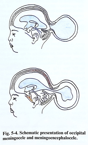
The most common group, however, is that of convexital lesions, and they can be frontal, parietal or occipital. This last subtype is very common and involves, separately or simultaneously, supra-, infratentorial and craniospinal structures (Figs. 5-3; 5-4).
The last group - atretic - comprise two of the more rarely encountered types that are smaller in size, in the parietal (for the 'alopecic) and the occipital (for the 'nodular') midline. They do not present any particular different surgical technical problems than do the convexity type. External encephaloceles are detected at birth, but the basal remain unrecognised until they become symptomatic with the presence of a pharyngeal or nasal mass, CSF rhinorrhea, or at hypertelorism or orbital mass investigation.
For surgical planning, detailed imaging studies are required in all three basic projections, as well as a careful local examination of the mass. This will clearly reveal the contents of the sac, its relation to the intracranial structures and the presence of ventricular dilatation or other congenital anomalies. Angiography is rarely needed, and it is performed preoperatively only when significant arterial or venous sinus compromise must be avoided. The size of menigocele or encephalocele is different and in rare cases surpasses the volume of the head. Occipital lesions tend to be the largest. The size of the bone defect can differ also, but ordinarily does not exceed a few centimetres. The aim of the operation is to close the defect. Operative treatment decreases the possibility of intrathecal infection, improves infant care and corrects the cosmetic defect. Surgical techniques include excision of the sac and repair of the anatomical layers in corresponding sequence - dura and epicranial tissue. Significant bone defects are covered with cranioplasty when necessary, but that can be done also at a late stage. Any coexisting hydrocephalus is treated before or simultaneously with the lesion by shunting.
The decision to operate depends on the risks related to the sac and the disfiguring effect it produces. Basal and sincipital meningoceles are repaired in conjunction with maxillofacial and plastic repairs of facial defects but the neurosurgeon precludes any ascending infection intrathecally by repairing the dural defect. Convexity encephaloceles are at times huge and associated with microcephaly, but surgery has to be considered in the great majority of them because:
1. The lesion interferes, especially if big, with infant care;
2. The lesion tends to expand with time, erode, rupture and bleed;
3. Some children are sensitive to the stimulation of the sac;
Development of hydrocephalus may create an additional difficulty in the treatment of the infant. External lesions are operated on as early as possible, but basal ones are not detected early and they are treated when the associated problems present clinically.
Hydrocephalus can have its onset at a later stage - in the postoperative period or later, and it is managed in accordance with the corresponding rules, usually with early shunting to avoid additional brain damage. Surgery is postponed if meningitis or local soft tissue infection has developed, and is only performed when clinical and laboratory data clearly indicate their complete control for more than 2 weeks.
Surgical planning contains the sequence of techniques to be applied in every individual case, and the most important factor to consider is whether or not there is a need to perform any plastic or maxillofacial procedure at the same session. At this stage the way to approach the area of the defect is decided with the expected line of amputation, and the proper tailoring and preservation of the tissue layers available to provide flaps for closure without wound edge tension. Following this principle, the incision is marked with the surgeon already having in mind a clear idea of how the flaps will be formed and turned at closure.
These are particular techniques related to the location and they vary to certain degree as shown further on.
ANTERIOR (SINCIPITAL) ENCEPHALOCELES
The exact diagnosis of the size and location of the bone defect and the relation of the hernia wall to the adjacent bone structures play a decisive role in the selection of the operative method. The bone defect in anterior lesions is most frequently found in the anterior cranial fossa near the foramen coecum. Beginning from there, the canal has a different exit according to the type of lesion. In the nasofrontal type the exit is situated exactly along the midline in the region of the nose root between the frontal and nasal bones. In frontoethmoidal cases the exit is a little more laterally, between the nasal process of the maxilla and the nasal bone. In the third group - the nasoorbital the canal of the herniation opens into the orbital cavity between the frontal, lacrimal and ethmoidal bones. The diameter of the entrance usually reaches 3.5 - 4.5 cm, with the external opening always being smaller than the internal.
The surgical methods for treatment of these types of lesions differ in the first stage of the operation - the approach to the area of the defect. This can be done either through the soft tissues overlying the defect or through a craniotomy. Accordingly, surgical methods applied in cases of anterior meningoencephalocele are divided into extracranial and intracranial.
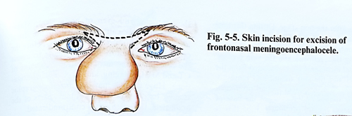
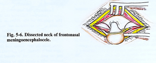
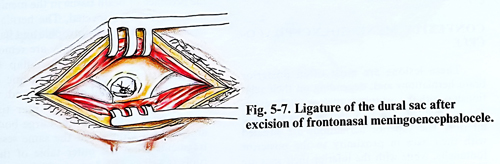
The extracranial methods are applied in cases of small bone defects and herniations. The shape and location of the skin incision depends on the size and the condition of the skin in the region. Frequently, a large part of the incision is made along the eyebrows to avoid visible scars on the face (Fig. 5-5). The excessive skin is resected, and the meningeal sac is dissected to the bone defect (Fig. 5-6). The meningeal sac is opened and its contents are carefully inspected, its neck is ligated at the bone defect level and is resected close to the ligature (Fig. 5-7). Small bone defects may be effectively closed by turning the edges of the periosteum inward and fixing them with a few sutures. For the same purpose in bigger defects, a bone graft or cranioplasty material may be used.
The intracranial methods require frontal craniotomy with either an extradural or intradural approach to the meningeal sac where the bone defect is reached. The dura is dissected inside the
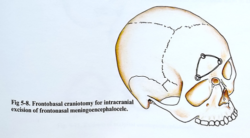
bone defect and closed with a flap of fascia. Narrow dural sacs can even be ligated extradurally if they do not contain brain tissue. Always when there is herniated brain tissue we recommend resection with an intradural approach and the use of microsurgery. The routine approach to such lesions with an anterior and, in some cases, a basal location is by a bifrontal skin incision along the anterior hairline and a flap turned anteriorly. The lower edge of the craniotomy must reach the plane of the anterior skull base (Fig. 5-8).
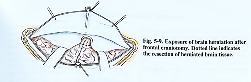
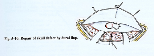
The dura mater is opened with a horizontal incision, parallel to the anterior skull base, with division of the falx between ligatures. With such malformations the falx can be found to be narrower than normal or even absent. The frontal lobes are retracted away from the defect, through which the meninges and the brain tissue protrude (Fig. 5-9). Arachnoid adhesions are torn and cysts opened and evacuated. The brain tissue protruding into the meningeal sac usually has a gliotic appearance, its consistency is harder than normal and must always be resected and removed at the bone defect level. Extradural ligation of the narrow dural neck is recommended. This requires extradural dissection of the neck immediately after the craniotomy. The neck of the herniation is well defined and ligated, dividing it distally from the ligation. This method is possible with a herniation whose neck is narrow and situated anteriorly. The advantage of this method is the watertight closure of the meningeal sack neck which and by that avoiding CSF leakage. If this is found to be impossible, the dura is opened and dissected from the anterior fossa, after which it is sutured again. The defect can also be closed without previous dissection of the dura from the anterior fossa. A dural flap from one of the orbital surfaces is folded along the midline and is sutured to the dura on the other side (Fig. 5-10). A flap of fascia can also be used. The sutures can be sealed with fibrin glue. The bone defect is repaired with a piece of bone taken from the temporal region or with cranioplasty material.
The plastic cosmetic repair can be done at a second stage. It is performed not earlier than 1- 2 months after the first stage. During this time the meningeal sac decreases its volume due to a lack of contents. The skin incisions are often made along the eyebrows and skin creases. The residual meningeal sac is dissected from the bone canal and the soft tissues and removed. Often the bone defect edges remain prominent anteriorly and cause widening of the base of the nose. The aesthetic correction of these changes is carried out within the same or another surgical session.
CSF leakage and subsequent meningitis are the most frequent and dangerous complications. This is explained by the close relationship of the meningeal sac with the lacrimal sac, nasal cavity, and paranasal sinuses where, as a result of the deformation, chronic infection frequently persists. For that reasons it is very important to make the closure of the dura watertight; antibiotic prophylaxis is also considered. Measures to reduce CSF pressure in the postoperative period might also be needed.
CONVEXITY MENINGOENCEPHALOCELE
These lesions are most often posterior brain herniations and depending on their relationship with the protuberantia occipitalis externa are divided into two groups: superior and inferior. The superior herniations are situated with their sacs in proximity to the posterior fontanelle, while with the inferior sacs are in proximity to the foramen magnum. The size of meningeal sacs and bone defects differ signifi-cantly. The content of the sack may be CSF, brain hemisphere or cerebellar tissue, parts of the ventricles and even of brain stem structure. Major venous channels can be related to the dural layer of the lesion. Because of all this, before the operation every effort must be made to assess the contents of the meningeal sac by means of CT and MRI - imaging, and by X-ray investigations. Huge lesions may require angiographic studies. In a case where it contains parts of the brain stem, the operation is contraindicated.
Technically the operation in convexity lesions is performed following the standard steps.
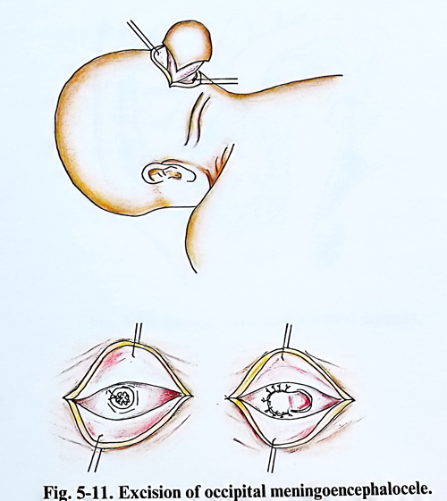
The exposure of the defect is done with an incision of the meningeal sac and soft tissues, taking care the incision lines to provide sufficient skin flaps to cover the defect without causing tension to the sutures (Fig. 5-11). The meningeal sac is dissected from the soft tissues and the bone defect edges, and is opened. The contents is evacuated and the neck is ligated or closed in a watertight manner with a dural flap to avoid CSF leakage through the operative wound. If brain tissue i present in the meningeal sac, it is caerfully inspected. The herniated brain tissue parts are usually atrophic and harder than the normal brain tissue; such contents is removed. The bone defect is closed with a flap from the pericranium and the subaponeurotic layer, taken from adjacent tisuues, which in many cases are thickened by scar tissue and allow easy defect closure. Large bone defects are closed at the same session with outer table of the convexital skull bone, as well as with bone grafts from elsewhere.
The postoperative complications and care are similar to those after surgery for meningomyelocele with any other cranial location.