Legacy: "Atlast of Neurosurgery" / L.Karaguiosov, A. Ramadan, K.Karaguiosov / Kiwait/ 1998
PREV < 4. Vascular Diseases of the Brain
5. Skull and Brain Malformations
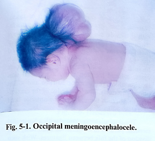
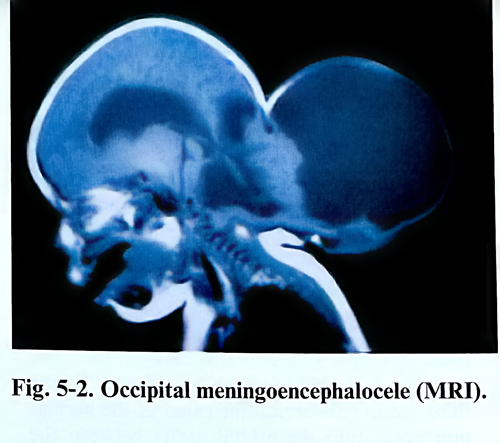
Cranium bifidum represents a congenital defect in the closure of the mesodermal layer covering the neural tube through which herniation of meninges and cerebrospinal fluid (meningocele) or meninges and brain tissue (encephalopcele) takes place. This is always at or near the middline (Figs. 5-1; 5-2).
Referring mainly to their location, these lesions are subdivided into basal, sincipital, convexital, and atretic. Defects in the basal type are found in different areas, named after the structures affected. Basal encephaloceles can be sphenophayngeal, intranasal, sphenoorbital and sphenomaxillary. All basal types lesions are internal. Sincipital encephaloceles can be found in a similar way as nasofrontal, frontoethmoidal or nasoorbital. They are all located in the anterior and upper part of the forehead.
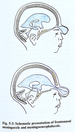
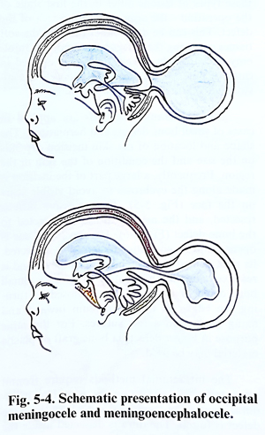
The most common group, however, is that of convexital lesions, and they can be frontal, parietal or occipital. This last subtype is very common and involves, separately or simultaneously, supra-, infratentorial and craniospinal structures (Figs. 5-3; 5-4).
The last group - atretic - comprise two of the more rarely encountered types that are smaller in size, in the parietal (for the 'alopecic) and the occipital (for the 'nodular') midline. They do not present any particular different surgical technical problems than do the convexity type. External encephaloceles are detected at birth, but the basal remain unrecognised until they become symptomatic with the presence of a pharyngeal or nasal mass, CSF rhinorrhea, or at hypertelorism or orbital mass investigation.
For surgical planning, detailed imaging studies are required in all three basic projections, as well as a careful local examination of the mass. This will clearly reveal the contents of the sac, its relation to the intracranial structures and the presence of ventricular dilatation or other congenital anomalies. Angiography is rarely needed, and it is performed preoperatively only when significant arterial or venous sinus compromise must be avoided. The size of menigocele or encephalocele is different and in rare cases surpasses the volume of the head. Occipital lesions tend to be the largest. The size of the bone defect can differ also, but ordinarily does not exceed a few centimetres. The aim of the operation is to close the defect. Operative treatment decreases the possibility of intrathecal infection, improves infant care and corrects the cosmetic defect. Surgical techniques include excision of the sac and repair of the anatomical layers in corresponding sequence - dura and epicranial tissue. Significant bone defects are covered with cranioplasty when necessary, but that can be done also at a late stage. Any coexisting hydrocephalus is treated before or simultaneously with the lesion by shunting.
The decision to operate depends on the risks related to the sac and the disfiguring effect it produces. Basal and sincipital meningoceles are repaired in conjunction with maxillofacial and plastic repairs of facial defects but the neurosurgeon precludes any ascending infection intrathecally by repairing the dural defect. Convexity encephaloceles are at times huge and associated with microcephaly, but surgery has to be considered in the great majority of them because:
1. The lesion interferes, especially if big, with infant care;
2. The lesion tends to expand with time, erode, rupture and bleed;
3. Some children are sensitive to the stimulation of the sac;
Development of hydrocephalus may create an additional difficulty in the treatment of the infant. External lesions are operated on as early as possible, but basal ones are not detected early and they are treated when the associated problems present clinically.
Hydrocephalus can have its onset at a later stage - in the postoperative period or later, and it is managed in accordance with the corresponding rules, usually with early shunting to avoid additional brain damage. Surgery is postponed if meningitis or local soft tissue infection has developed, and is only performed when clinical and laboratory data clearly indicate their complete control for more than 2 weeks.
Surgical planning contains the sequence of techniques to be applied in every individual case, and the most important factor to consider is whether or not there is a need to perform any plastic or maxillofacial procedure at the same session. At this stage the way to approach the area of the defect is decided with the expected line of amputation, and the proper tailoring and preservation of the tissue layers available to provide flaps for closure without wound edge tension. Following this principle, the incision is marked with the surgeon already having in mind a clear idea of how the flaps will be formed and turned at closure.
These are particular techniques related to the location and they vary to certain degree as shown further on.
ANTERIOR (SINCIPITAL) ENCEPHALOCELES
The exact diagnosis of the size and location of the bone defect and the relation of the hernia wall to the adjacent bone structures play a decisive role in the selection of the operative method. The bone defect in anterior lesions is most frequently found in the anterior cranial fossa near the foramen coecum. Beginning from there, the canal has a different exit according to the type of lesion. In the nasofrontal type the exit is situated exactly along the midline in the region of the nose root between the frontal and nasal bones. In frontoethmoidal cases the exit is a little more laterally, between the nasal process of the maxilla and the nasal bone. In the third group - the nasoorbital the canal of the herniation opens into the orbital cavity between the frontal, lacrimal and ethmoidal bones. The diameter of the entrance usually reaches 3.5 - 4.5 cm, with the external opening always being smaller than the internal.
The surgical methods for treatment of these types of lesions differ in the first stage of the operation - the approach to the area of the defect. This can be done either through the soft tissues overlying the defect or through a craniotomy. Accordingly, surgical methods applied in cases of anterior meningoencephalocele are divided into extracranial and intracranial.
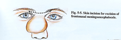
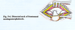
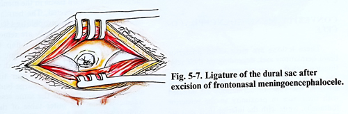
The extracranial methods are applied in cases of small bone defects and herniations. The shape and location of the skin incision depends on the size and the condition of the skin in the region. Frequently, a large part of the incision is made along the eyebrows to avoid visible scars on the face (Fig. 5-5). The excessive skin is resected, and the meningeal sac is dissected to the bone defect (Fig. 5-6). The meningeal sac is opened and its contents are carefully inspected, its neck is ligated at the bone defect level and is resected close to the ligature (Fig. 5-7). Small bone defects may be effectively closed by turning the edges of the periosteum inward and fixing them with a few sutures. For the same purpose in bigger defects, a bone graft or cranioplasty material may be used.
The intracranial methods require frontal craniotomy with either an extradural or intradural approach to the meningeal sac where the bone defect is reached. The dura is dissected inside the
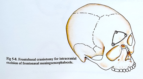
bone defect and closed with a flap of fascia. Narrow dural sacs can even be ligated extradurally if they do not contain brain tissue. Always when there is herniated brain tissue we recommend resection with an intradural approach and the use of microsurgery. The routine approach to such lesions with an anterior and, in some cases, a basal location is by a bifrontal skin incision along the anterior hairline and a flap turned anteriorly. The lower edge of the craniotomy must reach the plane of the anterior skull base (Fig. 5-8).
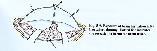
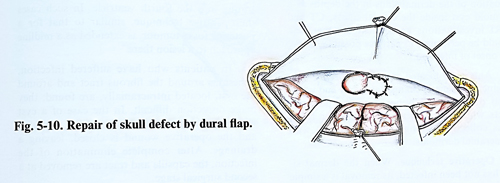
The dura mater is opened with a horizontal incision, parallel to the anterior skull base, with division of the falx between ligatures. With such malformations the falx can be found to be narrower than normal or even absent. The frontal lobes are retracted away from the defect, through which the meninges and the brain tissue protrude (Fig. 5-9). Arachnoid adhesions are torn and cysts opened and evacuated. The brain tissue protruding into the meningeal sac usually has a gliotic appearance, its consistency is harder than normal and must always be resected and removed at the bone defect level. Extradural ligation of the narrow dural neck is recommended. This requires extradural dissection of the neck immediately after the craniotomy. The neck of the herniation is well defined and ligated, dividing it distally from the ligation. This method is possible with a herniation whose neck is narrow and situated anteriorly. The advantage of this method is the watertight closure of the meningeal sack neck which and by that avoiding CSF leakage. If this is found to be impossible, the dura is opened and dissected from the anterior fossa, after which it is sutured again. The defect can also be closed without previous dissection of the dura from the anterior fossa. A dural flap from one of the orbital surfaces is folded along the midline and is sutured to the dura on the other side (Fig. 5-10). A flap of fascia can also be used. The sutures can be sealed with fibrin glue. The bone defect is repaired with a piece of bone taken from the temporal region or with cranioplasty material.
The plastic cosmetic repair can be done at a second stage. It is performed not earlier than 1- 2 months after the first stage. During this time the meningeal sac decreases its volume due to a lack of contents. The skin incisions are often made along the eyebrows and skin creases. The residual meningeal sac is dissected from the bone canal and the soft tissues and removed. Often the bone defect edges remain prominent anteriorly and cause widening of the base of the nose. The aesthetic correction of these changes is carried out within the same or another surgical session.
CSF leakage and subsequent meningitis are the most frequent and dangerous complications. This is explained by the close relationship of the meningeal sac with the lacrimal sac, nasal cavity, and paranasal sinuses where, as a result of the deformation, chronic infection frequently persists. For that reasons it is very important to make the closure of the dura watertight; antibiotic prophylaxis is also considered. Measures to reduce CSF pressure in the postoperative period might also be needed.
CONVEXITY MENINGOENCEPHALOCELE
These lesions are most often posterior brain herniations and depending on their relationship with the protuberantia occipitalis externa are divided into two groups: superior and inferior. The superior herniations are situated with their sacs in proximity to the posterior fontanelle, while with the inferior sacs are in proximity to the foramen magnum. The size of meningeal sacs and bone defects differ signifi-cantly. The content of the sack may be CSF, brain hemisphere or cerebellar tissue, parts of the ventricles and even of brain stem structure. Major venous channels can be related to the dural layer of the lesion. Because of all this, before the operation every effort must be made to assess the contents of the meningeal sac by means of CT and MRI - imaging, and by X-ray investigations. Huge lesions may require angiographic studies. In a case where it contains parts of the brain stem, the operation is contraindicated.
Technically the operation in convexity lesions is performed following the standard steps.
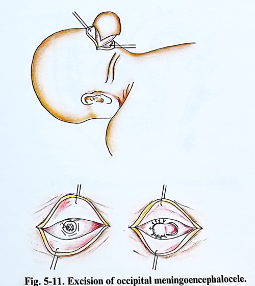
The exposure of the defect is done with an incision of the meningeal sac and soft tissues, taking care the incision lines to provide sufficient skin flaps to cover the defect without causing tension to the sutures (Fig. 5-11). The meningeal sac is dissected from the soft tissues and the bone defect edges, and is opened. The contents is evacuated and the neck is ligated or closed in a watertight manner with a dural flap to avoid CSF leakage through the operative wound. If brain tissue i present in the meningeal sac, it is caerfully inspected. The herniated brain tissue parts are usually atrophic and harder than the normal brain tissue; such contents is removed. The bone defect is closed with a flap from the pericranium and the subaponeurotic layer, taken from adjacent tisuues, which in many cases are thickened by scar tissue and allow easy defect closure. Large bone defects are closed at the same session with outer table of the convexital skull bone, as well as with bone grafts from elsewhere.
The postoperative complications and care are similar to those after surgery for meningomyelocele with any other cranial location.
DERMAL SINUS OF THE SKULL
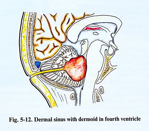
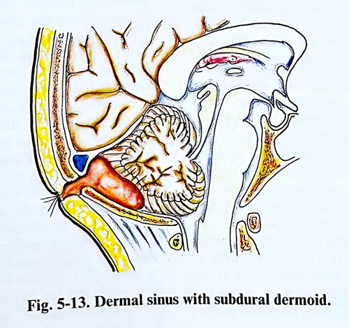
This abnormal communication between the skin and the intracranial space may traverse all anatomic layers or only a few of them. It can have the same location as do cranial encephaloceles. The main problem with this type of pathology is its role as a path for infection, formation of cysts or dermoids along its path. The option for treatment is its excision (Figs. 5-12; 5-13).
The removal of a dermal sinus should be performed when the infection is well controlled or before it has appeared. CT and MRI investigations are indicated to understand the eventual continuation of the dermal sinus into the deeper layers of tissues and structures, as it can be the case of a dermoid cyst inside the cranial cavity. Operation should be performed as early as possible, before the onset of infection.
Anteriorly located sinuses are occult (not patent), only producing cosmetic changes and they end in the basal dura or falx, opposite to the posterior tracts which often cause meningitis or infectious collections along the tract, or even end in an intracranial dermoid.
Operative technique. When the dermal sinus has not been infected, its removal is a simple excision. An elliptical incision is made around the skin changes in a vertical direction, which encloses the dermal sinus, and the fibrous tract is dissected into the depth to its end. Its extirpation concludes when it reaches the bony surface and ends there. However, if it continues and penetrates into the cranial cavity, the bone defect has to be adequately widened to inspect the pathological changes in the cranial cavity as well. In some cases the fibrous cord-like pathologica structure ends in the dura or is broadened into a dermoid cyst situated extradurally, intracerebellarly or penetrating into the fourth ventricle. In such cases the operative technique, similar to that for a posterior fossa tumour, is extended as a midline approach to a lesion there.
In patients who have suffered infection, adhesions around the fibrous tract and around the cyst, located intracranially are found; they make dissection difficult. In the case of a cyst with purulent contents, the latter is aspirated and the operative wound is closed, leaving a drainage. After complete elimination of the infection, the capsule and tract are removed at a second surgical procedure.
ARACHNOID CYSTS
These membrane-delineated collections contain fluid which in general is indistinguishable from CSF, and their walls are similar in structure and continuous to the surrounding arachnoid. They can be primary, without a known aetiology, congenital or secondary, as a result of trauma, surgery or haemorrhage. Primary cysts are often found located in the areas of normal cisterns.
Part of the arachnoid cysts tend to enlarge and exert pressure upon the surrounding structures, which results in an increased intracranial pressure either by the effect of the mass lesion, or by obstructing CSF outflow and producing hydrocephalus. With the advent, however, of the CT and MRI imaging, the number of accidentally discovered arachnoid cysts now appears to be significant. These clinically silent lesions are not indicated for surgery and deserve only follow-up imaging.
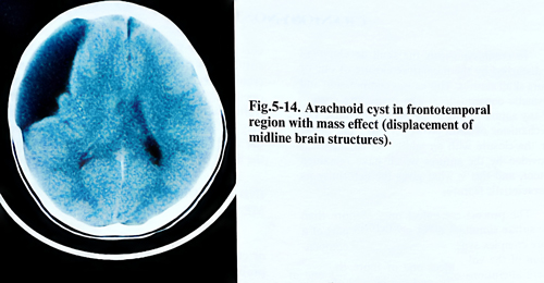
Presurgical work-up contains imaging of the lesion, with the best possible demonstration of its relationships with the surrounding structures (Fig.5-14). MRI has the advantage of giving more information about the contents of the cyst by measuring intensities of the fluid. For this last determination, there is no need to inject water-soluble contrast into the CSF spaces except in extremely particular circumstances.
Surgery must drain the fluid and prevent expansion of the lesion, tentatively forever. This is possible with a good percentage of success with the available three main surgical techniques: by fenestration of the cyst to adjacent cisterns by open surgery, by endoscopy or by drainage with a shunting device to the peritoneum. Up to the present no unanimous opinion exists concerning the choice of a method.
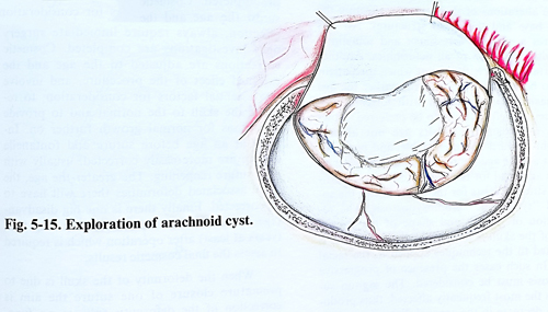
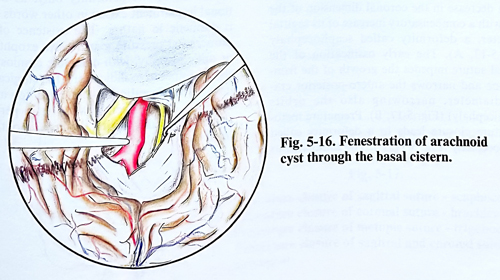
Fenestration of the cyst is made as wide as possible to their surrounding normal CSF spaces. The cyst is approached with a limited, but sufficient in size craniotomy (Fig.5-15). It is opened widely and as much as possible for that part of the wall which is unattached to brain structure and that part of the wall is excised. The places close to the basal cisterns are fenestrated under magnification and broad communication with all surrounding normal CSF spaces is established (Fig. 5-16). This is usually demonstrated by the pulsation (simultaneously with the ventilator movements and pulse) of the CSF level inside them. Haemostasis should be particularly strict because of the huge residual spaces left at the end of the procedure; the dura should be closed with special attention to water-tightness.
More recent advances in minimally invasive neurosurgery and the application of ventriculoscopy have made possible the endoscopic fenestration. A burr hole entrance to the cyst makes the exploration of the cyst and its multiple fenestration a less invasive procedure. The long-term advantage of the method still needs to be proved.
Shunting is the easiest option and is preferred by many surgeons. A shunting device is inserted to allow the cyst cavity to communicate (usually) with the peritoneum. If the cyst cavity is isolated from the CSF spaces, then to achieve its evacuation, it is drained using a device with an opening pressure that is lower than that of the intraventricular spaces. The absence of any constant flow through the valve is probably a factor which leads to more frequent shunt malfunction than in other cases. Hydrocephalus associated with a cyst also requires shunting and is common. Because of the existence of two compartments to be drained, if a second drainage tube exists in the cyst the opening pressure for the ventricular compartment should be slightly higher than that of the cystic compartment.
In general, cysts have some tendency to recur, regardless of the choice of method. We consider the choice to be related more to the skills and experience of the surgeon, and that determines the rate of complications to some extent. A thorough follow – up with imaging, therefore is a must for the further management of the patient after surgery.
CRANIOSYNOSTOSIS
In craniosynostosis the skull development is disturbed by the premature closure of one or more skull sutures. This causes compression and cosmetic consequences. The premature closure of any specific suture results in a typical deformity. The mechanisms of skull growth try to compensate for the closure with an additional enlargement provided by the sutures which have remained patent, and this is what gives the deformity its characteristic features.
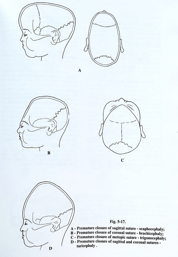
The process can affect one or more than one suture simultaneously, and can be part of a more complex syndrome. The need for compensation of the volume results in additional more severe alterations of skull growth, affecting the skull base and the facial structures. All this requires a complex assessment and sometimes stepwise correction of all deformities. Another important factor is the age at which the patient has to be treated. As a rule the earlier the age the easier the correction, with fewer consequences; therefore this has to be preferred. However, the cosmetic results are not always straight-forward and predictable, and reappraisals at a later age are recommended in order to assess any need for further corrections. A particular strategy exists for the treatment of the so-called "syndromic" craniosynostosis. The restoration of growth and shape of the cranial part of the skull is part of the treatment and is adjusted to the techniques applied to the facial part. In such cases the presence of progressive proptosis must be considered. The sagittal suture is the most frequently affected, thus producing a decrease in the coronal dimension of the skull with a compensatory increase of its sagittal proportion, a deformity called scaphocephaly (Fig. 5-17, A). The early ossification of the coronal suture impedes the growth of the frontal bone and reduces the antero-posterior cranial diameter, narrowing also the orbits (brachicephaly) (Fig. S-17, B). Premature metopic suture closure leads to a deformity called trigonocephaly (Fig. 5-17, C). The simultaneous closing of the sagittal and coronal sutures or all sutures sharply reduces the volume of the cranial cavity. If the anterior fontanelle remains open longer than usual, the skull grows upwards (turricephaly) (Fig, 5-17, D). A skull deformity may be accompanied by facial malformation in the syndrome of Cruson or by malformation of the limbs in the syndrome of Appert.
INDICATIONS FOR SURGICAL TREATMENT
All the existing problems - functional and/or cosmetic - must first be defined. Functional problems such as increased intracranial pressure, compression, exophthalmos or optic nerve affection, always require immediate surgery once investigations are completed. Cosmetic corrections are adjusted to the age and the expected effect of the procedure, and involve two essential factors for consideration: to reshape the skull to the normal and to provide conditions for normal growth further on. Infants at an age before suture and fontanelle closure are more easily corrected, usually with only suture reopening. The greater the age, the more associated deformities will have to be corrected. Finally, there is one big disadvantage to this surgery: it is the very long time (years at least) after operation which is required to assess the final cosmetic results.
When the deformity of the skull is due to premature closure of one suture the aim is correction of the deformity only, as no functional impairment exists; in other words, surgery is cosmetic in nature. The existence of raised intracranial pressure, progressive exophthalmos or optic atrophy, with complete craniosynostosis are imperative indications for surgical treatment. These surgical corrections are indicated at all ages, but it is most effective when it is performed earlier, preferably during the first 3 months after birth.
URGICAL TECHNIQUE
Many operations are recommended for the surgical treatment of craniosynostosis, but linear craniectomies 1,5 - 2 cm width running along the different sutures or in other directions are still applied. As the bone gaps tend to close again, the bone edges may be lined with impermeable synthetic material to retard reunion. There are also multiple variations for the width and the way to perform this technique. Suture reopening, where the case is not associated with other deformities, is effective up to the age of few months or so.
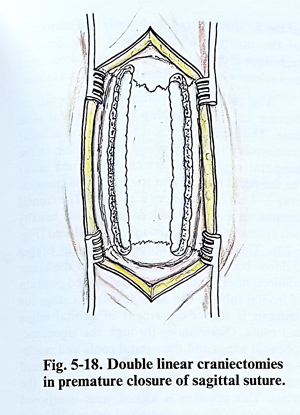
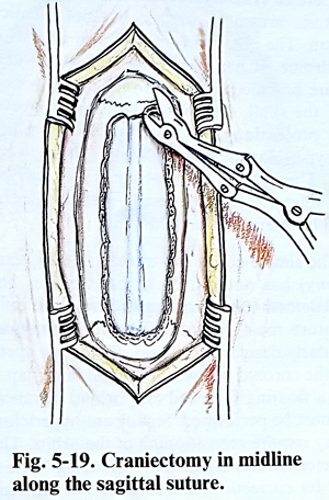
Sagittal craniosynostosis. For linear craniectomy of the sagittal suture, the patient is placed on the operative table in a supine, prone or lateral position with the head slightly elevated. The incision of the scalp is delineated exactly along the midline, beginning in the frontal region from the limit of the hairy skin and running to a point 3 - 4 cm behind the lambdoid suture. The skin, the subcutaneous tissue and galea aponeurotica are incised and are retracted together laterally 3 - 4 cm from the midline. The periosteum is also retracted and excised from the exposed bone surface. Two straight lines two centimetres on each side and parallel to the midline from the coronal to the lambdoid suture are marked. Burr holes are made at the ends of each line, and the bone between them is resected with a nibbler to produce a linear craniectomy 1,5 - 2 cm wide. The craniectomy must reach beyond the coronal and the lambdoid sutures at least 2 cm on each end, i.e. to also involve the frontal and occipital bones (Fig 5-18). Careful haemostasis is maintained throughout and at the end of the resection. As alternative, a single but wider craniectomy can be made in midline along the closed sagittal suture (Fig.5-19). The dura is fixed to the pericranium with single stitches either directly, or through nonabsorbable plastic material, wrapping the bony edges. Closure of epicranial tissues is as usual.
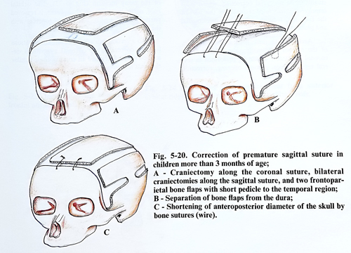
After the age of 3 months, scaphocephaly requires more extensive surgery, its aim being reduction of the antero-posterior diameter and separation of both parietal flaps (lateral expansion). As in all corrective surgery except for the minor ones, the skin incision is bitemporal. Bone segments are lifted on both sides of the sagittal suture including a small part across the lambdoid suture and a third big segment includes the coronal suture from temporal to temporal. Any sagittal bony ridge is smoothed out and the bony edge over the sagittal area is fixed to the frontal bone with non-absorbable sutures thereby shortening the antero-posterior diameter. Parietal flaps are trimmed to reshape the skull and sutured, remaining fixed with a thin bridge to the temporal bone (Fig. 5-20). Closure is as usual.
Elder children (1,5 - 2 y or more) need correction with multiple bone flaps on a rather individual basis, shortening and expanding different segments to remodel the skull.
Coronal craniosynostosis. Uni- and bilateral synostosis present a similar, but not identical problem. The bilateral form can also be part of a syndrome. It has several main features: children are brachicephalic with bitemporal widening of the skull, they have some degree of ocular protrusion due to orbital foreshortening with hypertelorism; the skull tends to be "towering". The onset of the problem is very early in life. Unilateral forms present with ipsilateral frontal bone and orbital flattening.
The simple coronal suturectomy (strip craniectomy) has not proven to be effective. The several deformities coexisting with the closure of this suture must be corrected with techniques particularly designed for them. Following opening of the coronal suture, the frontal bone flap is left as a floating forehead and orbital advancement must be performed. Significant hypertelorism may require repositioning of the orbits. The same refers to any significant "towering" deformities. Its correction can be effective with circumferential craniotomies, preferably done in stages, and under ICP monitoring. The last stage can be midfacial correction, which must improve the associated retrusion of this part of the face, and it is undertaken by maxillofacial and plastic surgeons.
According to these general characteristics of the related deformities, the surgery for a coronal synostosis should be performed in several regular steps: coronal opening, correction of the forehead (frontal squama) and correction of the position of the orbital rims. Surgical technique intends to combine these components and selectively adjust them for the patient's age.
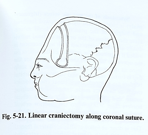
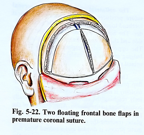
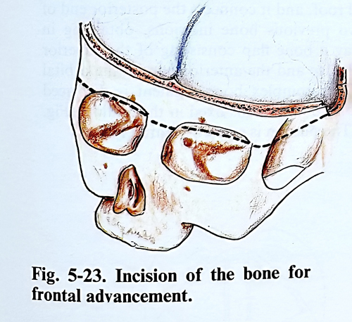
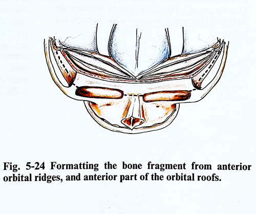
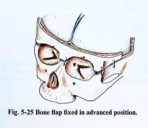
The incision is invariably bitemporal, reflecting an epicranial flap to the frontal skull base. The periosteum is separated and a linear craniectomy is made, whose ends remain in the temporal region, and which is widened under the temporal muscle as a round shaped craniectomy 3-4 cm in diameter (Fig. 5-21). The frontal squama must be moved forward, and usually two symmetrical frontal flaps are produced (Fig. 5-22). They can be left floating or in bigger children bone strips are interposed in the linear craniectomies to maintain the advanced position of frontal tubera. The most important stage for cosmesis is the advancement of orbital edges and roofs. Osteotomies through the zigomatic, sphenoid wings and the orbital roofs and reaching the superior orbital fissure allow for forward repositioning of the superolateral part of the orbits. This position is locked with interposed bone strips in the zigomatic and sphenoidal part of the bony incision (Fig. 5-23). The orbital roof, naturally shorter in these cases, is removed partially or part of it is advanced together with the orbital edges, the usual advancement not exceeding 10-15 mm (Figs. 5-24; 5-25).
To a certain extent, the different surgical techniques for the correction of coronal synostosis described in the literature all contain these common steps mentioned above.
The unilateral synostosis of the coronal suture produces a unilaterally flat or concave forehead involving also the superior orbital edge. The nose is deviated towards the affected side and there is exophthalmos on the same side. The opposite side compensates with frontal bulging and less detectable participation of the orbital structures.
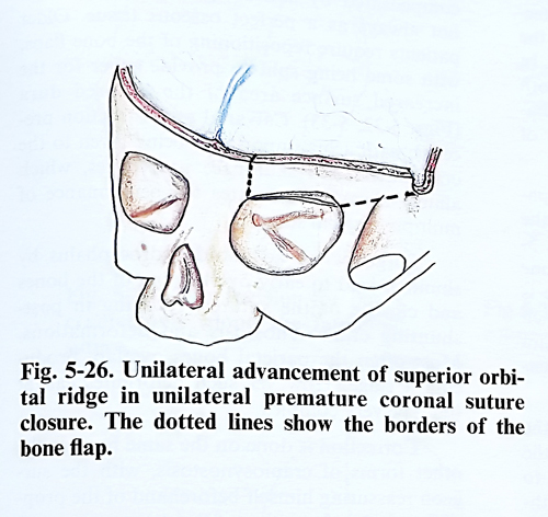
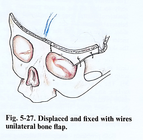
The surgical technique consists of a scalp flap produced by a bicoronal incision. A craniotomy is made medially to the midline, posteriorly reaching the coronal suture and laterally coming near to the pterion. After excision of the pterion, the orbital roof is resected in coronal plane, reaching the crista galli. The frontozigomatic process is divided and a second bone flap is produced with the pedicle containing the superior orbital border (Fig. 5-26). This second flap is displaced anteriorly and remains fixed with its medial end to the nasal root (Fig. 5-27). This fragment's curvature should produce an additional anterior convexity to correct the deformity.
The suegical technique can be performed in the same way as in bicoronal synostosis, but the frontal flaps and orbital edges are treated differently (one is advanced and the other is moved posteriorly if needed after been independently mobilized).
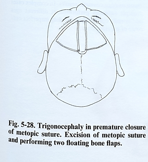
Synostosis of the metopic suture (trigonocephaly) is characterised by a narrowed forehead and bulging of the prematurely closed suture. Here the position of the orbit is assessed individually and their correction with advancement is added if required.
The position of the patient on the operative table and the skin incision are the same as in coronal craniosynostosis. The scalp flap is turned anteriorly and the superior orbital edges are stripped. Using linear craniectomies two bone flaps are made, removing the bulging suture, and they are fixed only on the orbital borders (Fig. 5-28).
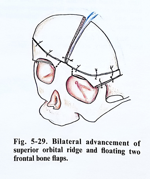
When the superior orbital borders are small and displaced posteriorly the "floating forehead" technique may be applied. This is done as follows: after a bilateral frontal craniotomy both frontal lobes are retracted extradurally to the crista galli. The orbital roofs are incised near and parallel to the crista galli starting at the orbital edge. Next the orbital roof is divided along the frontozigomatic suture from the cantus. A third section is transversal to the orbital roof, and it connects the posterior end of the two previous bone incisions, obtaining in this way a bone flap consisting of the superior orbital edge and the anterior part of the orbital roof. This complex bone segment is displaced anteriorly and remains fixed in the midline (Fig.5-29). The pterion is excised separately.
Synostosis of the lambdoid suture. In a case of unilateral lambdoid synostosis, the affected side of the skull is flattened in the occipital area. The auricle on the same side is situated posteriorly to the normal position, and the ipsilateral frontal bone can be found bulging. In bilateral synostosis the flattening affects both sides of the skull and the antero-posterior diameter is diminished with the posterior part of the skull also narrowed transversally.
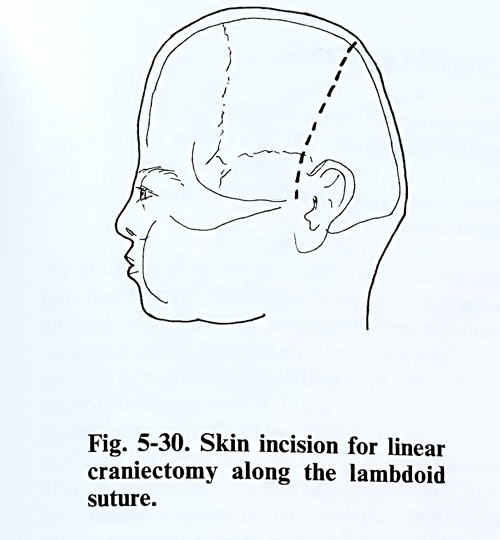
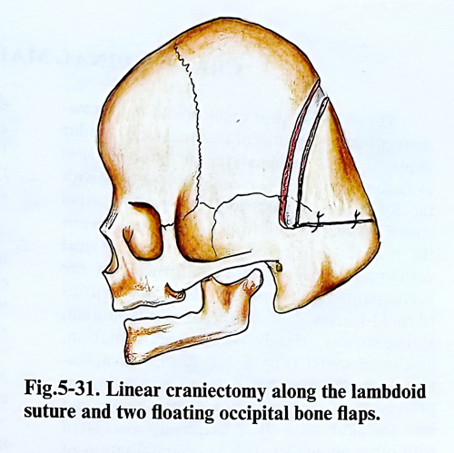
For lambdoid suturectomy, with the patient in a prone position, the incision of the scalp is made over the lambdoid suture (Fig. 5-30). The periosteum is stripped from the bone surface and a craniectomy 1.5 - 2 cm wide is made precisely to include the suture, and reach the asterion point. If the surgery has to attain more than this simple correction, two “floating” bone flaps can be made (Fig. 5-31) .
Both problems - occipital flattening and the associated deformities - must be treated at the same time. The closed lambdoid suture has to be reopened with a suturectomy, but the method is effective only in early infancy. At a later age the condition is evaluated individually and calvarial transfers are needed for the correction of the skull's shape. In more severe cases cranio-facial techniques are also needed.
In total craniosynostosis, when there are signs of raised inracranial pressure, linear craniectomies should provide the necessary expansion and accommodation of the intracanial contents.
Such total forms are seen in the "clover head" deformity, acrocephaly, and oxycephaly, which are included in the so-called syndromic forms.
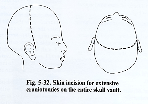
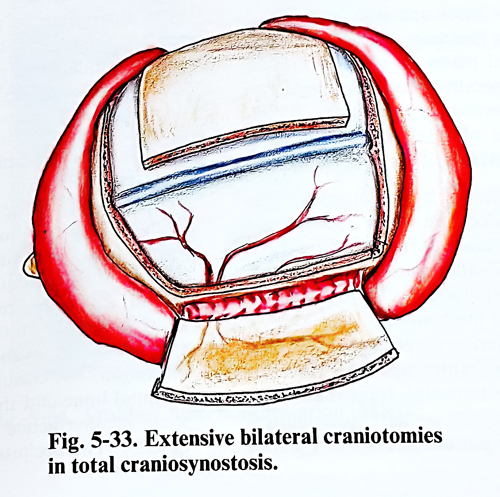
Extensive removal and/or repositioning of bone is required. In young infants the bone removal is compensated by active regeneration, although not always with a perfect osseous tissue. Older patients require repositioning of bone flaps, with some being split to provide cover for the increased surface area of the exposed dura (Figs. 5-32; 5-33). Calvarial reconstruction precedes , followed by the craniofacial and skull-base procedures, which almost always need the performance of multiple surgical steps.
Surgeries for advanced hydrocephalus by shunting lead to early overlapping of the bones and closure of the sutures, resulting in post-shunting craniosynostosis and deformations. Most often the parietal bones overlap, producing a ridge. However, such deformities can at times be very complex. Correction is done on the same basis as in the other forms of craniosynostosis, with the surgeon reassuring beforehand the proper function and position of the shunt.
Complications are rare in the postoperative period. The extensive potential space in these types of surgery can lead to accumulation of huge haematomas, what for an infant can be a serious treat of shock and acute posthaemorrhagic anaemia. Later complications have a wide range of variety and are associated to early bone union or poor cosmetic results. In complex cases multiple surgeries performed at different ages additionally correct the preceding imperfect outcome of the previous ones.
CRANIOSPINAL MALFORMATIONS
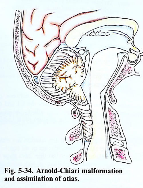
The most frequent craniospinal malformations, which need surgical treatment are: basilar impression and invagination, the different types of fusion of the first two cervical vertebrae with the foramen magnum, and the pathological effects which result from dislocation between the occipital bone and the first two cervical vertebrae. Mani of these malformations are accompanied by nervous tissue deformities (Arnold-Chiary malformation, syringobulbia, syringomyelia, Dandy-Walker malformation, arachnoid cysts) (Fig. 5-34).
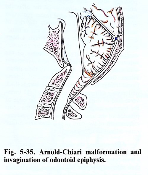
Basilar invagination is a developmental defect consisting of protrusion of the vertebral column's upper end into the skull base. It is frequently associated with other anomalies such as occipitalisation of the atlas, defective fusion and block vertebrae (Fig. 5-35). The incidence of associated neural malformation is also high.
Basilar impression is a secondary phenomenon due to softening of the bone tissue, as osteomalacia, hyperparathyroidism, Paget's disease of the bone, osteogenesis imperfecta and so on.
Platibasia expresses the presence of an obtuse angle between the planes of the anterior skull base and the clivus, but no dysfunction can by attributed directly to it; it occurs in association with basilar invagination.
The treatment of these type of lesions depends on the reducibility of the lesion, the mechanisms of compression and their direction, the aetiology of the lesion and associated neural anomalies, and the presence of ossification centres and epiphiseal plates in some congenital conditions. The surgical treatment should be based on an understanding of craniocervical dynamics, the site of involvement of neural elements and the stability of all the segments.
The primary goal of treatment is to obtain reduction of the dislocation of the craniovertebral junction if such exists. If the lesion proves to be reducible, stabilisation must follow. When the lesion remains irreducible, a thorough analysis must identify the mechanism of compression- its direction (anterior or posterior) and the influence of biomechanics upon it. Anterior decompressions are transoral, transpalatal, or lateral extrapharyngeal; posterior decompressions are midline or posterolateral. The effect of decompression upon stability must be considered as well in arriving at a choice of technique.
In this way every treatment consists of these three components; reduction, decompression, stabilisation.
Neuroradiological investigations must demonstrate the position of all craniobasal and craniocervical structures, as well as the medulla with the upper cord. In selected cases, an angiography may be required for evaluating the patency of the vertebral arteries. Craniospinal structures are visualised well on plain X-rays; multiplane conventional CT scans also provide excellent axial visualisation, which can be combined with intrathecal contrast, as these studies can also provide functional i.e. dynamic imaging of the segment. This is obtained with separate images in flexion, extension, and even under traction.
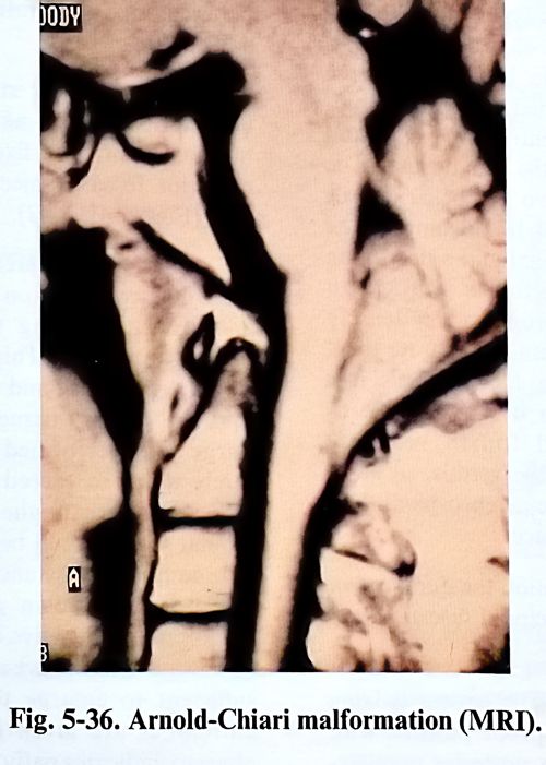
Recently such dynamic studies can also be obtained with the MRI. Otherwise, MRI remains the best means of neural imaging of this area (Fig. 5-36). Sometimes, when MRI is unavailable or impossible, the remaining option is CT with intrathecal contrast. All investigations should answer the questions: is there a need of reduction, is there instability, is there compression and what is its site and mechanism?
All cases in need of reduction, are put on traction, at present with a 'Halo' device. The effect of the traction is evaluated and if found to be sufficient in reposition, the patient is stabilised, most often by a posterior approach. That can be combined with posterior decompression in irreducible cases or preceded by an anterior decompression if the mechanic effects are from that direction.
Operative technique. Posterior decompression. The operation is performed with the patient in a sitting or prone position, the head being fixed in a pinholder or under traction. Placing the patient on the operating table can be difficult due to the reduced flexion in the upper (axial) cervical spine. It is recommended to have X-ray monitoring (C-arm) at the operating table to avoid distension and even dislocation after muscle relaxation. The closure of the space between the occipital bone and the cervical vertebrae causes some inconvenience in dissection of the neck muscles. The occipital bone is less convex and can even be concave, creating difficulties in dissection and drilling. The foramen magnum's posterior edge is also deeper and hidden, and when there is occipitalization of the atlas the osseous part to be removed is rather particular in shape.
With such an anomaly the risk of vertebral artery damage must be kept in mind, as it enters the cranial cavity through a bony canal in the osseous block of the foramen magnum and atlas. The laminectomy of the superior cervical vertebrae is also associated with some difficulties, as a result of their defor-mities, thickening or the presence of defects (spina bifida). In many patients the dura is thick and closely adherent to the bone. A tight transverse fold of dura can be found at the edge of the foramen magnum, contributing to the compression of the neural structures. The hypervascularization of the dura in this area also causes some difficulties in opening it.
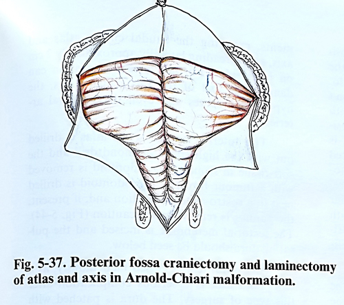
After opening the dura, the cerebellar tonsils are usually found elongated and herniated at a level below the foramen magnum (Fig. 5-37). If their inferior poles are not exposed, the laminas of one or two vertebrae caudally are additionally opened. The cleft between the two tonsils is opened and the foramen Magendie, cranial nerves and visible cord are identified along with their elements; the different types of malformative structural changes can also be detected. If the foramen of Magendie is closed with a membrane, it must be opened to create a communication between the ventricular cavity and the subarachnoid space of the spinal canal. The penetration between the two cerebellar tonsils may reveal a tunnel-shaped broadening of the initial part of the central canal and a deepening of the midline fissure of the fourth ventricle. In rare cases the herniated tonsils are densely adherent and in an attempt to separate them there is a risk of injuring blood vessels, so it is better not to insist. Then it is preferable to open the usually dilated fourth ventricle through the lower part of the vermis, so to establish a draining path of the cerebrospinal fluid toward the subarachnoid space.
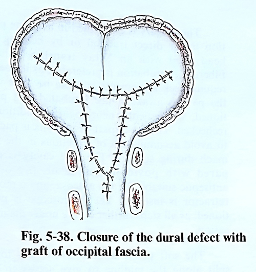
At the end of the operation the dural defect is closed with a graft of occipital fascia, rhomboid in shape (Fig. 5-38).
Posterior stabilisation. The patient is lying on the operative table in a prone position with support of the head as in a posterior fossa craniectomy. Skeletal traction is applied with a 2.5 - 3 kg weight. X-ray control in this position is necessary to verify that the alignment of the craniocervical joints is correct.
The midline incision is performed from the external occipital protuberance to the spinous process of the fourth cervical vertebra. The approach follows the midline along the septum nuchae and after that comes subperiosteal dissection of the muscles from the occipital squama and the posterior apophyses.
In patients with instability between the atlas and axis, fixation involves the Cl and C2 vertebrae. If instability also affects t
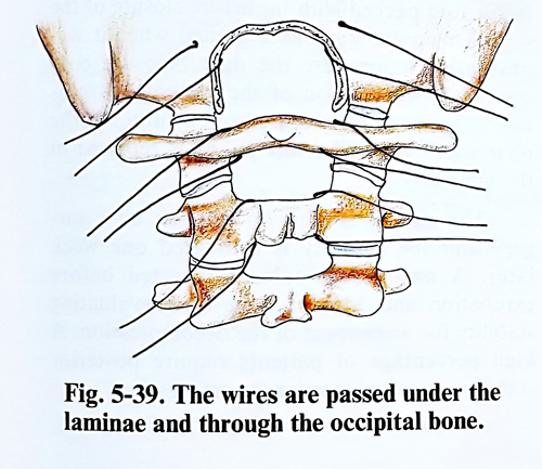
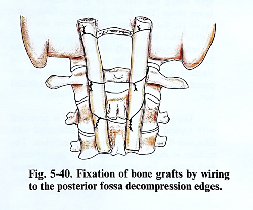
he atlanto-occipital junction, the fixation must include the occipital bone. The materials used for fixation are either special metallic instrumentation or an autobone graft. Metallic fixation has the advantage of early postoperative mobilisation with rehabilitation, and physiotherapy procedures start early after operation. Autobone graft for stabilisation is usually taken from a rib or the iliac crest. The fixation is by wiring or by special metallic plates and screws. The contact surfaces between graft and bone must be decorticated. In addition, small bone fragments are indented between bone and graft and around them (Figs. 5-39; 5-40).
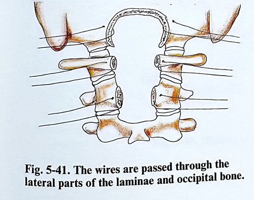
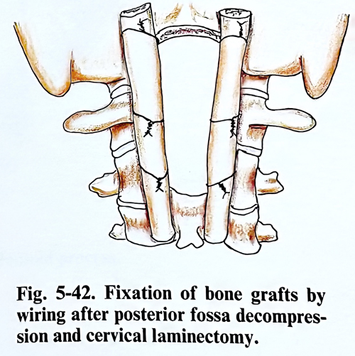
When posterior stabilisation is done after decompression, the metallic instrumentation or the bone grafts are fixed to the borders of the posterior fossa craniectomy and the laminectomy (Figs. 5-41; 5-42).
Anterior decompression. Irreducible lesions, where the compression is produced by the dens and its surrounding tissues, are most often treated transorally. This approach is intended to be only extradural and to relieve bony, ligamentous or scarring tissue (pannus) compression.
Surgery is performed under traction, which rarely needs to exceed 3 - 3.5 kg. To provide sufficient access to the area, the temporomandibular joint should be mobile enough to open the mouth to a distance of 2.5 - 3 cm between the incisors. Median glossotomy and midline mandibular splits have been added to the technique in some cases when mouth opening is not sufficient to enlarge the access. Preoperative antibiotics are given if the culture from the pharynx indicates pathogens.
The surgery is performed in a supine position under direct traction or by keeping the head fixed with an X-ray translucent halo. Fiberoptic intubation is preferable, as it doesn't require neck movements and can be done with the patient awake. Once intubated and positioned, the patient's neurological condition is rechecked. The lower part of pharynx is packed to avoid accumulation of secretions in the stomach during surgery. The oral cavity is prepared with povidone iodine or any other antiseptic suitable for the mucosa, and a mouth retractor is inserted. The microscope is positioned, as all steps further on are under magnification.
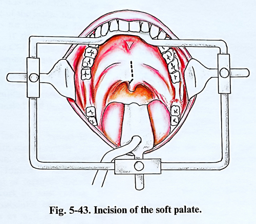
The soft palate almost always has to be split along the midline to give access to the lower clivus (Fig. 5-43). Its incision follows the raphe at the midline starting from the hard palate and just before reaching its edge, is turned slightly laterally to avoid the uvula. The flaps produced by the incision are lifted on traction sutures and in selected cases the exposed edge of the hard palate can also be drilled out.
The posterior pharyngeal wall is prepared with topical cocaine 2.5% and infiltrated with lidocaine 0.5% with 1/200,000 adrenaline. The midline is incised and both flaps are retracted with sutures. The prevertebral fascia and longus colli muscles are dissected from bone and ligaments, exposing the caudal clivus, atlas and axis, and spreading laterally approximately 2 cm on both sides. Further lateral separation can damage the exit of the Eustachian tubes, the hypoglossal nerves and even the vertebral arteries.
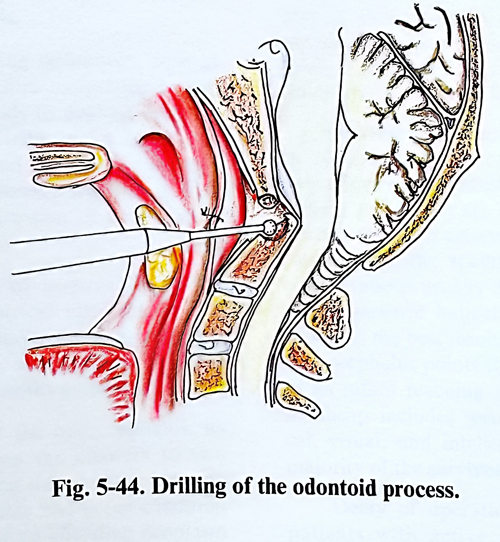
The anterior lamina of the atlas is drilled out with the high speed diamond drill and the soft tissue in front of the odontoid is removed with a tumour forceps. The odontoid is drilled next in a rostro-caudal direction and, if present, the pannus is removed with caution (Fig. 5-44). The tectorial membrane is incised and the pulsating dura should be seen below.
Any dural tear is closed meticulously, as a CSF fluid leak is a serious complication after this type of surgery. The dura is patched with fascia, and packed with fat before closure of the wound. Pannus removal is limited when it will obviously compromises the dura. Closure consists of approximation of the pharyngeal muscles with absorbable sutures and a suture of the pharyngeal wall. The soft palate is repaired in the same way.
The patient is kept on traction after surgery and the stability is evaluated one week later. A nasogastric tube is inserted before extubation and MRI is done after evaluating stability for assessment of the decompression. A high percentage of the patients require posterior stabilisation after anterior decompression.