
Legacy: "Atlast of Neurosurgery" / L.Karaguiosov, A. Ramadan, K.Karaguiosov / Kiwait/ 1998
Part One
SKULL AND BRAIN SURGERY
GENERAL SURGICAL TECHNIQUES IN CRANIAL AND INTRACRANIAL LESIONS
As a separate surgical speciality, neurosurgery has much in common with all other branches of surgery. However, the basic techniques of general surgery - division of tissues, haemostasis, etc. performed in neurosurgery are of relatively little use. This is because the anatomical objects in neurosurgery - the skull, spine and their contents - present specific requirements for working on such extremely complex structures. During a gradual development over the past century or so, with innovations evolving into standard procedures, some widely applied surgical techniques have become accepted as basic to the practice of this speciality.
These surgical techniques, which have paralleled and reflected the growing status of technology in instrumentation, have not really altered the basic approach. Even at present, to gain access to the intracranial structures we perform craniotomy (preserving the bone from the place of opening) or craniectomy (progressive excision of bits of bone); both these procedures are firmly based on the ideas of the pioneers in neurosurgery. Cerebral resection, haemostasis and other procedures on the brain have been also designed as a principle in the "classical" period of neurosurgery. Modern neurosurgical instrumentation however, offers significantly less trauma and greater efficiency in the performance of all cranial and cerebral procedures. These technical achievements are essential when the objects of manipulation are intracranial structures of vital importance.
Every surgical operation on the cranial part of the central nervous system and adjacent related anatomical structures has some common steps, amongst which are the invariable approach through the skull and its subsequent closure. The approach should be thoroughly planned, aiming the best access to the desired structures at the cost of the least surgical trauma and an optimal aesthetic effect. The decision for the approach should be taken in the full context of the general condition of the patient and local condition of tissues, accounting for the possible perioperative and postoperative complications.
The plan should include a study of exactly which part of the skull bones should be removed (and how), and to this end, where the incision of the epicranial tissues should be made and how they should be retracted. For the different types of intracranial pathology there are multiple and widely accepted transcranial approaches applied in current neurosurgical practice, but all too often modifications are required. One can justifiably describe planning such an approach as constituting a synthesis of surgical and anatomical knowledge and imagination (as an ability to
"see" through the intact structures and thereby prevent unexpected disappointments). For the precise description of the approach are used anatomical structures and points, among which landmarks on the scalp and skull, and the average projection of certain cortical structures on the cranial surface are very important.
LANDMARKS ON THE SCALP AND SKULL
BASIC ANTHROPOLOGICAL POINTS
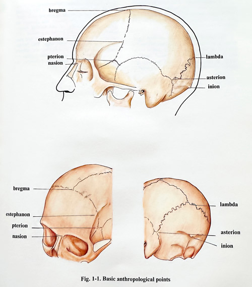
Some of the essential anthropological points are very useful references for the precise description of the planned approach. The essential advantage comes from the feature that they are seen at the same time on the patients head as well as on the imaging studies showing the intracranial pathology found (Fig. 1-1).
1) Nasion. A point situated on the base of the nose, in the middle of the nasofrontal suture.
2) Bregma. A point, where the coronal and sagittal sutures join; on the intact skin it can be determined as the intersection of the two perpendicular lines, drawn up from the midpoints of both zygomatic arches.
3) Lambda. The point, where the lambdoid and sagittal sutures join.
4) Inion. This point corresponds to the external occipital protuberance.
5) Estephanon. Symmetrical points on both sides of the skull at the place, where the coronal suture crosses the superior temporal line.
6) Pterion. Approximately symmetrical points on both sides of the skull at the place, where the frontal, parietal, sphenoidal and temporal bones join the large wing of the sphenoid bone.
7) Asterion. Similar to the previous bilateral points at the place, where the occipital and parietal bones join with the mastoid part of the temporal bone.
EXTERNAL PROJECTIONS OF THE BRAIN HEMISPHERES
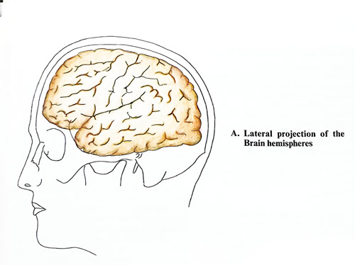
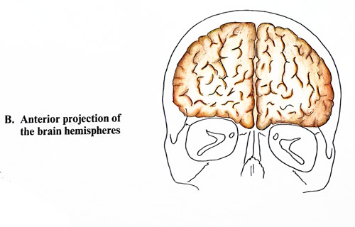
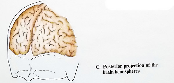
The sagittal border of the brain hemisphere corresponds more or less to the midline of the
cranial vault, bearing in mind the midline location of the superior sagittal sinus. This line begins at nasion and ends at inion.
The lateral border of the cerebral hemisphere is uneven and its projection follows the floors of the anterior and middle cranial fossae and the tentorium. In the frontal region it begins at the nasion, directed laterally but slightly upwards. The place where the border of the frontal lobe reaches the temporal, is 1.5 cm behind the zygomatic process of the frontal bone and 2 cm above the zygomatic arch level. From that point the border line changes its direction, being a curved line turning downwards to the external auditory meatus and passing 1 cm above its upper edge. This border reaches the asterion and further on, already corresponding to the occipital lobe reaches the external occipital protuberance following the nuchal line (Fig 1-2).
TOPOGRAPHY OF THE MAIN CEREBRAL SULCI
The lateral and central fissures are the two most important sulci on the convex surface of the cerebral hemisphere, serving as a reference for the localisation of other sulci and convolutions (gyri) of the hemispheres. However, when planning craniotomies, we should consider also the displacement produced by mass lesions, as after the planned cortical exposure, gyri and sulci can be found to be not exactly at their expected places. Although these localising methods may be simplified, they can nonetheless provide useful information to the surgeon undertaking a procedure, not preceded by 3D CT or MRI imaging.
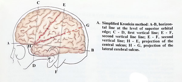
According to the simplified Kronlein method, a horizontal line is drawn at the level of the upper orbital edge. Then two vertical lines are superimposed: the first one through the midpoint of the zygomatic arch and the second - through the posterior border of the auricle. The line connecting the crossing point between the horizontal and the first vertical line with the point, where the second vertical line crosses the midline, corresponds to the projection of the central sulcus. If the angle, formed by the projection of the sulcus centralis and the horizontal line is bisected, this last line will correspond to the lateral sulcus (Fig. 1 - 3, A).
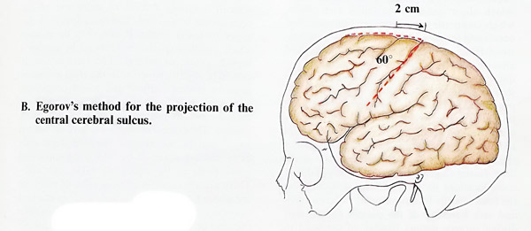
After Egorov's method the central sulcus projection corresponds to the line, which begins 2 cm behind the midpoint of the nasion-inion distance on the sagittal line and forms a 60 degrees angle with it, open in an anterobasal direction (Fig. 1 - 3, B).
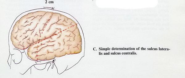
The topography of the two basic fissures of the cerebral hemisphere (the lateral and central sulci) can be determined in the following simple way: the lateral sulcus lies along the length of the line connecting the zygomatic process of the frontal bone with a point at 3/4 of the length of the nasion-inion distance on the sagittal line starting from the nasion. The central sulcus corresponds to the upper half of the line, connecting the midline (2 cm behind its nasion-inion midpoint) and reaches the middle of the zygomatic arch (Fig. 1- 3, C).
APROACHING THE INTRACRANIAL STRUCTURES
PATIENT POSITIONING ON THE OPERATING TABLE
The proper position of the patient on the operating table is a very important step when undertaking surgery. It should allow the easiest performance of the surgical technique as required with no functional disturbances of the patient's physiology due to the prolonged positioning under general anaesthesia. Therefore, in general, we can indicate two essential groups of requirements for proper positioning: those which allow technical feasibility and others, which create the best intracranial physiological conditions for the patient and prevent complications from the long lasting positioning and anaesthesia. Regarding the first group of requirements, it is essential to foresee all technical steps and devices that will be required for the procedure as planned. These include the location and type of craniotomy, the needs of the microsurgical technique, as well as the presence of any additional equipment. It is preferred that the operative field be the uppermost part of the head during surgery; however, it is acceptable in some locations for it to be located laterally on the head after its final positioning. Microsurgery and safe handling of the patient by the staff during surgery require fixation of the head to avoid disturbances during micromanipulation.
For this purpose the three pin holder, which fixes the head within the outer table of the skull to the operating table by means of a special clamp armed with pins, is widely accepted.
Moreover, if the operative field is not strictly horizontal, it allows easier drainage of fluids from it during surgery (Figs. 1-4; 1-5).
Once the position of the skull is decided, it becomes necessary to complete the second group of requirements. As a rule, there should not be strains in different parts of the body when it is placed in the final position. Any major or requiring even minor force rotation, flexion or extension of the neck should be avoided. The same applies to the upper and lower girdle. Only then it can be decided which position of the body is to be chosen - supine, prone or lateral. The patient's head should be slightly superior to the right atrium to improve venous outflow and avoid congestion intracranially. Lifting the lower limbs will have the same benefit and will increase the circulating volume. These requirements will give additional angulation of the already positioned patient's body and are achieved by lifting the upper and lower parts of the operating table separately.
There are also certain general rules of positioning which have to be followed, such as allowing free chest respiratory movements, avoiding compression of superficial arteries, veins and peripheral nerves unprotected by muscles; for instance the most vulnerable cubital nerve at the elbow and the peroneal at the fibular neck.


Since the introduction of modern anaesthesia and perfection of intraoperative functional control, the semi-sitting and sitting positions of the patient during surgery became possible. It can be easily noticed, that these positions can be applied as an option in certain, but not all neurosurgical approaches (mainly posterior cranial fossa) instead of the horizontally positioned patient and some additional implications should be considered. The additional elevation of the head significantly decreases the venous and CSF pressures making the approach and drainage of fluid easier, but it may increase the risks of venous air embolism and it does also present additional requirements for cardiac and circulatory monitoring.
When the operation is in the frontal, fronto-parietal or fronto-temporal region the patient is placed supine, the head being slightly turned to the opposite side. If the anterior skullbase surface and sellar region have to be approached, the head should also be slightly extended. For approaches in the parietal, temporal and occipital regions the lateral position is usually the most convenient. In occipital approaches and in some lesions in the posterior fossa, especially in the cerebellopontine angle region lesions, the lateral position is combined with different degrees of rotation and flexion of the head (Fig. 1-6).
The prone position is that most commonly used in operations on the posterior fossa. In this position the patient's head is placed on a special headrest if not in a pin holder, and this should allow the anaesthetist to have a adequate access to the face. The shoulders, the chest and the pelvis should be supported only in their lateral parts, allowing the middle part of the chest and the abdomen to be free for respiratory movements.
The sitting position has great advantages in operations on the posterior cranial fossa, especially in cases of increased intracranial pressure, when venous pressure inside the cranial cavity is high (Fig. 1-7). The access to the fourth ventricle, the upper surface of the cerebellum and foramen magnum is easier. Because of the potential risks of air embolism and other complications, the "classical" sitting position is not very often applied, being substituted by the semi-seated position, in which the legs are well flexed. Several surgical technical rules should be followed when operating on a patient in the sitting position. It is important that the anaesthetist increases the central venous pressure at the dissection of every layer of the operative wound. The opened veins can at once be noticed and be occluded, the bleeding bone surface can be waxed or packed with oxidised cellulose. This increase of the central venous pressure should continue for not more than 3 - 4 min. It is advisable to maintain the ventilation with 10 mm H2O positive pressure at expiration.


BURR HOLE APPROACH
This is an essential basic technique for approaching the intracranial cavity safely. Performing a burr hole can be a separate procedure in certain conditions requiring such a limited approach, or a step in the techniques of craniotomy or craniectomy. Here it is considered as a separate procedure. Some of the goals of the burr hole technique can be achieved at present by "twist drill" or other smaller drill perforation techniques, especially in intracranial pressure monitoring or intraventricular drainage placements, but a burr hole also permits visual control over the dural and cerebral surface for better haemostasis.
Indications. Burr holes may be used for exploration in patients with suspected extracerebral collections and the drainage of those which are fluid, such as chronic subdural haematomas and hygromas, for the aspiration of intracerebral fluid collections such as abscesses, for needle biopsy of a cerebral tumour, or to aspirate intracerebral haematomas.
Anaesthesia. Burr holes may be made under local anaesthesia in a conscious, cooperative adult patient and in unconscious patients who are not restless. In all other circumstances general anaesthesia is recommended.
Surgical technique. After preparing the shaved skin surface with antiseptic solutions, an incision 2 - 2,5 cm long is done by transsecting all layers of soft epicranial tissues in one. A small self-retaining retractor is inserted and opened widely for control of bleeding and better visualisation. Any remaining bleeding is controlled by electrocoagulation while preserving the skin edges. The pericranium is incised in the same direction as the skin and detached to each side with a small periosteal elevator. Perforation of the bone is performed by electric or manual drill. Using a manual drill, the surgeons learns to sense a characteristic change of feeling in the drilling resistence after reaching and perforating the inner skull table. However, until they acquire this 'feeling', for safety of dura, beginners should frequently take out the blade to check to what depth it has penetrated, and once dura visible, to change it with the burr. An automatic drill will stop by itself reaching the dura. If bleeding appears from the bone, because of the opening of diploic vessels, haemostatic wax is pressed on the bleeding point. If the bleeding is evoked from an injury of the dura, the hemostasis is achieved by tamponade with cottonoids, soaked in saline, or, if this is not effective, with bipolar coagulation. The diameter of the bone opening must be 10 - 15 mm (and with a thicker cranial vault, even larger). A larger diameter provides better access for manipulation through the burr hole.
If the dura must be opened, it is elevated with a sharp hook and delicately incised with a small scalpel blade. Care must be taken to avoid injury to the underlying brain. Once the incision is done it can be extended crosswise. Any bleeding from the edge of the dura is controlled with bipolar coagulation.
Closure. When closing the wound, the dura may not be sutured, but may be covered with gel-foam. The galea is closed with a few interrupted sutures, the knots being inverted and the thread ends cut short. The skin is closed with interrupted sutures that will control all but major bleeding (Fig. 1 - 8).
Postoperative care and complications. The sutures can be removed on the fifth day; usually the dressing needs not be disturbed until that time. Complications after a burr hole procedure are usually rare. Intracranial haematoma is unusual unless bleeding from the dura or brain has not been properly stopped. However, as in all intracranial procedures, infection of the wound may have serious consequences.

SUPRATENTORIAL CRANIOTOMY
SKIN INCISIONS AND SCALP FLAPS
After optimal positioning of the patient and preparation of the scalp with antiseptic solutions, the skin incision is marked out with a sterile marking pen. When the patient is under general anaesthesia, as is common, a marking scratch can be made along the skin incision with a scalpel or a needle, affecting mainly the epidermis, until a tiny red line of blood is obtained; however, no drops should be seen. Transversal lines at a distance 5 - 6 cm along the skin incision are also marked. They indicate at closure the proper position of the scalp flap, avoiding its rotation or distension of the wound edges. Scalp incisions have most commonly horseshoe shapes, open to the cranial base and intended to preserve the neural and vascular supply (Fig. 1 - 9). In the frontal region the incisions have the same shape; however, for aesthetic reasons, they should appear on the forehead only exceptionally. To avoid visible scars in the frontal region incisions most often go along the anterior hairline in a coronal or bitemporal shape. Such a bilateral incision is very often followed by a standard unilateral craniotomy. The temporal muscle usually occupies one part of the region enclosed by the incision. It can serve in the following stage of the operation as a pedicle for the bone flap if it is required.
At the next stage local anaesthetic can be injected. The majority of operations are performed under general anaesthesia, however, local infiltration (to 40 cc of local anaesthetic,
1 cc of 1% epinephrine is added) has not lost its value due to the action of the added epinephrine which considerably reduces the bleeding at the time of incision. Additionally, this infiltration is very helpful for the easy separation of galea from periosteum (pericranium). The infiltration of the skin with local anaesthetic is done in the following way: first the skin and the subcutaneous tissue along the course of the incision is injected until the surface acquires an 'orange rind' appearance. At the time of this infiltration the needle should not penetrate through the galea and usually strong resistance is felt, due to the firm attachment of the epicranial aponeurosis to the skin. After that, at different points along the skin incision, the needle is inserted beyond the galea and the supraperiosteal space (subaponeurotic areolar tissue) is copiously infiltrated over the complete operative field. This tissue is loose and the infiltration is easily done, without particular resistance. At the end, as a last stage, infiltration is done in the temporal muscle in the region of the craniotomy. This is accomplished by vertical penetration in one or two places and the needle is inserted until the resistance of the bone is felt.

The initial incision involves the skin, the hypodermic fatty tissue and galea aponeurotica.
The pericranium and the subaponeurotic areolar connective tissue remain intact. The incision is better performed in three to four steps along its full length for easier control of bleeding. At the time of the incision the surgeon and his assistant are firmly pressing the edges of the wound to the underlying bone to occlude the bleeders and to retract wound edges for better control in the depth. The galea is attached intimately to the skin and they can be retracted only together; if the galea is not cut, the wound will not open (Fig. 1-10). After completing every step of the incision, the pressing fingers are released one by one applying in their place haemostatic clips or artery forceps. When applying an artery forceps, it is better to occlude the bleeding artery itself, but when this presents difficulties, the galea should be fixed and stretched or pulled by the forceps, pressing the adjacent bleeders with it.
However, technically it is much easier to apply special haemostatic clips (for instance Raney clips) which have the same action (pressing the galea to the skin and by that occluding the blood vessels). The big vessels penetrate through the aponeurosis from the subaponeurotic areolar tissue, and there they are better controlled by coagulation. Once the incision and the haemostasis are completed, if a soft tissues flap was prepared, it is elevated, separating galea from periosteum (pericranium); this is the easiest layer to separate due to the areolar tissue (Fig. 1-11). The elevated soft flap is fixed by sutures or hooks on the galea, so that it will not move during surgery and thereby disturb subsequent manipulation. Then it is better to cover it by wet gauzes or towels.


BONE FLAPS
Bone flaps are planned before surgery and their limits are described clearly according the basic anthropological points of the skull. Size and limits depend on the needs of the surgeon with respect to viewing and manipulation spaces. With the advent of the surgical microscope the dimensions of craniotomies have become smaller. As the surgeon intends to perform certain techniques intracranially, he needs to guarantee sufficient space for safe manipulation and an additional angle for observing simultaneously the field. He should be able to sufficiently expose the lesion or provide space for brain retraction with the selected craniotomy size and location.
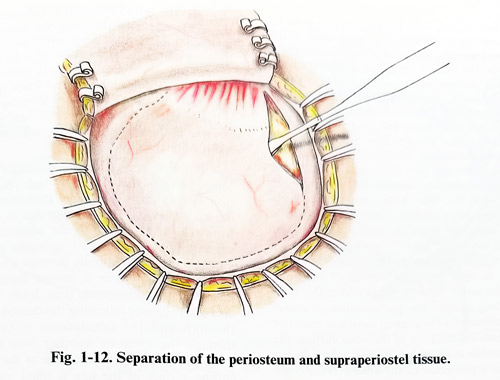
Every craniotomy is performed in a certain standard technical manner. It includes bone exposure at the preselected cutting line, positioning of burr holes, bone cutting and elevation of the flap. Incision of the periosteum is done over the presumed areas of burr holes and bone cutting. Basal burr holes often require incision of temporal fascia and muscles to expose the drilling and cutting places (Fig. 1-12). Eventual bleeding is controlled. Then one, two or more burr holes are produced. The placing of burr holes is decided according to the needs of the surgeon in introducing the bone cutting device and for better control under the flap before lifting it. Therefore burr holes should be placed where bone cutting starts and ends, when the flap reaches the skull base and where difficulties are or can be encountered in turning the bone
cutting line. Some burr holes play "key" roles for the proper shaping of the flap and thence for proper exposure. They are placed with regard to some anatomical structure laying below. Such burr holes are the pterionial, the sinus angle (transverse to sigmoid), burr hole in trigeminal neuralgia, the burr holes at the edge of the sagittal sinus and torcular and others. Their misplacement is a clear surgical mistake and special attention must be paid to determining their proper location. They usually stay on the path of the approach and have given ground to contemporary key-hole surgery. Other burr holes can vary slightly in their positions for purposes of easier performance of the craniotomy.
Once the burr hole places have been determined they are drilled in such an order that the one which is expected to bleed most is done last.
The drilling is done in the manner already described in the section on burr hole techniques.
The bottoms of the burr holes are packed with topical haemostatics and cottonoids.
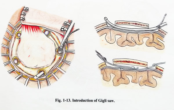
Bone cutting can be performed with the traditional Gigli saw or with the more recently designed air or electrically powered craniotomes. However, for both techniques it is essential to separate the dura from the inner table with dissectors, as the cutting instrument's edges and bone laminae can injure the dura (Fig. 1-13).
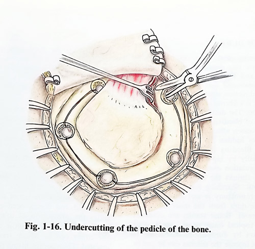
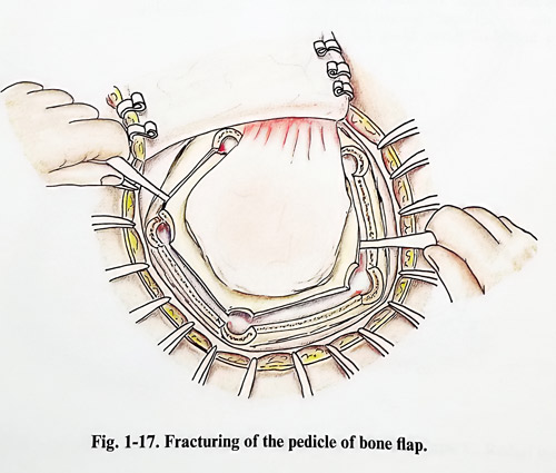
There are two options in removing the flap: to keep it attached to a pedicle of periosteum or temporal muscle during surgery, or to remove it and fix it again at the end of the surgery. If the flap is to be kept attached, the bone cutting line under the attachment will not be accessible and it is fractured after proper undercutting with Gigli saw, craniotome or nibbler (Fig. 1-16; 1- 17). The removed flap is either kept in a pot of saline or covered with wet gauze, and the temporal muscle or periosteum are retracted. Lifting the flap provokes bleeding and the first task that follows is to control it. Some craniotomies for special approaches may require the lifting of two flaps or drilling out additionally of some portions of the skull base.
OPENING OF DURA
The opening of the dura can be made in different ways: this depends on the place of craniotomy and the pathological process. It should be mentioned that the incision of the dura, in any direction whatever, does not evoke necrosis, as it is a fibrous structure and is rich in vascularization. The dura is opened after complete control of bleeding. It is caused either by visible dural vessels affected by bone flap lifting or by sources hidden under the rim of the craniotomy. Bleeding from exposed dura is controlled easily by bipolar coagulation. No extensive coagulation over larger areas is applied as that shrinks the dura and makes its closure difficult. Searching for bleeders under the rim of craniotomy is not efficient as it detaches more dura from the inner table and bleeding increases. Better results are obtained by inserting topical haemostatic agents and lifting the dura by duroperiosteal stitches. If bleeding is not intense the stitches can be put in after closure of the dura.
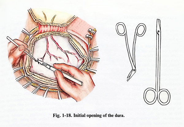
Dural incision should not cause additional cortical injury, which is prevented by lifting the dura with a hook when it is tense and with fine forceps when it is lax. The hook is inserted only below the external dural layer and gently pulled, while a fine blade incises along 2-3 mm both layers beside the hook until the arachnoid seen. Sometimes, if the arachnoid is torn there is a leakage of CSF. Bleeding is temporarily covered by a cottonoid, but it can occur despite the fact that vessels are carefully avoided (Fig. 1-18).
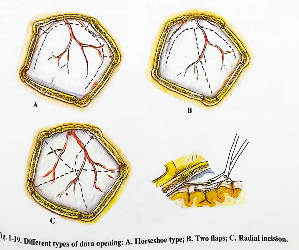
The incision lines are decided after some principles are considered. They should not cross dural sinuses and lacunae, they have to fold conveniently and should not reach close to or under the bone edges. The most simple pattern of incision lines has to be chosen, as well as that one associated with easier closure and better water-tightness. The incision is done with scissors having blunt tips and edges. A small cottonoid is inserted under the tip of the scissors, as the arachnoid and cortex need protection. Sometimes the protection of a narrow spatula is needed. Once the incision is 1 - 2 cm long and the edges can be lifted, traction sutures are inserted. They permit lifting the edges and safer continuation of the incision. When the incision is completed the flaps produced by the incisions are turned out of the craniotomy and covered by wet gauzes or cottonoids. Delicate bipolar coagulation can be used for bleeders of the dural edges (Fig. 1-19).
CLOSING A CRANIOTOMY
As with all surgical wounds, the craniotomy is closed strictly following the anatomical layers, in the reverse order of that adhered to when opening.
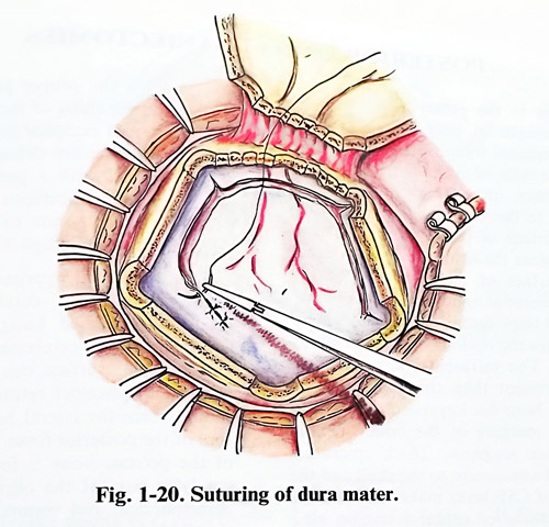
Dural closure is done first, the intention being the production of a complete and watertight cover of the cerebral surface. Because the dura may have been turned and pulled for many hours, the dural flaps must be well repositioned. The properly oriented flap position is fixed with interrupted stitches. Then the incisions are closed with continuous or interrupted sutures, avoiding any minimal gapping and guaranteeing a watertight closure. Mild tension of edges is acceptable at closure. Gaps have to be repaired with plastic techniques. Several techniques are used routinely. Dural layers (they are two) can be split and the upper turned out of the dural flap to cover the defect of closure. If it is impossible, then a free flap of periosteum and supraperiosteal fibrous tissue, fascia lata or other commercially available materials for this purpose can be used. Whatever it is, closure should assure protection of the brain and prevention of CSF leakage (Fig. 1-20).
Before replacing the bone flap care is taken to perform haemostasis. Epidural bleeding is controlled with topical haemostatic agents and duroperiosteal stitches. We prefer to leave epidural drainage tubes under mild negative (suction) pressure in the great majority of supratentorial craniotomies, taking out the tube through a burr hole and aside from the epicra-nial tissues incision. The already positioned tube is fixed to the skin with a stitch. Then the bone flap is repositioned. Free flaps are fixed to the bone edges of the craniotomy by silk or wire.
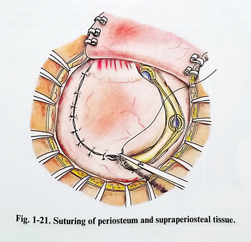
Flaps attached to the periosteum or temporal muscle are turned back and also fixed. Periosteum is repaired with interrupted sutures. Temporal muscle is sutured if long and gaping incisions remain, but only to overcome the gap.
When muscle fascicles are only split and not transsected, sutures are rarely needed. Temporal fascia is closed with interrupted stitches. Before proceeding to the more superficial layer and attachment of the temporal muscle to the skull, stable bone flap fixation is assured (Fig. 1-21).
Epicranial flap is usually closed in two layers - galea and skin. Haemostatic clips and forceps are gradually removed and major bleeders controlled by coagulation. In conditions of significant diffuse bleeding a second suction drain can be left below the galea. The galea is sutured with inverted interrupted stitches avoiding any misorientation of the flap. Therefore at the beginning three to four stitches are placed at the points already marked at the skin before incision and only then is the entire layer completed. Galeal sutures are intended to properly reposition the galea and ensure coincidence of the edges of this layer for efficient fibrous union. Last to be sutured is the skin; this is also accomplished with interrupted stitches at a distance of 1 to 2 cm between sutures. If the bone flap is replaced, the dressing can be used to apply mild compression on the wound by using a stretching bandage.
POSTERIOR FOSSA CRANIECTOMIES
Approaches to the posterior fossa differ from the supratentorial ones because of the anatomical differences found in the area at all levels. Below the external occipital protuberance the skin is thicker, muscles are much larger, firmly attached to the bone and as a rule they cover all the surface of the approach through the bone. Therefore, the skull opening is deeper beneath the surface of the skin than in the common supratentorial approach. In these conditions attached flaps cannot be turned. Therefore, remaining options are a craniectomy or a free flap. The infratentorial space is a smaller compartment than the supratentorial and can require bone decompression at the end of surgery as well as merging of the bone opening with foramen magnum opening. Thick epicranial layers with good attachment to the skull and the increased risks of CSF leaks make very real the danger of placement of epidural suction tube drainage; hence, such drainage is hardly ever used. All these different conditions require different techniques in posterior fossa approaches.
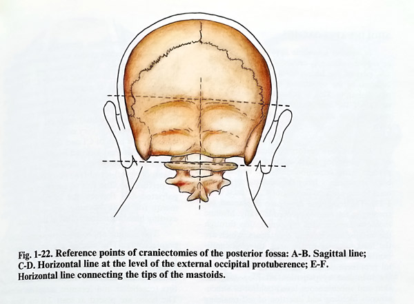
Other specific characteristics are the presurgical planning of the approach and the choice of location for the craniotomy. The two main types of approach - the midline and the lateral, have proved to provide access to the majority of extra- and intraaxially located lesions as well as to a very extensive part of the internal skull base. Some essential anatomical reference points are used for the proper placement of craniectomies and planning of incisions. The positions of the external occipital protuberance, mastoid tips and midline are defined by palpation and better marked with a pen. Next the external projection of the posterior arch of Cl is established; it corresponds to the horizontal line through the mastoid tips (Fig. 1-22).
Infratentorial approaches differ according to which surface of cerebellum or brainstem needs to be reached. Lateral surface of cerebellar hemispheres, cerebellar peduncles, pontine and medullar surface can be approached only after hemispherial retraction; skull opening should reach the lateral border of the posterior wall of the posterior fossa where the medial wall of the petrous bone is in continuity to the condylar part of the occipital bone. Posterior hemispherial and vermian surfaces as well as the IVth ventricle are reached caudally after median and paramedian exposures through occipital squama with different width. Standard approaches have been designed for the needs of treatment in different types of posterior fossa lesions by the eminent pioneers of neurosurgery. Some of them have survived the severe verdict of our rapidly innovating times, with small modifications due to microsurgery. Because of their anatomical properties, they naturally can be either midline or lateral.
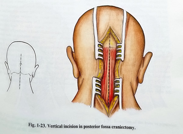
MIDLINE APPROACHES
In general a midline approach is intended to expose sufficiently cerebellar vermis and tonsils, part of the hemispheres and the posterior columns of the uppermost spinal cord for safe manipulation. This is achieved by a bone removal of the mid-occipital squama, foramen magnum rim and very often the posterior arch of C1.
Incisions are performed along the midline of the posterior fossa with a length which permits, after retraction, sufficient exposure of bony structures - from the occipital protuberance to the spinous process of C2 at least (Fig. 1-23). The transverse dimension is planned according to the need to proceed laterally. Midline incisions are intended for penetration through the midline structures of soft tissues, which are the least vascular in the area, thereby significantly reducing blood loss. The external occipital protuberance and the midline of the fascia are identified by palpation after opening the skin and subcutaneous layer, control of subcutaneous bleeding and insertion of self-retaining retraction. A small transverse incision of the muscles immediately below the external occipital protuberance and medial part of the upper nuchal line is performed, if the planned craniectomy does not need to reach the transverse sinus. The muscle strip that remains attached to the bone facilitates the suturing at the end of the operation (Fig. 1-24). The identification of the midline is crucial and only after that is it incised caudally in the direction of the C2 spinous process if this is palpable. The bone of the occipital squama is reached first in its most superficial part - below the external occipital protuberance. Self retaining retractors are advanced and muscles are detached from bone with periosteal elevators (Fig. 1-25). With advancement of the incision the step is repeated again and so on several times until the posterior arch of the atlas can be reliably palpated. For better access it is sometimes necessary to expose the C2 spinous process at this stage thereby making its arch palpable. The incisions are done with electric cautery to reduce bleeding. When the occipital squama is completely exposed along the midline and the gap between squama and Ci has been well located, it is carefully approached (but not quite reached) with the midline incision. The midline is very cautiously incised over the posterior arch of Cl and the arch stripped of soft tissues at least 1 cm on both sides of midline with a small periosteal elevator. The posterior atlantooccipital membrane is dissected avoiding its perforation, the rim of foramen magnum is palpated and thereby the minimum requirements for bone opening are achieved. Any existing bleeding is controlled at this stage.
The midline approach is routinely made by craniectomy. The size of the craniectomy is preplanned and can vary, but as a rule, it is started by drilling two burr holes on both sides of the midline avoiding the dural venous collectors (cerebellar and foramen magnum sinuses).
The holes are placed at least 2.0 cm from the midline and at the mid-distance between the foramen magnum and the superior nuchal line (Fig. 1-26). Both burr holes are enlarged sufficiently and after gaining space the midline is crossed; one must of course expect venous bleeding at times. The enlargement continues cranially to the lower edge of the transverse sinuses and caudally until it reaches the foramen magnum, where venous bleeding is also commonly encountered. In this area, the atlanto-ocipital membrane is thoroughly dissected from the dura (Fig. 1-27). The foramen magnum rim opening is enlarged laterally to no more than 2 cm on each side. Following the dural plane, the inner surface of the Cl arch is dissected and removed with a fine nibbler or bone punch to an extent not more than 1.5 - 2 cm from the midline if its removal has been preplanned.

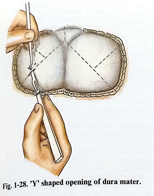
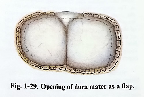
Dural and epidural bleeding is controlled by bipolar coagulation and topical haemostatic agents; bone bleeding is waxed. The dura is inspected for the position of dural sinuses and the control of intracranial pressure. Opening is undertaken only when intracranial pressure is within normal values; otherwise a disastrous brain prolapse, shifts and tentorial herniation can occur. Increased intracranial pressure is controlled by osmotic agents or ventricular puncture. The dura can be opened in many different ways, most commonly in a Y shaped or in a horseshoe form (Figs. 1-28; 1-29). The Y shaped opening of the dura avoids as much as possible dural sinuses and has conveniently turned flaps. Dural incisions have to be done on both sides of the midline over the hemispheres; both incisions converge to the midline at the dural fold corresponding to the foramen magnum. The midline is approached and transsected (it is the lower part of the falx cerebelli) only after the dural edges are well held by traction sutures. The lifting of dural edges will permit easy control of the expected bleeding from cerebellar and foramen magnum sinuses. The cerebellar sinus is best ligated; coagulation is ineffective (increased intravenous pressure opens the sealing by the bipolar coagulation) and shrinks the dura, making closure difficult. If bleeding is sudden, dural edges containing venous sinuses and lacunae can be clipped with big or giant temporary Sugita or Yasargil clips.
When the surgical field is dry, haemostasis is easily secured with ligatures or permanent titanuim clips, as the permanent clips are small and they do not obstruct vision and manipulation.
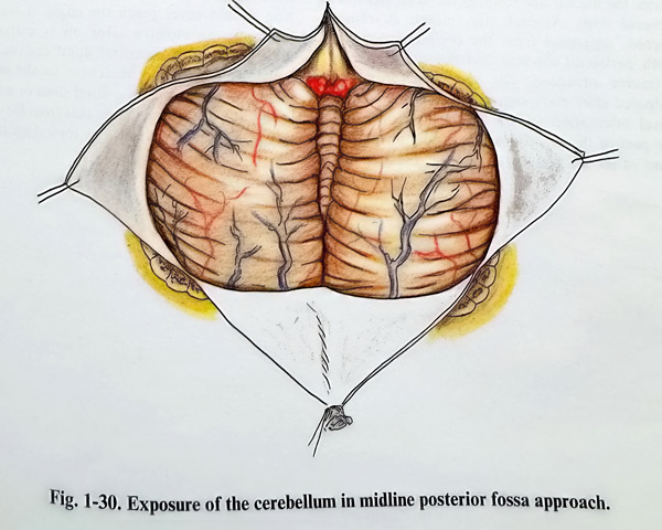
Then incision of the dura is completed along the spinal posterior midline. Dural flaps are lifted and, additional haemostasis is done (releasing the dural tension sometimes provokes epidural oozing). If needed, tension of dural flaps is restored by stitching them to the muscles during intracranial work (Fig. 1-30).
LATERAL APPROACHES
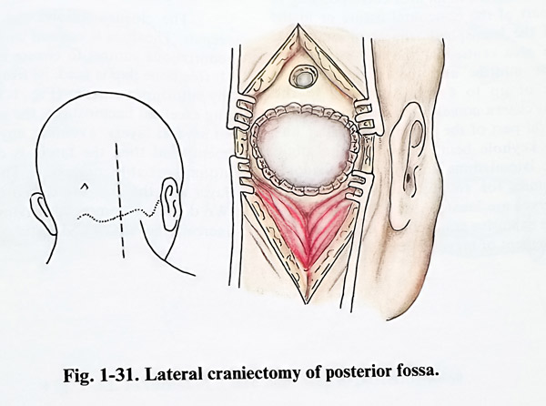
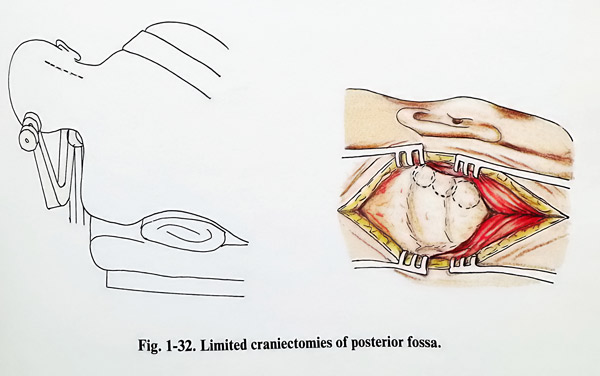
These approaches can vary in size and location. They give access to hemispherial lesions or retraction of the hemisphere. After the hemisphere retraction, the cisterns laterally and ventrally to the pons and medulla oblongata can be reached. This makes these approaches also convenient for lateral extraaxial lesions or intraaxial manipulation after lateral penetration. For hemispherial lesions, bone removal is performed over the lesion itself or the location of the intended cerebellar incision. To penetrate after cerebellar retraction, the bone opening should reach the lateral edge of the hemisphere. This corresponds to the inferior edge of the transverse sinus and the medial edge of the sigmoid. The transverse-sigmoid angle is located under the base of the mastoid. Therefore, cranial openings are oriented in accordance with the line connecting the external protuberance with the base of mastoid and the tip of the mastoid.
Straight or slightly curved incisions are used for lateral approaches at a certain distance from the midline and approximately parallel to it. The distance from the midline corresponds to the centre of the bone removal and its incision length - to its major diameter. If the lower edge of the transverse sinus has to be reached, 2-3 cm of the incision is cranial to the superior nuchal line. For reaching the medial edge of the sigmoid sinus, the incision should be able to expose the posterior mastoid surface (Fig. 1-31).
Incision is done along the preplanned distance and opening the subcutaneous layer is done with the same strokes to reach the fascia. Bleeding is controlled by bipolar coagulation and special attention is paid to the occipital artery, usually crossing the field from lateral to medial, and just below the fascia in the region near the superior nuchal line. The artery should be carefully coagulated along a few millimetres, otherwise it can bleed suddenly at any time during the next stage of the surgery. Cutting with the diathermy is efficient for the subsequent transsection of the fascia and muscles. Self-retaining retractors are positioned to maintain proper access to the depth of the wound. As in midline approaches, splitting and transsection of muscles proceeds from the nuchal line towards the skull base, in several steps. At each step, muscle is transsected, separated from the occipital squama with periosteal elevator and the self-retaining retractor advanced. The bone structures are palpated after every step to preserve the anatomical orientation and to ensure awareness on the part of the surgeon of the proper advancement of the incision. Little by little, the area of bone removal is exposed. Its exposure is completed with careful haemostasis. Craniectomy starts with a burr hole, placed within the preplanned bone removal, but away from the dural sinus margins and preferably in a thinner part of the occipital squama. The burr hole is enlarged by nibbling, advancing the craniectomy to the sinus margins and to the preplanned size. Special care is taken to nibble safely the margins of the dural sinuses if retraction of the hemisphere is intended. This leads at times to the exposure of mastoid cells, but they must be sealed with wax to prevent postoperative CSF leaks and infection. The mastoid emissary vein usually bleeds during separation of muscle from bone in its area and later - on during nibbling. On both occasions it is easily occluded with bone wax. The edges of the craniectomy are smoothed and bleeding is controlled. Some lateral bone exposures are done as craniotomies, inserting the craniotome through the burr hole and producing a free flap. The dura is opened with radial incisions, after ensuring that any abnormal rise of the intracranial pressure is controlled. Osmotic agents and even a lateral ventricular puncture may precede the dural opening if intracranial pressure was preoperatively increased. Dural flaps are turned out after incision and held by traction sutures. The incision should never reach the edges of the cra-niectomy as closure later on is extremely difficult, so after a central dural opening, the incisions run from the centre to the edges, taking care not to open a dural sinus or lacuna.
Epidural bleeding, including that from the emissary vein area, is controlled by topical agents.
SUPRA-INFRATENTORIAL APPROACHES
Two types of lesions require this type of approach, those which are superficial, but under the dura, over and under the tentorium, and those which are in the depth, and require a much wider angle of view and manipulation between delicate neural and vascular structures.
All approaches open supra- and infratentorially or middle and posterior cranial fossae with or without modifications enlarging the exposure on the account of the petrous bone. The bone bridge, left in previous times, is no longer left for protection of the lateral sinus, as refined and delicate bone removing instruments have been introduced. The extent of bone removal is planned according to the needs of width, angle and direction of the approach.
These approaches can consist of three parts or separate exposures: middle cranial fossa (or supratentorial occipital area, posterior cranial fossa and an optional petrous bone approach.
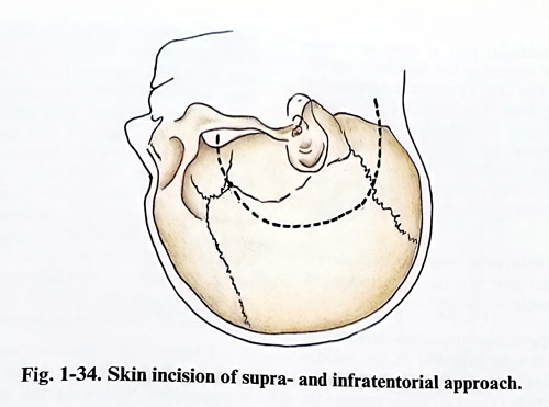
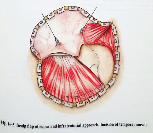
The three exposures can form a common dural field. This field is more often a lateral approach
- involving lateral suboccipital and temporal or temporooccipital areas with or without the petrous bone, but it can be also medial - occipital, supra- and infratentorial (Fig. 1-34; 1-35).
The supratentorial part is always a craniotomy reaching the temporobasal plane or continuing towards the inion - the upper margin of the transverse sinus. Its limits in anterior, posterior and superior directions depend on which part of the temporal and/or occipital cortex has to be retracted and which part of the free tentorial edge has to be reached. Important points for the temporal and occipital basal cortical surface retractions are the eminentia arcuata (corre-sponding to the superior semicircular canal), the superior petrosal sinus and the straight sinus.
The eminentia arcuata provokes a natural indentation on the cortex permitting easy retraction to the interpeduncular cistern. The superior petrosal sinus indicates the posterior edge of the superior petrosal surface and therefore the natural continuity of the supra- and infratentorial spaces along the skull base. The superior petrosal sinus can be transected in some combined approaches, allowing further complete splitting of the tentorium. The straight sinus is an important path determining structure position in the posterior, close to the midline approaches, where complete tentorial division is impossible.
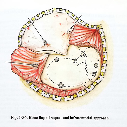
The infratentorial part is done as a cra-niectomy or by extending the flap from supra-tentorial area. The area of special attention for craniectomy or bone flap lifting is the transverse sinus, where proper dural separation from the inner table is crucial. Burr holes, placed on both sides of the sinus allow the introduction of a blunt dissector to separate sinus wall from the bone before cutting it with the craniotome (Fig.1-36).
Bone flap is lifted in one and two pieces, and the temporal part can remain attached to the temporal muscle.
In lateral approaches the supra-infratentor-ial craniotomy can be extended in the middle and posterior fossa by additional petrous bone removal extradurally (Fig. 1-37). For the middle fossa a small flap mainly including the roof of the tympanic cavity can be helpful for increasing the angle of subtemporal access. Another option is presented by a posterior petrosectomy, drilling an exposure to the intrapetrosal internal carotid and internal acoustic meatus. A posterior fossa approach can be also enlarged, most often by a partial mastoidectomy, for purposes of presigmoid dural exposure. This is achieved by drilling out the posterolateral mastoid cells, carefully skeletonizing the labyrinth and the Fallopian canal. The internal acoustic meatus can be widely exposed from a posterior direction. Additional partial mastoidectomy completely exposes the dural sinus angle with the walls of the lateral, sigmoid and superior petrous sinuses. This allows the division of the superior petrous sinus and Labbe vein drainage.
Although this sinus division seems technically feasible, any decision to undertake it should be weighted against some complications, mainly associated with brain venous drainage impairment. Therefore a detailed angiographic study should precede the surgical planning, and the step is taken only when its benefits are obvious.
Closures in supra-infratentorial craniotomies don't differ from those in the other types of craniotomies, following the above mentioned layer supra- and infratentorially. However, special care is taken for the bone protection of sinus walls which have been exposed, guarding aginst their accidental coincidence with bone gaps. We also prefer leaving epidural drains and careful fixed dura to the edges of the craniot-omy, as these approaches are associated with long lasting intracranial work and massive CSF drainage.
GENERAL RULES OF WORK ON BRAIN TISSUE
The methods used for performing punctures, incisions, haemostasis, excisions and all other standard neurosurgical techniques differ from those in general surgery. Despite the extensive use of microsurgical technique over the past three decades, some manipulations are still performed macroscopically.
Once the dura is opened, the flaps produced are turned over the edges of the cranial defect. They are covered with wet cottonoids together with all other exposed tissues. The cortical surface should be protected from drying by irrigation with saline and cottonoids. The saline solution used to irrigate the brain should be at body temperature, i.e. about 38 degrees C to prevent neuronal injury. It also has a haemostatic effect upon minor bleeding. Before use, the saline must be checked by the surgeon on his glove (a useful custom to prevent thermal brain injury), independently of the thermometer in the scrub nurse's pot.
Brain tissue and the lateral ventricles can be punctured through the exposed cortical sur-face. This puncture is performed with special cannulas having blunt tips and side openings after coagulating the preselected point of penetration. Precision of these techniques has increased significantly with the introduction of the intraoperative ultrasonic B-scan, localising beforehand the location of the lesion or the ventricle.
Incisions of the cortex are usually made on the most convex part of a gyrus, avoiding major blood vessels. The pia-arachnoid is coagulated and cut at the place where the incision is intended. Then the surgeon penetrates to the depth of the tissue by transsecting the white matter with fine tip forceps, microdissectors and a fine suction tube. The border of the incision is protected with wet cottonoids.
Haemostasis during the incision and penetration into the brain tissue is achieved through bipolar coagulation, with small pieces of oxidised cellulose or, if the bleeding is from bigger vessels, by clipping.
Irrigation with saline while the brain is being resected is also useful. The liquid serves to clean the operative field and, when the site where the bleeding is occurring is filled, the source of bleeding in the resected area can be established by observing the thin spurt that is clearly visible in the transparent liquid. The usually available warm saline also has some haemostatic action as it evokes vascular spasm and thereby facilitates blood coagulation.
If the ventricular system should be penetrated, the opening is closed with small wet cottonoids to prevent blood or tissue debris from entering the ventricular system.
MICROSURGICAL TECHNIQUE AND INSTRUMENTATION
Microsurgery is the most essential part of the surgical work on the nervous system. With its application a number of new operative methods have been introduced and many others have been improved upon. Microsurgery - the performance of micromanipulation under optic magnification - has significantly improved neurosurgery.
Surgical microscopes have certain main characteristics, and much similarity is found among the different commercial types available in the market. The main characteristics include the range of the magnification power, the type and power of illumination and the mechanical characteristics of the stand. A number of accessories are also considered essential such as the photo and video equipment, and facilities for observation by assistants.
The magnification power at present usually ranges much wider than that necessary. It is between 4X and 20X for the experienced surgeon, but special conditions during surgery can require more. It is controlled by a manually or electronically operated zoom. Contemporary optics permit a relatively wider field and depth of focus despite the increase of magnification.
The placement of the optical part of the microscope should not disturb access to the surgical field. Therefore a minimal focus distance is usually required (200-250 mm), which should be able to be increased in a gradual way up to 350 to 400 mm (for deep skull base, transsphenoidal surgery and so on). Eyepieces that can change their angle relative to the front lens axis are convenient as they permit a much more relaxed position for the surgeon regardless of the position of the surgical field.
To perform micromanipulation on magnified objects is required greater light intensity per unit of surface than durinng routine surgery. At the same time to avoid object shadowing, illumination must follow the axis of observation. Powerful illumination must not lead to heating of the surgical field. At present this is achieved by powerful halogen sources of several hundred watts conveyed to the field by fiberoptics.
Safe micromanipulation and stability of the image require firm fixation (to the ceiling of the operating room) or a heavy stand for the microscope. With all tools affixed, microscopes have now become very heavy, with the result that mechanical balancing of the system is now essential. A well-balanced microscope now requires minimal effort from the surgeon and 1-2 seconds to be repositioned, even in a difficult and tense moment of the operation.
Microsurgical skills differ significantly from the macrosurgical ones. Although all essential manoeuvres such as dissection, suturing, artery occlusion, irrigation/suction, retraction and others are the same, the decreased pressure on neural structures and tissue disruption leads to less traumatic manipulation. However, to achieve this special instrumentation and is required training.
As the types of essential manoeuvres are the same, the types of instruments are the same too, but designed to handle easily microobjects.
In a microsurgical set we will find scissors, forceps, needleholders, knives, hooks, dissectors with correspondingly smaller tips, blades and branches. However, because of their tiny size they are made of much harder and more durable materials. Their handles must permit easily controlled fine movements, and are therefore best if they have rounded shapes. The instrument should be not heavy and its weight should be centred in the hand of the surgeon, not at the tips. Handling the instruments should not obstruct vision through the microscope, so bayonet shaped branches are preferred (Fig, 1-38;1-39).
The suction and irrigation systems are an important adjunct to everyday microsurgical technique. Ordinary suction tubes are not convenient for microsurgery. Microsurgical suction tubes should be of finer diameter, the edges of the tip opening should not be sharp, and the distal part of the tip should be more malleable.
The negative pressure of the suction tip should be controllable (too high negative pressure can injure by sucking up neural tissue and fine vessels). Suction is intended to drain the fluids from the operative field that obstruct micromanipulation and vision, but it should be applied cautiously. The inexperienced should not approach important objects with the tip without the protection of a cottonoid. Moreover, the structures have the tendency to dry as a result of the fluid suction and the illumination; therefore intermittent saline irrigation is applied. This also prevents sticking of the instruments to tissues (especially the bipolar forceps), and eases the removal of cloths. Combining suction and irrigation in one handpiece is sometimes a convenient solution.
Manual retraction has proven to be inadequate because the association of physiological tremor of assistant and surgeon makes micro-manipulations of the microstructures very difficult and unsafe. Retraction of brain and other important structures, performed usually with spatulas, should rely on firm fixation and arms with plasticity when locked. Firm fixation guarantees against added movements from outside and relieves the assistant, and an arm with shape adjustable properties (usually controlled by lock) prevents excessive pressure on the retracted object. Retractors are firmly fixed on the edge of the craniotomy (but the key reduces the exposed surface), on the three pin holders or additional devices (rings, arches) attached to it in more convenient position to the operative field, or even to the operating table (Fig. 1-40).
Bipolar coagulation is also an essential tool for microsurgery. Its convenience comes from its basic properties: the effect is confined between the branches of the forceps (its electrodes). Spread of heat and current is minimal and control of the electrical power is much more precise than with monopolar coagulation. Morphologically it provokes shrinkage of tissues and occlusion of vessels and is, therefore, a very effective haemostatic tool. Its tips should be fine but not pin point sharp (some surgeons prefer very sharp tips for simultaneously using the same instrument for dissection). Its tips have limited active surface (non insulated). The forcepses have different lengths and are straight or bayonet in shape.
When haemostasis is intended for bigger arteries or veins, when the vessels are under relatively higher pressure, when the shrinkage of the wall by bipolar coagulation is unable to occlude them or the walls have to be preserved, then the vessels must be clipped. Clipping in microsurgery is an essential method which assures safety of haemostasis. Many types of cl to ips have been designed. Every removable clip has branches to be closed encroaching the vessel, a key and spring providing stability and predefined force of closure and ends where the application forceps is fitted when they are placed and removed The best types have a great variety of shapes and lengths of the branches: straight, curved, bayonet and so on. If the closing force of a clip is reduced to a level that does not seriously damage the endothelium, it can be used only for temporary occlusion. Clips are placed and removed with a special forceps designed for each clip type, which has also been designed as a micro-instrument.
Some other microinstruments are also of significant value. One is the microdrill permitting removal of bone and hard tissues. The diamond drills, particularly the airpowered types, have less vibration and are less likely to destry adjacent tissues. Specila attention must be paid to starting and stopping the drill. Small mirrprs, mounted on bayonet-shaped handles and set at various angles, are useful in inspecting narrow spaces located outside the surgeon’s direct visibility, as the is the space under the anerior clinoids, interpeduncular fossa or internal auditory meatus.
However, as with every other specific surgical technique, microneurosurgery cannot be embarked upon immediately to without previous training. That is needed until a certain basic level of skills is ac hieved. There some widely accepted types of experimental technical procedures to be performed by a trainee before basic training in microsurgery is recognized. These techniques include the performance of patent nerve and vascular anastomoses on rat’s carotids, jugular veins and sciatic nerves in different variations. The surgeon should also adapt to lengthy work under the microscope without tension and have control of everything needed for the work in the operative field without looking outside it (switches, pedals, standard placement of instruments outside, etc.). Microsurgery is performed at its best with hands already adapted to the instruments and the operative field, not tired and with the surgeon in sitting position (there a special chairs with armrest support). Microsurgery is improved with practice and it is very effective for the trainee to be an apprentive to the master for a while (Fig. 1- 41).
Lasers combined with microsurgical technique are a very effective tool in brain surgery. The action of the laser does not require manipulation with an instrument on the brain and removal of tissues, coagulations and incisions are touchless.
Two main types of laser sources are used at present in neurosurgery: the CO2 and the NgYAG.
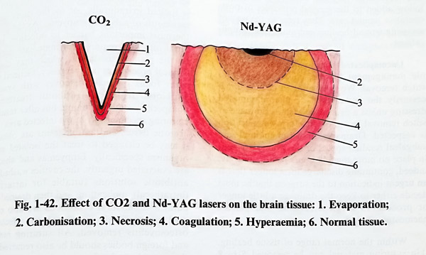
CO2 lasers lack significant penetration and their energy is completely absorbed by water. Therefore this action is suitable for the removal of tissues without damage on the underlaying structure and for incisions with minimal border necrosis. Its hemostatic efficiency is not so good, as blood vessels do not sgrink after coagulation.
NdYAG lasers have much deeper penetration than the CO2. Their power can be increased much more than the CO2 and the beam can be conveyed easily through fiberoptics. This type of laser coagulates and carbonises the tissues without easily vaporising them. Hemastasis with it is easier and even small and diffuse bleeding can be stopped by it (Fig. 1-42).
Both frequencies of radiation are on the infrared side of the spectrum and therefore invisible, so they need a visible light source with them to be guided. This is achieved with a visible “pilot” laser beam completely coinciding with the main beam. Most suitable for that is the He-Ne low powered laser wich emits in the red part of the spectrum.
Another inportant problem involves the technique of manipulating with the laser beam. There can be two options for microsurgical use – through the optical system of the microscope or by a fine fibreoptic channel.
The laser beam can be incorporated to the microasugical field of the microscope by a special micromanipulator attached to the front lens of the microscope. This micromanipulator contains a semi-transparent mirror under hand control, which reflects the beam from the laser onto the surgical field without disturbing the binocular vision through the microscope. With a small joystick, the surgeon can easily move the red spot of the pilot beam to the required point over the target and only after that is pressed the main laser footswitch. There is additional control for the focus (at the frontlens attachment) that can permit the use of a focused and defocused beam.


The other option is the fine fiberoptics path. Being also a fine instrument, it can be inserted deep in the operative field and handled under the microscope. The fiberoptics channel tip focuses the beam close to its end and is used defocused enlarging the distance to the object. The fiberoptics cord has certain flexibility and can be inserted through small openings. Both methods have their own conveniences. The micromanipulator must have direct visibility of the object under the microscope, but it is absolutely touchless. The fiberoptics can reach hidden and deep parts of the microsurgical field (for example debulking of small deep tumours), but can be not absolutely touchless.
Another important microsurgical tool is the ULTRASONIC ASPIRATOR. Its active part is an ultrasonic generator (24 to 38 kHz) applying its energy to a fine tip usually 2-3 mm in diameter. The size of the tip has its limitations in its miniaturisation because of the wavelength of the frequency used. The vibrating tip is the active end of a handpiece, designed as a microinstrument. Technical limitations permit bending of the handpiece to 15 and 30 degrees, but preclude a bayonet shape. The ultrasonic energy is designed to destroy tissue by cavitation. The power can be regulated so as to control the extent of tissue destruction. However tissue removal is achieved by the suction and irrigation systems attached to the hand piece. These systems also protect the active tip (and surrounding tissue) from overheating.
Ultrasonic aspiration reduces much of surgical trauma during manipulation, as pulling and displacement of normal structures is not needed for tissue removal. All tissues are not prone to ultrasonic aspiration easily. Those which have higher water contents are more easily aspirated.
On the other hand, all tissues with much fibrous collagen content cannot be fragmented, even with high power. Therefore some tumours, as some meningiomas, may sometimes not be suitable objects for ultrasonic aspiration. Despite this it can be beneficial for preserving major blood vessels, as they always contain much more connective tissue that the surrounding tumour tissue, so their walls are spared. The fragmented tissue obtained with the ultrasonic aspirator can be even studied with histo- and cyto-pathological methods.
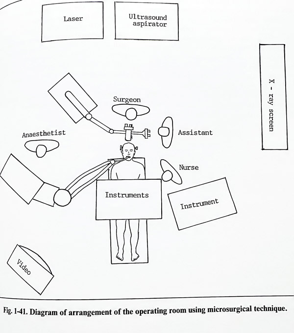
As a matter of principle, the assistants are not too busy while the work is proceeding under the microscope. They do however have the important task of following everything that is occurring outside the operative field. When the surgeon, watching through the microscope, manipulates with the self-retaining retractors, the assistant must be experienced in handing the instruments. Another important task of the assistants is to protect the exposed brain over the entire operative field beyond the view of the microscope. Assistants must sound an alarm should there be any danger of this part of the brain being injured. The role of the scrub nurse must not be undervalued, and it includes cleaning the tips of the forceps used for bipolar coagulation after every use, constantly maintaining the irrigation system runnung with saline and pass the surgeon small pieces of cotton whenever these are re-quired and positioning them conveniently for the surgeons in the operative field. It is also necessary for the scrub nurse to know the requirements for every stage of the operation and to be ready to be of support immediately a serious complication arises, for instance, in case of sudden bleeding.
POSTOPERATIVE WOUND CARE
Postoperative wound care after a craniotomy or craniectomy has few specific rules to assessing wound healing and managing complications.
Uncomplicated wound care consists of simple management by dressing, drainage and stitch removal. We prefer not to change unnecessarily the undamaged or uncontaminated dressing unless a procedure such as drainage or stitch removal has to be done or a complication is suspected. Drainages from the epidural and subgaleal residual spaces of the wound are kept in place no more than 48 hours postoperatively; indeed, continuing discharge for a longer time is an urgent indication to the surgeon that he must decide either to keep the drainage a little longer or proceed to surgical revision and perfect haemostasis.
Within the normal range of tissue healing, skin suturing material can be removed 5 to 8 days after surgery. Even with apparently healed wound incision the presence of separating tension of the edges may require leaving the stitches up to the eighth day. Posterior fossa wounds are kept with stitches in longer due to the higher tension applied by neck muscles.
However we do not recommend keeping them more than 12 days, except under very special circumstances.
Wound complications require specific care depending on the complication. The most important of them is bleeding in the operative wound. Bleeding can occur in any of the residual cavities after surgery. The intracranial blood cloth acts as a space occupying lesion with acute brain compression. It is detected by clinical monitoring criteria and imaging studies and requires the utmost attention of the surgeon. If a cloth is producing deterioration in the neurological condition, increase in the intracranial pressure or is significant in size, it must be removed urgently by wound revision. The dura and all overlaying levels are once again closed with the same care as previously. Cloths located outside the bone flap and exerting tension on the flap and sutures must also be removed.
Wound infection affects different layers to a different extent. It can lead to the formation of an abscess, osteomyelitis or a fistulous path if it is limited extradurally, or to the more severe meningitis or subdural empyema. As the dura is the most important barrier to infection, intra-dural infection requires an immediate decision on the plan of treatment, which may contain surgical procedures in emergency. General measures focus on massive cover with systemic antibiotic treatment. Antibiotic treatment is nonspecific at the beginning, but after results of culturing specimens from the infected area - pus, necrotic tissue, and antibiograms are obtained it may be changed to treat the isolated microorganismс specifically. Empyemas and abscesses are evacuated urgently, the cavities washed with antibiotic solutions suitable for intracranial (intrathecal) application, and then drained. Epidural and subdural collections are also evacuated and drained, and any bone affected by osteomyelitis removed. All suturing material and foreign bodies should be also removed from the wound. A fistula persisting more than 2-3 weeks, despite local and systemic treatment indicates some underlying cause - infected necrotic parts, suturing or plastic materials, or sequestrated osteomyelitic bone. The infected nidus can then only be identified and removed through a thorough surgical revision.
CSF leak can be observed at different stages of wound healing. The most common cause is the gapping dura, usually associated with a certain degree of increase in CSF pressure. Dural closure, even with a graft, is the best preventive measure against a leak. A persisting CSF fistula after the complete healing of the wound much reduces its chances of spontaneous closure, and it must be closed surgically. The surgical revision of the wound is undertaken to identify the dural defect and perform its most thorough and watertight closure. Before decision for surgery is taken, closure of the fistula by stitching and lumbar drainage can be attempted. A decision on surgical closure should not be delayed more than 2-3 weeks because of the risk of meningitis. However, if the fistula is already associated with meningitis, closure should be postponed until the complete resolution of the intrathecal inflammation (and CSF hypersecretion) is achieved.
Subgaleal fluid collections can be observed during wound healing, but there is an established policy always to avoid unnecessary punctures and evacuations due to the risk of subsequent abscesses formation or meningitis (if the collection is CSF). Follow-up has demonstrated reabsorption in a high percentage of such cases without any specific measures.
Necrosis and poor healing are treated cautiously, sparing as much vital tissue as possible from the wound. Necrotic parts are excised to facilitate resuture or granulation formation.
Resuturing is undertaken only after complete control of secondary infection. Some graftings and flap rotations have improved the results of secondary treatment of complicated wounds.
Postoperative radiotherapy is delayed until a complete control of wound complications is established.
TRIDIMENTIONALLY ORIENTED NEUROSURGICAL TECHNIQUE
THE IDEA AND THE PRINCIPLES
The need for tridimensional (3D) representation of the intracranial structures during surgery has been a persistently perceived requirement through the whole history of neurosurgery. The high complexity of structures and the inability to sacrifice tissues with few exceptions has increased the demand for surgical routes, which are minimally invasive and functionally compatible with normal function.
Microsurgery has only satisfied these demands in part. When dealing with structures accessible by brain retraction, microsurgery alone has dramatically reduced surgical tissue compromise. This is not the case, however, when treating intrinsic brain lesions. Microsurgery of the skull base and within CSF spaces always provides and follows well definable anatomical reference points, but once the brain tissue is penetrated, these extracerebral reference points become much less reliable. Neurosurgical techniques have provided different solutions for this problem that are concurrent with the present level of technology. Through all these decades, irrespective of the solutions achieved, the main goal has always been the same: to provide a precise tridimensional description of the intracranial space and to translate this information to identification and guidance in a quantitative manner with respect to the real surgical procedure. In simple terms this permits the surgeon to know which structure he has reached and estimate in precise measure how to reach the structure he wants.
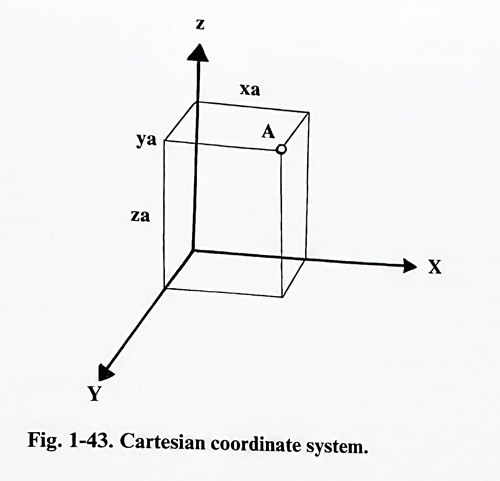
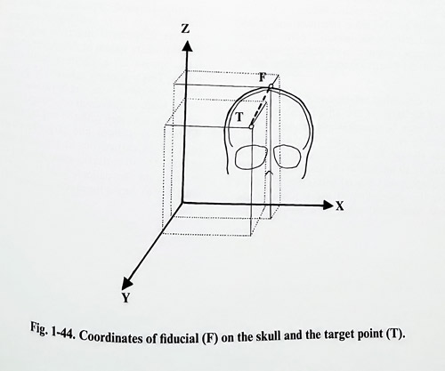
Localization in space is described mathematically by co-ordinate systems. Every point in space can be defined by its relation to another point (point of reference), when placed in a coordinate system. The Cartesian coordinate system defines in space every point by its distances to three intersecting orthogonal planes. The point of orthogonal intersection of the planes is assigned as the zero point (x = 0;y=0;z=0;). In this way every point is described by its X, Y and Z coordinates (Fig. 1-43). With this principle as a cornerstone, all contemporary imaging information for the intracranial space is organized in coordinate systems, thence having numeric expression for every point of the structure investigated. Therefore it is essential to describe reference points for every imaging investigation of the brain and to define the measuring unit which will permit proper measurements on the images and valid conversions of them from one type of investigation to another. Computer processed images permit the surgeon to do these procedures easily and quickly. Next, the intracranial space of the real patient must also be described within a coordinate system. Considering the practical reasons inferred by a planned 3D oriented neurosurgical procedure, reference points are chosen based on anatomical landmarks of the skull, on or inside the brain or especially selected skull points, which are fixed markers, called fiducials (Fig. 1-44).
The 3D oriented neurosurgical procedures aim at different types of targets: structures or functional areas, either normal or pathological, depending on the objective of the surgical procedure. This permits the use of the 3D image guided procedure in virtually all areas and types of pathology in neurosurgery.
THE METHOD
The method of intracranial space localization was created on a basis of the geometrical concept of the quantitative description of space.
It was born at the beginning of the century when Horsley (a medical doctor) and Clark (an engineer) designed localizing equipment for application to animals, termed stereotactic. Its technical construction contained a targeting electrode holder moving along bars 'materializing' the three main coordinate plane intersections. At present, the technical solutions for localization in humans are implementing advanced technological methods. Regardless of the technological solutions required, the same method stands with minimal alterations and contains the same logical steps.
Every 3D localization procedure is planned first by the selection of a target. Targets can be morphological and functional, and visible or not on the imaging studies. Morphological targets are lesions that have to be approached for diagnostic or therapeutic reasons. Diagnostic problems may require tissue or fluid sampling. This can have an important bearing on further steps such as therapeutic methods, i.e., tumor and fluid collection removals, implantation of sources of interstitial radiotherapy, tubes, and other devices. Functional targets are the objective in functional neurosurgery, whereby alteration of function of a normal structure (activation or suppression) is used to treat certain neurological disorders of movement, intractable pain and so on. As a next step the intracranial space is imaged and the target visualized. This can be done with different imaging methods, but must reveal all the information necessary for defining the lesion or structure of interest and all other structures that will be important in relation to the target, or to avoid complications. Some functional targets are not visible well even on MRI. They are reproduced on the imaging studies by calculating their position from visible ones. The precise basis for such calculation can be taken from stereotactic atlases. These atlases are valuable morphologic studies that demonstrate the human brain in an ordered sequence of stained sections in the three essential planes: the axial, the coronal and the sagittal. They are oriented and have reference measurements (in the form of grids) to the most common and widely accepted intracranial reference points such as the anterior commissure, posterior commissure, floor of the fourth ventricle, fastigium and so on.
As mentioned before, essential for the imaging of the target is that all the acquired information must be quantitatively defined in space regarding reference points. These reference points should be used consistently and be present on all imaging investigations (CT, MRI, angiography, and others) permitting, for every study, the description of every point of the imaged intracranial space by a set of coordinates. The digital transformation of all data permits further operation with this information in exact mathematical measurements and with the use of all available methods of digital processing. There is no doubt that the preselected target already has digital expression, at this stage. This digital expression however must be translated to the coordinate system of the patient. This is easily performed by using the same reference points at the time of 3D localization surgery. Performing the appropriate measurements to the reference points makes possible reproduction of the target and the other structures of interest within the patients' coordinate system.
THE TECHNIQUE
The method has been brought to reality by different schools and neurosurgeons creating ingenious innovations for materializing the coordinate system at the patients' head with maximum precision of data translation from the imaging studies and minimum burden for patient and surgeon. Following the idea of Horsley and Clark, Spiegel and Wycis constructed the first stereotactic instrument for humans. It became clear, that for the requirements of loca-lization, the extracranial skull reference points are not reliable for the normal brain. Subsequently designed stereotactic systems had to translate data from radiological investigations imaging intracranial structures and lesions to the coordinate system of the patient (the stereotactic apparatus) using intracranial reference points. This wás easily achieved by performing all investigations with the apparatus mounted on the head, and, without removing it, proceeding to the next step with the surgical approach.
When investigations and target localizations couldn't be carried out at the same surgical session, the stereotactic apparatus had to be mounted on the patients head in exactly the same way as during the previous session.
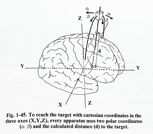
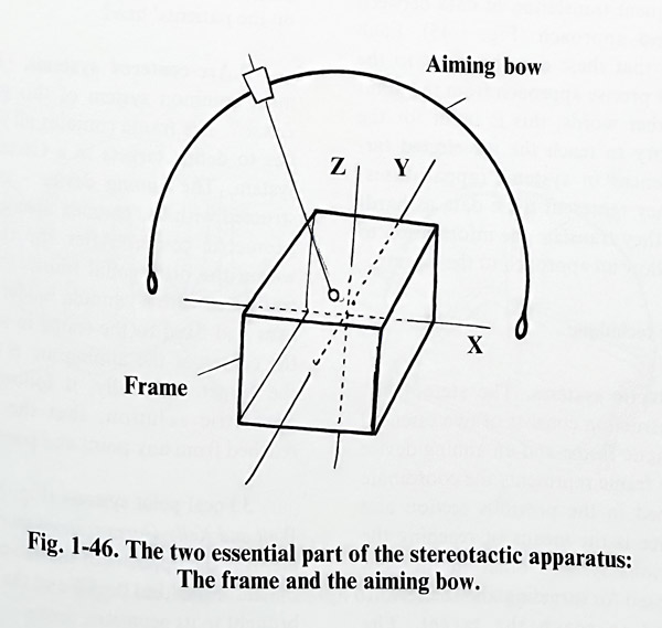
CONTEMPORARY STEREOTAXIS.
Many different systems have passed through several stages of innovation and perfection, evolving to contemporary universal systems, suitable for many different applications after the localizing part of the procedure has been done. Three of these are most commonly used in their latest generation: the Lecksell system, the Riechert system and the BRW system.
For the different stereotactic systems the method is applied in the following way:
Main components of a stereotactic apparatus
The stereotactic apparatus is a technical representation of a coordinate system that can be easily and reliably fixed in a stable manner to the patients' skull. Its elements represent the different axes (perpendicular to each other) and angles (as polar coordinates on parts of circles), to which are attached fixators to the skull.
Every apparatus uses a Cartesian coordinate system and a set of two polar coordinates at least somewhere in its technical construction and the subsequent translation of data between localization and approach (Fig. 1-45). Each target requires that these data be input to the apparatus for a precise approach from the point of entry. In other words, this is input for the specific trajectory to reach the preselected target. All differences in systems (apparatuses) refer to how they represent these data as hardware and how they translate the information to the patient to allow an approach to the target.
The stereotactic technique
A. Stereotactic systems. The stereotactic apparatus construction consists of two essential parts: a stereotactic frame and an aiming device (Fig. 1-46). The frame represents the coordinate system mentioned in the previous section and the aiming device is the means of reaching the target. The various systems differ in the geometric method used for targeting and the technical device used to reach the target. The stereotactic surgical procedure defines the position of the target with respect to the frame and puts the aiming device in correct position to reach the target. Three main approaches for obtaining a geometric solution have been implemented in the construction of the stereotactic apparatuses; they may be used for their classification. They are:
Systems using polar coordinates. The best known members of this group are the Riechert-Mundinger and the BRW systems (Fig. 1- 47).
The aiming device is a bow with a cannula holder which has rotational movements defined by polar coordinates in two planes. The third defining parameter is the depth to the target along the trajectory. The position of the target is given with reference to the geometric center of the frame. To avoid the complicated calculations of angles needed to reach the target, its position is given on another frame, not mounted on the patients head, called a phantom. The aiming device is adjusted with the appropriate angles and depth with reference to the model of the target on the phantom, and without changing its settings is transferred back to the frame on the patients' head.
Arc-centered systems. (Fig. 1-48) The most common system of this group is that of Leksell. The frame contains all required properties to define targets in a Cartesian coordinate system. The aiming device - an arc - is constructed with the cannula always pointing to its geometric center. After the target is defined within the orthogonal frame, the aiming arc is translated with a cannula holder along the three axes and fixed to the frame in such a way, that the center of the aiming arc is coincident with the target. Logically, as a result from such a geometric solution, the target will be reached from any point and position of the arc.
Focal point systems (Fig. 1-49). The Todd-Well and Kelly-Goers systems work on this principle. It is the opposite of the arc-centered systems: the arc is the fixed device and the defined target is brought to its geometric center.
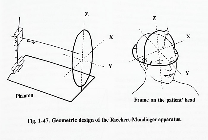
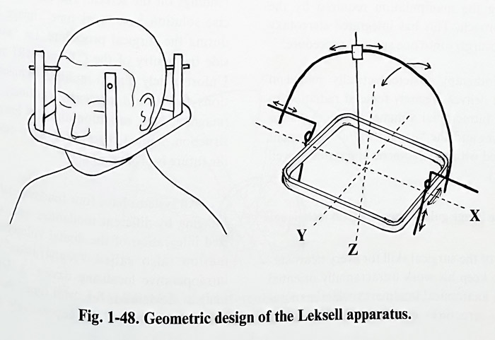
All major contemporary stereotactic systems were adapted to localization on CT and MRI images. For this, they required adapters for their frames. A precondition is that the adapter or any part of the frame on the head of the patient during imaging should not produce artifacts and decrease the quality of the study.
Scanning devices have the advantage that they provide axial images of the intracranial content that can be exactly defined with respect to the adapters position and by this they allow the measurement for the target along one of the coordinate system axis. The other two measurements can be obtained directly from the 'cuts' imaged where the target is visible or reproduced (Fig. 1-50).
With the constant improvement of software, the acquisition of information from scanning imaging techniques naturally leads to the more realistic volume representation of some targets and generated volume stereotaxis. From this moment on, the combination of open and stereotactic methods was made possible and opened new horizons for the method. The stereotactic localization procedure can be used also to perform subsequent "open" surgery.
During the "open" procedure is required that at least part of the stereotactic apparatus to be mounted on the patient's head to provide localization. This requirement has induced further redesign of the major stereotactic systems in such a way as to permit access for a craniotomy without disturbing the manipulation required by the open approach. This has integrated stereotaxy and microsurgery into one surgical procedure.
Radiotherapy is stereotactically guided in units that deliver precisely focused radiation to a limited intracranial volume of tissue. For that purpose linear accelerators and the "gamma knife" equipment are coupled with a stereotactic apparatus (Fig. 1-51).
INTERACTIVE IMAGE GUIDED NEUROSURGICAL TECHNIQUE
Part of the surgical skills for every neurosurgeon is to keep his work intracranially oriented to certain anatomical landmarks while manipulating the structures of interest and avoiding unnecessary tissue damage. This “navigational” experience is acquired with many years of practice and participation in a great number of operative procedures. That this knowledge cannot be obtained on the anatomical specimens only is obvious, because during surgery structure is uniquely distorted by pathology, anatomical variation, limited fields of view and non-standard approach directions. Following the principle of the 3D oriented neurosurgical technique and the possibility of having a tridimensional volumetric representation of the intracranial space in any desired viewing projection, intracranial "navigation" became possible with the help of localizing devices. This avoids the inconvenience of the stereotactic frame and the aiming bow obstructing the view at manipulating space during"open" surgery.
Based on these principles, every localizing system uses its own method for digital registration of the position of a special instrument (pointing arm) or a conventional one (forceps, dissector) with a special small attachment to permit its detection in space (by transmission or reflection). With the aid of this system, the surgeon approaches the structures following the position of his instruments not only within the surgical field itself, but also on the screen where the target is visible on the imaging studies. Registration reference points (fiducials) are usually placed on the skull to avoid any brain shift due to the evacuation of CSF or cystic fluid that would provoke erroneous readings on the screen. The ultimate, most precise localization solution is a "real time" image acquired during the surgical procedure, i.e. "surgery inside the gantry of the CT or MRI machine" Unfortunately due to multiple technical limitations this is not presently routinely possible (magnetic field, radiation, time for image reconstruction, limited space, and many others), but the future is promising.
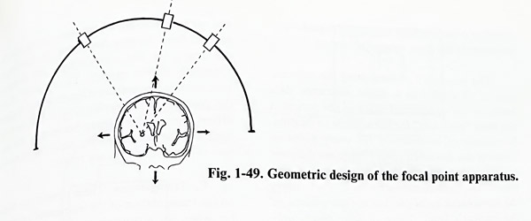
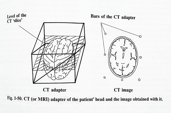
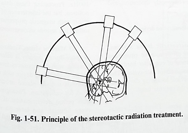
All localization “navigational”systems have four fundamental steps. 1.Imaging by different modalities and computerized integration of the digital volumetric information (also called "registration"). 2. An intraoperative localizing device setup. 3. Display of registered images. 4. Methods for "real time" feedback of information derived intraoperatively by ultrasound or other methods permitting digital representation in the same volumetric system.
The possibility of integrating the information of all types of studies (CT, MRI, radionuclide and so on) in a single volumetric data bank for the intracranial space of the patient is the ideal "roadmap" for intracranial navigation.It consists of information for every point or voxel of the described space. There are different ways to describe this space in quantitative terms. The most commonly used way is to apply the stereotactic principle, but not use a frame on the patient's head. Three or four points only (called fiducials too) are used as reference for the description of space. All investigations are registered with regard to these points, where all are clearly detectable on the images and their positions are determined first. External, artificially fixed markers or anatomical points on the skull can serve as fiducials. Giving every one of the fudicials the same values of all coordinates (same voxel) on all imaging studies integrates the images in a data bank. Another way of integrating the images is to calculate the corresponding points on the different studies with respect to some internal (usually skull) land-marks. This method requires a massive amount of mathematical computer operations and may be not precise when imaging the landmarks in different modalities (MRI, CT, radionuclide scanning, and so on). This last method has its clinical application based on the possibility of analysing images (curves and surfaces of the skull) and superimposing them. The rest of the existing techniques have only theoretical and research value.
INTRAOPERATIVE LOCALIZATION DEVICES
A/Modified stereotactic frame systems.
These are elements of stereotactic equipment that, with the addition of computer hardware, reproduce a stereotactic coordinate system. This simplifies the mechanical localizing device that must be attached to the patients' head.
B/Localization arms. These mechanical arms can be active (with independent motion) or passive (moved by the surgeon). They are calibrated initially with respect to the fiducials and next are actively or passively driven into the surgical field. Screens displaying the position of the arm's tip are observed by the surgeon constantly during surgery. The "joints" of the arm contain transducer elements that register the position of the device (Fig. 1-52). Actively driven arms (more clumsy because of the mo-tors) are the contemporary basis for robotization of neurosurgery in the future.
C/ Triangulation. These devices are based on the triangulation of the instrument's pointing tip when an ultrasonic or infrared signal is emitted from it. The source on the instrument emits the signal and several receivers (micro-phones or infrared cameras) detect it. The differences in the position of the instrument are estimated by computer software according to the triangulation data (angles and transmission time to the receivers) (Fig. 1-53). Direct visibility between the source and the receivers should be maintained throughout the surgery. An emission source can be added to the surgical microscope and with consideration of its focal distance, the position can be integrated precisely in these localizing systems.
D/Magnetic field guidance. The principle used is that of the measurement of the magnetic field strength from a source around the head of the patient during surgery. Magnetic impulses emitted along the three main axis (X, Y, Z) of the coordinate system are registered in the corresponding main planes at a frequency of 100Hz. Processing these data gives the tip's position, but the interference of all metal instruments with magnetic properties that create distortion is a serious inconvenience.
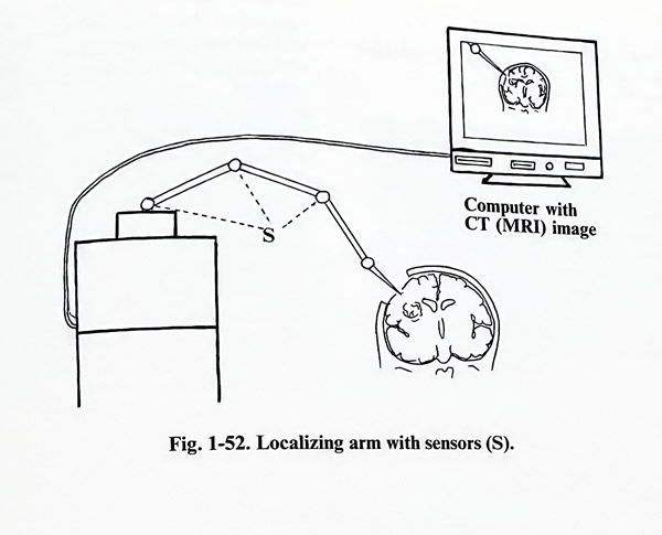
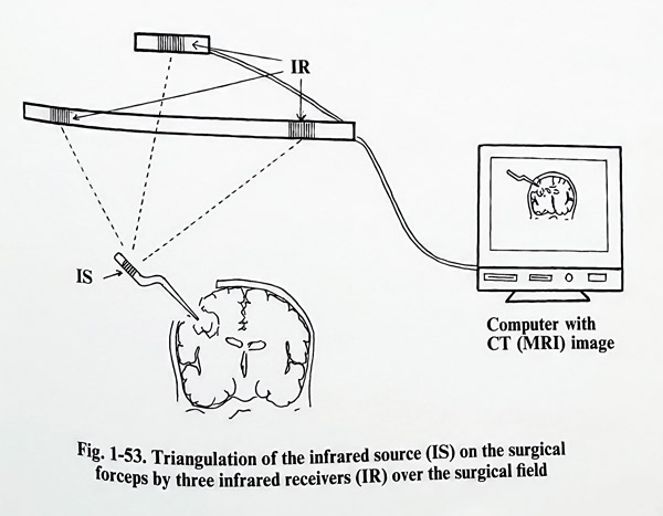
E/ Machine vision. Tridimensional position in space during surgery is determined by video imaging with two cameras from two different points. A localizing device with multiple fiducial points is initially placed around the patients' head for calibration. Photogrammetric techniques permit determination of the position of every fiducial, and later on during surgery of every point inside the surgical field, provided the patients' head and the cameras are not moved. This is based and related to
the preoperative imaging studies. Surgical instruments are tracked by attaching fiducials to them. Naturally, this method requires complete constant visibility and powerful computer hardware.
All these technologically advanced surgical methods, regardless of being presently reserved for the more specialized centers, represent the direction of progress and are harbingers of the expected new horizons of neurosurgical technique.