LANDMARKS ON THE SCALP AND SKULL
BASIC ANTHROPOLOGICAL POINTS
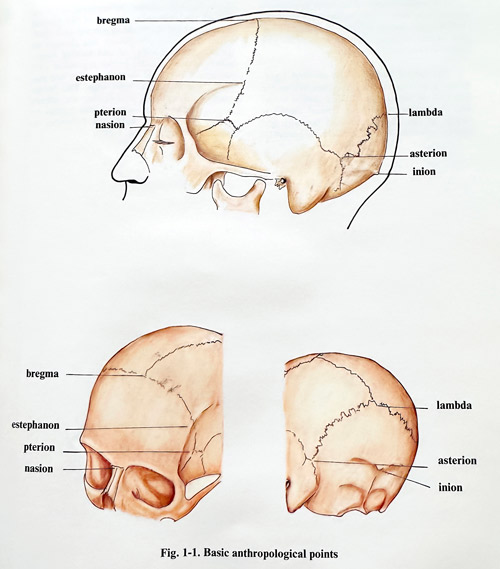
Some of the essential anthropological points are very useful references for the precise description of the planned approach. The essential advantage comes from the feature that they are seen at the same time on the patients head as well as on the imaging studies showing the intracranial pathology found (Fig. 1-1).
1) Nasion. A point situated on the base of the nose, in the middle of the nasofrontal suture.
2) Bregma. A point, where the coronal and sagittal sutures join; on the intact skin it can be determined as the intersection of the two perpendicular lines, drawn up from the midpoints of both zygomatic arches.
3) Lambda. The point, where the lambdoid and sagittal sutures join.
4) Inion. This point corresponds to the external occipital protuberance.
5) Estephanon. Symmetrical points on both sides of the skull at the place, where the coronal suture crosses the superior temporal line.
6) Pterion. Approximately symmetrical points on both sides of the skull at the place, where the frontal, parietal, sphenoidal and temporal bones join the large wing of the sphenoid bone.
7) Asterion. Similar to the previous bilateral points at the place, where the occipital and parietal bones join with the mastoid part of the temporal bone.
EXTERNAL PROJECTIONS OF THE BRAIN HEMISPHERES
The sagittal border of the brain hemisphere corresponds more or less to the midline of the
cranial vault, bearing in mind the midline location of the superior sagittal sinus. This line begins at nasion and ends at inion.
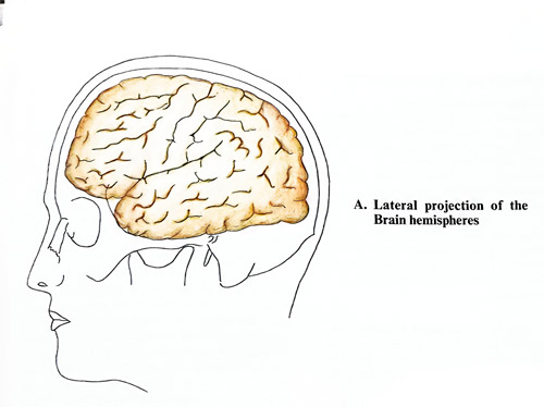
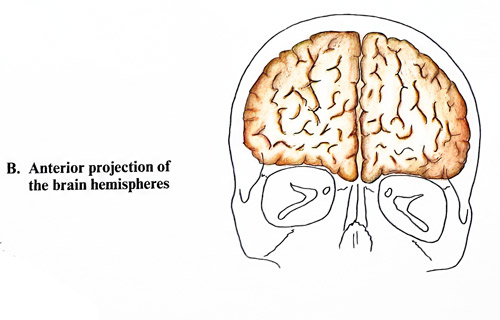
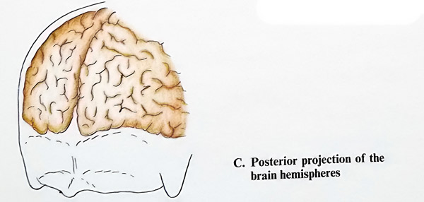
The lateral border of the cerebral hemisphere is uneven and its projection follows the floors of the anterior and middle cranial fossae and the tentorium. In the frontal region it begins at the nasion, directed laterally but slightly upwards. The place where the border of the frontal lobe reaches the temporal, is 1.5 cm behind the zygomatic process of the frontal bone and 2 cm above the zygomatic arch level. From that point the border line changes its direction, being a curved line turning downwards to the external auditory meatus and passing 1 cm above its upper edge. This border reaches the asterion and further on, already corresponding to the occipital lobe reaches the external occipital protuberance following the nuchal line (Fig 1-2).
TOPOGRAPHY OF THE MAIN CEREBRAL SULCI
The lateral and central fissures are the two most important sulci on the convex surface of the cerebral hemisphere, serving as a reference for the localisation of other sulci and convolutions (gyri) of the hemispheres. However, when planning craniotomies, we should consider also the displacement produced by mass lesions, as after the planned cortical exposure, gyri and sulci can be found to be not exactly at their expected places. Although these localising methods may be simplified, they can nonetheless provide useful information to the surgeon undertaking a procedure, not preceded by 3D CT or MRI imaging.
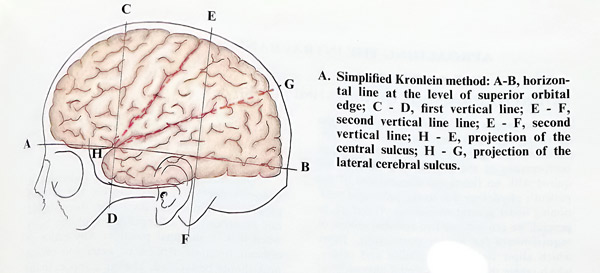
According to the simplified Kronlein method, a horizontal line is drawn at the level of the upper orbital edge. Then two vertical lines are superimposed: the first one through the midpoint of the zygomatic arch and the second - through the posterior border of the auricle. The line connecting the crossing point between the horizontal and the first vertical line with the point, where the second vertical line crosses the midline, corresponds to the projection of the central sulcus. If the angle, formed by the projection of the sulcus centralis and the horizontal line is bisected, this last line will correspond to the lateral sulcus (Fig. 1 - 3, A).
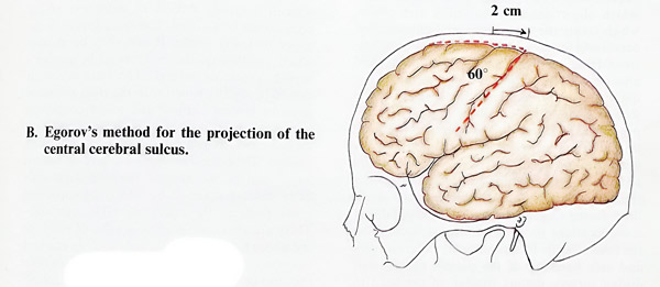
After Egorov's method the central sulcus projection corresponds to the line, which begins 2 cm behind the midpoint of the nasion-inion distance on the sagittal line and forms a 60 degrees angle with it, open in an anterobasal direction (Fig. 1 - 3, B).
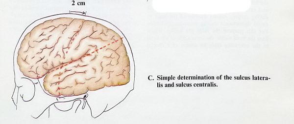
The topography of the two basic fissures of the cerebral hemisphere (the lateral and central sulci) can be determined in the following simple way: the lateral sulcus lies along the length of the line connecting the zygomatic process of the frontal bone with a point at 3/4 of the length of the nasion-inion distance on the sagittal line starting from the nasion. The central sulcus corresponds to the upper half of the line, connecting the midline (2 cm behind its nasion-inion midpoint) and reaches the middle of the zygomatic arch (Fig. 1- 3, C).