MICROSURGICAL TECHNIQUE AND INSTRUMENTATION
Microsurgery is the most essential part of the surgical work on the nervous system. With its application a number of new operative methods have been introduced and many others have been improved upon. Microsurgery - the performance of micromanipulation under optic magnification - has significantly improved neurosurgery.
Surgical microscopes have certain main characteristics, and much similarity is found among the different commercial types available in the market. The main characteristics include the range of the magnification power, the type and power of illumination and the mechanical characteristics of the stand. A number of accessories are also considered essential such as the photo and video equipment, and facilities for observation by assistants.
The magnification power at present usually ranges much wider than that necessary. It is between 4X and 20X for the experienced surgeon, but special conditions during surgery can require more. It is controlled by a manually or electronically operated zoom. Contemporary optics permit a relatively wider field and depth of focus despite the increase of magnification.
The placement of the optical part of the microscope should not disturb access to the surgical field. Therefore a minimal focus distance is usually required (200-250 mm), which should be able to be increased in a gradual way up to 350 to 400 mm (for deep skull base, transsphenoidal surgery and so on). Eyepieces that can change their angle relative to the front lens axis are convenient as they permit a much more relaxed position for the surgeon regardless of the position of the surgical field.
To perform micromanipulation on magnified objects is required greater light intensity per unit of surface than durinng routine surgery. At the same time to avoid object shadowing, illumination must follow the axis of observation. Powerful illumination must not lead to heating of the surgical field. At present this is achieved by powerful halogen sources of several hundred watts conveyed to the field by fiberoptics.
Safe micromanipulation and stability of the image require firm fixation (to the ceiling of the operating room) or a heavy stand for the microscope. With all tools affixed, microscopes have now become very heavy, with the result that mechanical balancing of the system is now essential. A well-balanced microscope now requires minimal effort from the surgeon and 1-2 seconds to be repositioned, even in a difficult and tense moment of the operation.
Microsurgical skills differ significantly from the macrosurgical ones. Although all essential manoeuvres such as dissection, suturing, artery occlusion, irrigation/suction, retraction and others are the same, the decreased pressure on neural structures and tissue disruption leads to less traumatic manipulation. However, to achieve this special instrumentation and is required training.
As the types of essential manoeuvres are the same, the types of instruments are the same too, but designed to handle easily microobjects.
In a microsurgical set we will find scissors, forceps, needleholders, knives, hooks, dissectors with correspondingly smaller tips, blades and branches. However, because of their tiny size they are made of much harder and more durable materials. Their handles must permit easily controlled fine movements, and are therefore best if they have rounded shapes. The instrument should be not heavy and its weight should be centred in the hand of the surgeon, not at the tips. Handling the instruments should not obstruct vision through the microscope, so bayonet shaped branches are preferred (Fig, 1-38;1-39).
The suction and irrigation systems are an important adjunct to everyday microsurgical technique. Ordinary suction tubes are not convenient for microsurgery. Microsurgical suction tubes should be of finer diameter, the edges of the tip opening should not be sharp, and the distal part of the tip should be more malleable.
The negative pressure of the suction tip should be controllable (too high negative pressure can injure by sucking up neural tissue and fine vessels). Suction is intended to drain the fluids from the operative field that obstruct micromanipulation and vision, but it should be applied cautiously. The inexperienced should not approach important objects with the tip without the protection of a cottonoid. Moreover, the structures have the tendency to dry as a result of the fluid suction and the illumination; therefore intermittent saline irrigation is applied. This also prevents sticking of the instruments to tissues (especially the bipolar forceps), and eases the removal of cloths. Combining suction and irrigation in one handpiece is sometimes a convenient solution.
Manual retraction has proven to be inadequate because the association of physiological tremor of assistant and surgeon makes micro-manipulations of the microstructures very difficult and unsafe. Retraction of brain and other important structures, performed usually with spatulas, should rely on firm fixation and arms with plasticity when locked. Firm fixation guarantees against added movements from outside and relieves the assistant, and an arm with shape adjustable properties (usually controlled by lock) prevents excessive pressure on the retracted object. Retractors are firmly fixed on the edge of the craniotomy (but the key reduces the exposed surface), on the three pin holders or additional devices (rings, arches) attached to it in more convenient position to the operative field, or even to the operating table (Fig. 1-40).
Bipolar coagulation is also an essential tool for microsurgery. Its convenience comes from its basic properties: the effect is confined between the branches of the forceps (its electrodes). Spread of heat and current is minimal and control of the electrical power is much more precise than with monopolar coagulation. Morphologically it provokes shrinkage of tissues and occlusion of vessels and is, therefore, a very effective haemostatic tool. Its tips should be fine but not pin point sharp (some surgeons prefer very sharp tips for simultaneously using the same instrument for dissection). Its tips have limited active surface (non insulated). The forcepses have different lengths and are straight or bayonet in shape.
When haemostasis is intended for bigger arteries or veins, when the vessels are under relatively higher pressure, when the shrinkage of the wall by bipolar coagulation is unable to occlude them or the walls have to be preserved, then the vessels must be clipped. Clipping in microsurgery is an essential method which assures safety of haemostasis. Many types of cl to ips have been designed. Every removable clip has branches to be closed encroaching the vessel, a key and spring providing stability and predefined force of closure and ends where the application forceps is fitted when they are placed and removed The best types have a great variety of shapes and lengths of the branches: straight, curved, bayonet and so on. If the closing force of a clip is reduced to a level that does not seriously damage the endothelium, it can be used only for temporary occlusion. Clips are placed and removed with a special forceps designed for each clip type, which has also been designed as a micro-instrument.
Some other microinstruments are also of significant value. One is the microdrill permitting removal of bone and hard tissues. The diamond drills, particularly the airpowered types, have less vibration and are less likely to destry adjacent tissues. Specila attention must be paid to starting and stopping the drill. Small mirrprs, mounted on bayonet-shaped handles and set at various angles, are useful in inspecting narrow spaces located outside the surgeon’s direct visibility, as the is the space under the anerior clinoids, interpeduncular fossa or internal auditory meatus.
However, as with every other specific surgical technique, microneurosurgery cannot be embarked upon immediately to without previous training. That is needed until a certain basic level of skills is ac hieved. There some widely accepted types of experimental technical procedures to be performed by a trainee before basic training in microsurgery is recognized. These techniques include the performance of patent nerve and vascular anastomoses on rat’s carotids, jugular veins and sciatic nerves in different variations. The surgeon should also adapt to lengthy work under the microscope without tension and have control of everything needed for the work in the operative field without looking outside it (switches, pedals, standard placement of instruments outside, etc.). Microsurgery is performed at its best with hands already adapted to the instruments and the operative field, not tired and with the surgeon in sitting position (there a special chairs with armrest support). Microsurgery is improved with practice and it is very effective for the trainee to be an apprentive to the master for a while (Fig. 1- 41).
Lasers combined with microsurgical technique are a very effective tool in brain surgery. The action of the laser does not require manipulation with an instrument on the brain and removal of tissues, coagulations and incisions are touchless.
Two main types of laser sources are used at present in neurosurgery: the CO2 and the NgYAG.
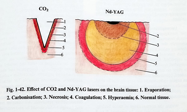
CO2 lasers lack significant penetration and their energy is completely absorbed by water. Therefore this action is suitable for the removal of tissues without damage on the underlaying structure and for incisions with minimal border necrosis. Its hemostatic efficiency is not so good, as blood vessels do not sgrink after coagulation.
NdYAG lasers have much deeper penetration than the CO2. Their power can be increased much more than the CO2 and the beam can be conveyed easily through fiberoptics. This type of laser coagulates and carbonises the tissues without easily vaporising them. Hemastasis with it is easier and even small and diffuse bleeding can be stopped by it (Fig. 1-42).
Both frequencies of radiation are on the infrared side of the spectrum and therefore invisible, so they need a visible light source with them to be guided. This is achieved with a visible “pilot” laser beam completely coinciding with the main beam. Most suitable for that is the He-Ne low powered laser wich emits in the red part of the spectrum.
Another inportant problem involves the technique of manipulating with the laser beam. There can be two options for microsurgical use – through the optical system of the microscope or by a fine fibreoptic channel.
The laser beam can be incorporated to the microasugical field of the microscope by a special micromanipulator attached to the front lens of the microscope. This micromanipulator contains a semi-transparent mirror under hand control, which reflects the beam from the laser onto the surgical field without disturbing the binocular vision through the microscope. With a small joystick, the surgeon can easily move the red spot of the pilot beam to the required point over the target and only after that is pressed the main laser footswitch. There is additional control for the focus (at the frontlens attachment) that can permit the use of a focused and defocused beam.


The other option is the fine fiberoptics path. Being also a fine instrument, it can be inserted deep in the operative field and handled under the microscope. The fiberoptics channel tip focuses the beam close to its end and is used defocused enlarging the distance to the object. The fiberoptics cord has certain flexibility and can be inserted through small openings. Both methods have their own conveniences. The micromanipulator must have direct visibility of the object under the microscope, but it is absolutely touchless. The fiberoptics can reach hidden and deep parts of the microsurgical field (for example debulking of small deep tumours), but can be not absolutely touchless.
Another important microsurgical tool is the ULTRASONIC ASPIRATOR. Its active part is an ultrasonic generator (24 to 38 kHz) applying its energy to a fine tip usually 2-3 mm in diameter. The size of the tip has its limitations in its miniaturisation because of the wavelength of the frequency used. The vibrating tip is the active end of a handpiece, designed as a microinstrument. Technical limitations permit bending of the handpiece to 15 and 30 degrees, but preclude a bayonet shape. The ultrasonic energy is designed to destroy tissue by cavitation. The power can be regulated so as to control the extent of tissue destruction. However tissue removal is achieved by the suction and irrigation systems attached to the hand piece. These systems also protect the active tip (and surrounding tissue) from overheating.
Ultrasonic aspiration reduces much of surgical trauma during manipulation, as pulling and displacement of normal structures is not needed for tissue removal. All tissues are not prone to ultrasonic aspiration easily. Those which have higher water contents are more easily aspirated.
On the other hand, all tissues with much fibrous collagen content cannot be fragmented, even with high power. Therefore some tumours, as some meningiomas, may sometimes not be suitable objects for ultrasonic aspiration. Despite this it can be beneficial for preserving major blood vessels, as they always contain much more connective tissue that the surrounding tumour tissue, so their walls are spared. The fragmented tissue obtained with the ultrasonic aspirator can be even studied with histo- and cyto-pathological methods.
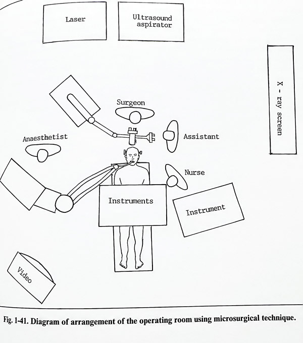
As a matter of principle, the assistants are not too busy while the work is proceeding under the microscope. They do however have the important task of following everything that is occurring outside the operative field. When the surgeon, watching through the microscope, manipulates with the self-retaining retractors, the assistant must be experienced in handing the instruments. Another important task of the assistants is to protect the exposed brain over the entire operative field beyond the view of the microscope. Assistants must sound an alarm should there be any danger of this part of the brain being injured. The role of the scrub nurse must not be undervalued, and it includes cleaning the tips of the forceps used for bipolar coagulation after every use, constantly maintaining the irrigation system runnung with saline and pass the surgeon small pieces of cotton whenever these are re-quired and positioning them conveniently for the surgeons in the operative field. It is also necessary for the scrub nurse to know the requirements for every stage of the operation and to be ready to be of support immediately a serious complication arises, for instance, in case of sudden bleeding.