
TRIDIMENTIONALLY ORIENTED NEUROSURGICAL TECHNIQUE
THE IDEA AND THE PRINCIPLES
The need for tridimensional (3D) representation of the intracranial structures during surgery has been a persistently perceived requirement through the whole history of neurosurgery. The high complexity of structures and the inability to sacrifice tissues with few exceptions has increased the demand for surgical routes, which are minimally invasive and functionally compatible with normal function.
Microsurgery has only satisfied these demands in part. When dealing with structures accessible by brain retraction, microsurgery alone has dramatically reduced surgical tissue compromise. This is not the case, however, when treating intrinsic brain lesions. Microsurgery of the skull base and within CSF spaces always provides and follows well definable anatomical reference points, but once the brain tissue is penetrated, these extracerebral reference points become much less reliable. Neurosurgical techniques have provided different solutions for this problem that are concurrent with the present level of technology. Through all these decades, irrespective of the solutions achieved, the main goal has always been the same: to provide a precise tridimensional description of the intracranial space and to translate this information to identification and guidance in a quantitative manner with respect to the real surgical procedure. In simple terms this permits the surgeon to know which structure he has reached and estimate in precise measure how to reach the structure he wants.
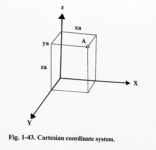
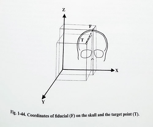
Localization in space is described mathematically by co-ordinate systems. Every point in space can be defined by its relation to another point (point of reference), when placed in a coordinate system. The Cartesian coordinate system defines in space every point by its distances to three intersecting orthogonal planes. The point of orthogonal intersection of the planes is assigned as the zero point (x = 0;y=0;z=0;). In this way every point is described by its X, Y and Z coordinates (Fig. 1-43). With this principle as a cornerstone, all contemporary imaging information for the intracranial space is organized in coordinate systems, thence having numeric expression for every point of the structure investigated. Therefore it is essential to describe reference points for every imaging investigation of the brain and to define the measuring unit which will permit proper measurements on the images and valid conversions of them from one type of investigation to another. Computer processed images permit the surgeon to do these procedures easily and quickly. Next, the intracranial space of the real patient must also be described within a coordinate system. Considering the practical reasons inferred by a planned 3D oriented neurosurgical procedure, reference points are chosen based on anatomical landmarks of the skull, on or inside the brain or especially selected skull points, which are fixed markers, called fiducials (Fig. 1-44).
The 3D oriented neurosurgical procedures aim at different types of targets: structures or functional areas, either normal or pathological, depending on the objective of the surgical procedure. This permits the use of the 3D image guided procedure in virtually all areas and types of pathology in neurosurgery.
THE METHOD
The method of intracranial space localization was created on a basis of the geometrical concept of the quantitative description of space.
It was born at the beginning of the century when Horsley (a medical doctor) and Clark (an engineer) designed localizing equipment for application to animals, termed stereotactic. Its technical construction contained a targeting electrode holder moving along bars 'materializing' the three main coordinate plane intersections. At present, the technical solutions for localization in humans are implementing advanced technological methods. Regardless of the technological solutions required, the same method stands with minimal alterations and contains the same logical steps.
Every 3D localization procedure is planned first by the selection of a target. Targets can be morphological and functional, and visible or not on the imaging studies. Morphological targets are lesions that have to be approached for diagnostic or therapeutic reasons. Diagnostic problems may require tissue or fluid sampling. This can have an important bearing on further steps such as therapeutic methods, i.e., tumor and fluid collection removals, implantation of sources of interstitial radiotherapy, tubes, and other devices. Functional targets are the objective in functional neurosurgery, whereby alteration of function of a normal structure (activation or suppression) is used to treat certain neurological disorders of movement, intractable pain and so on. As a next step the intracranial space is imaged and the target visualized. This can be done with different imaging methods, but must reveal all the information necessary for defining the lesion or structure of interest and all other structures that will be important in relation to the target, or to avoid complications. Some functional targets are not visible well even on MRI. They are reproduced on the imaging studies by calculating their position from visible ones. The precise basis for such calculation can be taken from stereotactic atlases. These atlases are valuable morphologic studies that demonstrate the human brain in an ordered sequence of stained sections in the three essential planes: the axial, the coronal and the sagittal. They are oriented and have reference measurements (in the form of grids) to the most common and widely accepted intracranial reference points such as the anterior commissure, posterior commissure, floor of the fourth ventricle, fastigium and so on.
As mentioned before, essential for the imaging of the target is that all the acquired information must be quantitatively defined in space regarding reference points. These reference points should be used consistently and be present on all imaging investigations (CT, MRI, angiography, and others) permitting, for every study, the description of every point of the imaged intracranial space by a set of coordinates. The digital transformation of all data permits further operation with this information in exact mathematical measurements and with the use of all available methods of digital processing. There is no doubt that the preselected target already has digital expression, at this stage. This digital expression however must be translated to the coordinate system of the patient. This is easily performed by using the same reference points at the time of 3D localization surgery. Performing the appropriate measurements to the reference points makes possible reproduction of the target and the other structures of interest within the patients' coordinate system.
THE TECHNIQUE
The method has been brought to reality by different schools and neurosurgeons creating ingenious innovations for materializing the coordinate system at the patients' head with maximum precision of data translation from the imaging studies and minimum burden for patient and surgeon. Following the idea of Horsley and Clark, Spiegel and Wycis constructed the first stereotactic instrument for humans. It became clear, that for the requirements of loca-lization, the extracranial skull reference points are not reliable for the normal brain. Subsequently designed stereotactic systems had to translate data from radiological investigations imaging intracranial structures and lesions to the coordinate system of the patient (the stereotactic apparatus) using intracranial reference points. This wás easily achieved by performing all investigations with the apparatus mounted on the head, and, without removing it, proceeding to the next step with the surgical approach.
When investigations and target localizations couldn't be carried out at the same surgical session, the stereotactic apparatus had to be mounted on the patients head in exactly the same way as during the previous session.
CONTEMPORARY STEREOTAXIS.
Many different systems have passed through several stages of innovation and perfection, evolving to contemporary universal systems, suitable for many different applications after the localizing part of the procedure has been done. Three of these are most commonly used in their latest generation: the Lecksell system, the Riechert system and the BRW system.
For the different stereotactic systems the method is applied in the following way:
Main components of a stereotactic apparatus
The stereotactic apparatus is a technical representation of a coordinate system that can be easily and reliably fixed in a stable manner to the patients' skull. Its elements represent the different axes (perpendicular to each other) and angles (as polar coordinates on parts of circles), to which are attached fixators to the skull.
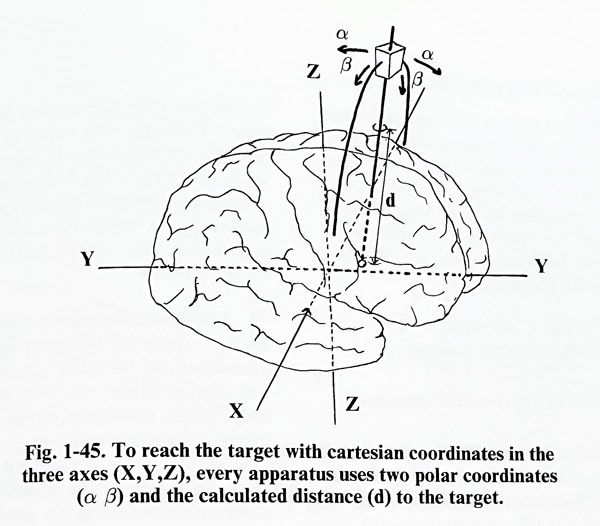
Every apparatus uses a Cartesian coordinate system and a set of two polar coordinates at least somewhere in its technical construction and the subsequent translation of data between localization and approach (Fig. 1-45). Each target requires that these data be input to the apparatus for a precise approach from the point of entry. In other words, this is input for the specific trajectory to reach the preselected target. All differences in systems (apparatuses) refer to how they represent these data as hardware and how they translate the information to the patient to allow an approach to the target.
The stereotactic technique
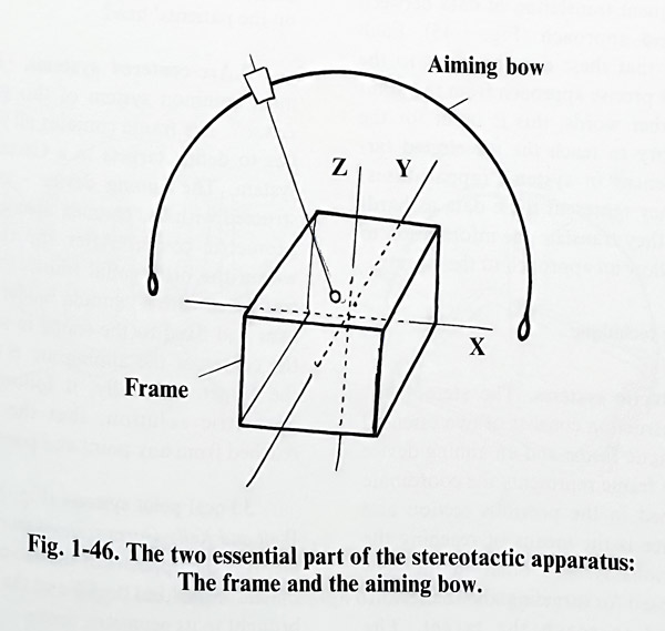
A. Stereotactic systems. The stereotactic apparatus construction consists of two essential parts: a stereotactic frame and an aiming device (Fig. 1-46). The frame represents the coordinate system mentioned in the previous section and the aiming device is the means of reaching the target. The various systems differ in the geometric method used for targeting and the technical device used to reach the target. The stereotactic surgical procedure defines the position of the target with respect to the frame and puts the aiming device in correct position to reach the target. Three main approaches for obtaining a geometric solution have been implemented in the construction of the stereotactic apparatuses; they may be used for their classification. They are:
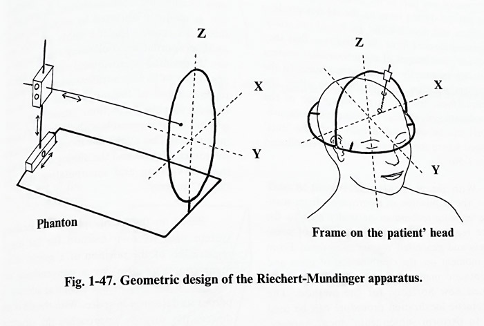
Systems using polar coordinates. The best known members of this group are the Riechert-Mundinger and the BRW systems (Fig. 1- 47).
The aiming device is a bow with a cannula holder which has rotational movements defined by polar coordinates in two planes. The third defining parameter is the depth to the target along the trajectory. The position of the target is given with reference to the geometric center of the frame. To avoid the complicated calculations of angles needed to reach the target, its position is given on another frame, not mounted on the patients head, called a phantom. The aiming device is adjusted with the appropriate angles and depth with reference to the model of the target on the phantom, and without changing its settings is transferred back to the frame on the patients' head.
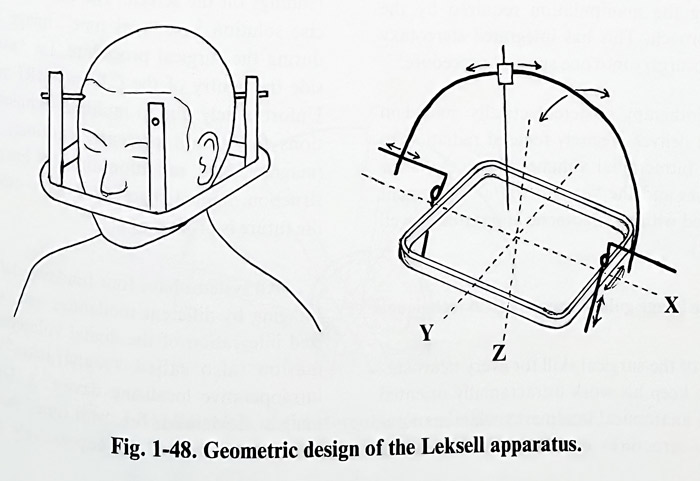
Arc-centered systems. (Fig. 1-48) The most common system of this group is that of Leksell. The frame contains all required properties to define targets in a Cartesian coordinate system. The aiming device - an arc - is constructed with the cannula always pointing to its geometric center. After the target is defined within the orthogonal frame, the aiming arc is translated with a cannula holder along the three axes and fixed to the frame in such a way, that the center of the aiming arc is coincident with the target. Logically, as a result from such a geometric solution, the target will be reached from any point and position of the arc.
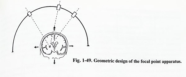
Focal point systems (Fig. 1-49). The Todd-Well and Kelly-Goers systems work on this principle. It is the opposite of the arc-centered systems: the arc is the fixed device and the defined target is brought to its geometric center.
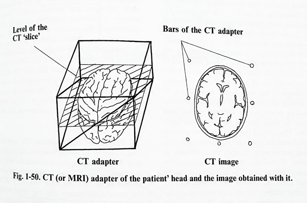
All major contemporary stereotactic systems were adapted to localization on CT and MRI images. For this, they required adapters for their frames. A precondition is that the adapter or any part of the frame on the head of the patient during imaging should not produce artifacts and decrease the quality of the study.
Scanning devices have the advantage that they provide axial images of the intracranial content that can be exactly defined with respect to the adapters position and by this they allow the measurement for the target along one of the coordinate system axis. The other two measurements can be obtained directly from the 'cuts' imaged where the target is visible or reproduced (Fig. 1-50).
With the constant improvement of software, the acquisition of information from scanning imaging techniques naturally leads to the more realistic volume representation of some targets and generated volume stereotaxis. From this moment on, the combination of open and stereotactic methods was made possible and opened new horizons for the method. The stereotactic localization procedure can be used also to perform subsequent "open" surgery.
During the "open" procedure is required that at least part of the stereotactic apparatus to be mounted on the patient's head to provide localization. This requirement has induced further redesign of the major stereotactic systems in such a way as to permit access for a craniotomy without disturbing the manipulation required by the open approach. This has integrated stereotaxy and microsurgery into one surgical procedure.
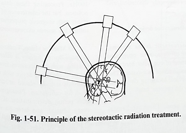
Radiotherapy is stereotactically guided in units that deliver precisely focused radiation to a limited intracranial volume of tissue. For that purpose linear accelerators and the "gamma knife" equipment are coupled with a stereotactic apparatus (Fig. 1-51).
INTERACTIVE IMAGE GUIDED NEUROSURGICAL TECHNIQUE
Part of the surgical skills for every neurosurgeon is to keep his work intracranially oriented to certain anatomical landmarks while manipulating the structures of interest and avoiding unnecessary tissue damage. This “navigational” experience is acquired with many years of practice and participation in a great number of operative procedures. That this knowledge cannot be obtained on the anatomical specimens only is obvious, because during surgery structure is uniquely distorted by pathology, anatomical variation, limited fields of view and non-standard approach directions. Following the principle of the 3D oriented neurosurgical technique and the possibility of having a tridimensional volumetric representation of the intracranial space in any desired viewing projection, intracranial "navigation" became possible with the help of localizing devices. This avoids the inconvenience of the stereotactic frame and the aiming bow obstructing the view at manipulating space during"open" surgery.
Based on these principles, every localizing system uses its own method for digital registration of the position of a special instrument (pointing arm) or a conventional one (forceps, dissector) with a special small attachment to permit its detection in space (by transmission or reflection). With the aid of this system, the surgeon approaches the structures following the position of his instruments not only within the surgical field itself, but also on the screen where the target is visible on the imaging studies. Registration reference points (fiducials) are usually placed on the skull to avoid any brain shift due to the evacuation of CSF or cystic fluid that would provoke erroneous readings on the screen. The ultimate, most precise localization solution is a "real time" image acquired during the surgical procedure, i.e. "surgery inside the gantry of the CT or MRI machine" Unfortunately due to multiple technical limitations this is not presently routinely possible (magnetic field, radiation, time for image reconstruction, limited space, and many others), but the future is promising.
All localization “navigational”systems have four fundamental steps. 1.Imaging by different modalities and computerized integration of the digital volumetric information (also called "registration"). 2. An intraoperative localizing device setup. 3. Display of registered images. 4. Methods for "real time" feedback of information derived intraoperatively by ultrasound or other methods permitting digital representation in the same volumetric system.
The possibility of integrating the information of all types of studies (CT, MRI, radionuclide and so on) in a single volumetric data bank for the intracranial space of the patient is the ideal "roadmap" for intracranial navigation.It consists of information for every point or voxel of the described space. There are different ways to describe this space in quantitative terms. The most commonly used way is to apply the stereotactic principle, but not use a frame on the patient's head. Three or four points only (called fiducials too) are used as reference for the description of space. All investigations are registered with regard to these points, where all are clearly detectable on the images and their positions are determined first. External, artificially fixed markers or anatomical points on the skull can serve as fiducials. Giving every one of the fudicials the same values of all coordinates (same voxel) on all imaging studies integrates the images in a data bank. Another way of integrating the images is to calculate the corresponding points on the different studies with respect to some internal (usually skull) land-marks. This method requires a massive amount of mathematical computer operations and may be not precise when imaging the landmarks in different modalities (MRI, CT, radionuclide scanning, and so on). This last method has its clinical application based on the possibility of analysing images (curves and surfaces of the skull) and superimposing them. The rest of the existing techniques have only theoretical and research value.
INTRAOPERATIVE LOCALIZATION DEVICES
A/Modified stereotactic frame systems.
These are elements of stereotactic equipment that, with the addition of computer hardware, reproduce a stereotactic coordinate system. This simplifies the mechanical localizing device that must be attached to the patients' head.
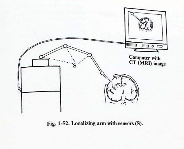
B/Localization arms. These mechanical arms can be active (with independent motion) or passive (moved by the surgeon). They are calibrated initially with respect to the fiducials and next are actively or passively driven into the surgical field. Screens displaying the position of the arm's tip are observed by the surgeon constantly during surgery. The "joints" of the arm contain transducer elements that register the position of the device (Fig. 1-52). Actively driven arms (more clumsy because of the mo-tors) are the contemporary basis for robotization of neurosurgery in the future.
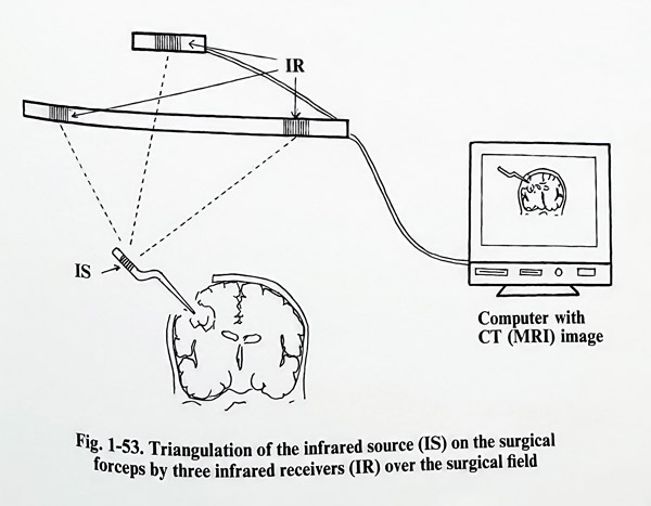
C/ Triangulation. These devices are based on the triangulation of the instrument's pointing tip when an ultrasonic or infrared signal is emitted from it. The source on the instrument emits the signal and several receivers (micro-phones or infrared cameras) detect it. The differences in the position of the instrument are estimated by computer software according to the triangulation data (angles and transmission time to the receivers) (Fig. 1-53). Direct visibility between the source and the receivers should be maintained throughout the surgery. An emission source can be added to the surgical microscope and with consideration of its focal distance, the position can be integrated precisely in these localizing systems.
D/Magnetic field guidance. The principle used is that of the measurement of the magnetic field strength from a source around the head of the patient during surgery. Magnetic impulses emitted along the three main axis (X, Y, Z) of the coordinate system are registered in the corresponding main planes at a frequency of 100Hz. Processing these data gives the tip's position, but the interference of all metal instruments with magnetic properties that create distortion is a serious inconvenience.
E/ Machine vision. Tridimensional position in space during surgery is determined by video imaging with two cameras from two different points. A localizing device with multiple fiducial points is initially placed around the patients' head for calibration. Photogrammetric techniques permit determination of the position of every fiducial, and later on during surgery of every point inside the surgical field, provided the patients' head and the cameras are not moved. This is based and related to
the preoperative imaging studies. Surgical instruments are tracked by attaching fiducials to them. Naturally, this method requires complete constant visibility and powerful computer hardware.
All these technologically advanced surgical methods, regardless of being presently reserved for the more specialized centers, represent the direction of progress and are harbingers of the expected new horizons of neurosurgical technique.