Legacy: "Atlast of Neurosurgery" / L.Karaguiosov, A. Ramadan, K.Karaguiosov / Kiwait/ 1998
PREV> 11. Principles of Spinal Cord Surgery
Next < 13. Tumors of The Spine and Spinal Cord
12. SURGICAL TREATMENT OF SPINAL INJURY
When the structures of the spine fail to dissipate an impact sufficiently, this results in damage of bony, muscular and ligamentous structures and possible affection of the spinal cord. Several types of forces can provoke excessive flexion, extension, rotation, compression, dislocation and shearing. As a consequence, bone fractures, ligamentous and disc ruptures, dislocations or subluxations may occur. Secondary impact on the cord can be induced by displaced vertebras, fragments of bone and discs or by the primary impact. After the mechanical cord injury, additional damage can follow through intrinsic mechanisms, leading to ischaemia, calcium influx, lipid peroxidation and free radical activation. As a whole, the existing pathological changes can be summarized under two major groups: biomechanical for the spine and neurological for cord and roots. They determine the principles of surgical treatment: alignment of the spine, stabilization, and decompression of neural structures.
Diagnostic evaluation should include assessment of these three major problems to solve: alignment, stability and the presence of compression. Radiological diagnosis is crucial. Initiating the work up with X-ray of the presumably affected area on admission, imaging is extended to CT scan visualization in different planes and MRI or myelography to detect neural damage. Spinal trauma most often affects segments bordering mobile to relatively immobile areas, such as the cervical and thoracolumbar segments. Immobilization is the first and most important temporary measure to undertake when spinal injury is suspected; it must be maintained until instability is not ruled out. If alignment is found compromised and instability is possible, a strategy for restoration must be established.
The presence of any neurological deficit requires the exclusion of neural compression, and if such is found, appropriate decompression must be undertaken. Alignment can be obtained by external or internal repositioning, or simply by axial skeletal traction. Stabilisation is either internal or external. The latter is relying on natural stabilization by healing, especially bone repair. Decompression is adjusted to the existing type of compression in such a way, that minimal additional destabilization is inflicted by the approach, and any compressing agent is removed reducing to a minimum manipulation of neural tissue. Often decompressive and stabilizing techniques are combined.
OPERATIONS ON THE CERVICAL SPINE
The cervical spine has two specific areas of interest in injury: the Cl - C2 area and the lower cervical spine (sub-axial); these areas differ regarding the techniques of decompression and internal fixation. The external method of immobilisation and repositioning (traction) is more or less the same for both areas of affection.
CERVICAL TRACTION OF THE SPINE
The application of external skeletal traction and immobilization is used in all injuries inducing axial deformation of the cervical spine, irrespective of whether they are fractures, fracture-dislocations or pure dislocations in the cervical region. This method corrects spinal deformity, and at times relieves the existing compression on the spinal cord and roots.
The patient's neck on admission is usually already immobilised and cervical traction is applied as early as possible to replace it. In some delayed cases this can be done within one week after the injury. Cervical traction is preferably followed by long-term immobilization or fixa-tion, which will permit early mobilisation and rehabilitation of the patient.
Traction devices have been designed for several decades; all follow the principle of applying a traction force to an instrument fixed to the skull with pins through the outer table of the calvaria. The last two decades have allowed a gradual replacement of these by the halo. This metal ring fixed with 4 pins can be used for traction and after repositioning, for immobilization connecting it with rigid bars to a vest. This permits the patient to be ambulatory without compromise of his immobilisation. The instrument of Crutchfield is one of the first to have been employed and is still occasionally in use.
Many other instruments, those of Gardner, Winke, Barton, Ramandier, etc, can also be used; they all have similar modes of application.
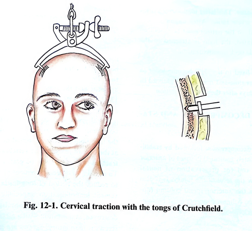
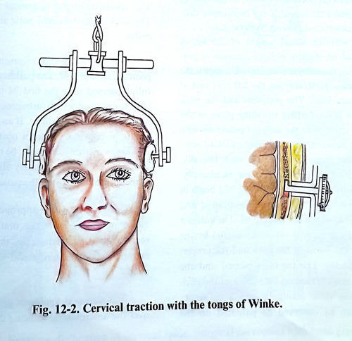
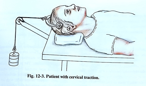
The midline of the cranium is marked. The points of fixation of the tongs are chosen in the plane of transverse processes of the cervical vertebrae symmetrically to the midline. The supposed pin penetration areas are infiltrated with local anaesthetic. Small skin perforations are made that reach and also incise the periosteum. The outer table may or may not be perforated, according to the design of the pins, and the instrument is applied (Fig. 12-1). The instrument of Winke specifically has the advantage of being firmly fixable to the cranium; it cannot be pulled out easily. At the end of the hook there is a special eccentric disc, which after perforation of the outer table is well fixed in the diploe (Fig. 12-2). Traction begins with small weights from 2 - 4 kg, which over a week are gradually increased and reach up to 10 - 12 kg if required for alignment. Daily follow-up of the neurological symptoms can be done and X-ray control if needed can follow. After the dislocation is corrected, the weight is gradually decreased to a level which maintains the spine in the correct position and at the same time allows the patient to feel comfortable; alternatively, the patient is immobilized in a halo vest. The traction may remain for long time if the patient is in a special bed, permitting position changes without removing the traction (Fig. 12-3).
The halo has four pins which screw through the ring. Insertion points are at the level below the frontal tubera and the external occipital protuberance and the screws are adjusted to the prescribed force of resistance in a crosswise order. This latter manoeuvre avoids leaving any screw loose. After alignment has been achieved, or the patient must be mobilized and rehabilitated, the halo is attached to the vest to continue neck immobilization. All pins fixed to the skull become loose in their beds over time and need to be tightened in the next few days after their initial insertion.
ANTERIOR DECOMPRESSION AND STABILISATION
Anterior decompression followed by stabilisation is absolutely indicated in cases of anterior compression and for preservation of neural function below the level of the injury. It is also required when the anterior spinal column (applying the “three column” concept) must be reinforced to avoid kyphosis.
The patient is placed in a supine position on the operating table, with the neck elevated with a small support roll cushion, so that during the operation the lateral X-ray monitoring cannot be obstructed. Direct skeletal traction is applied with an initial weight of 2,5 kg. A horizontal or slightly oblique incision is performed on the anterior cervical skin along the skin creases, preferably on the left side and at the fracture level. The platysma and the neck fascia are divided along the same plane of the incision, after which is penetrated into the space between the sternocleidomastoid muscle and the great vessels of the neck laterally, and oesophagus, thyroid and pharynx medially.
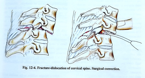
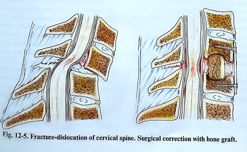
We recommend the penetration should begin in the angle between the sternocleidomastoid and the superior belly of the omohyoid. The anterior surface of the spine is exposed, covered by the deep cervical fascia of the neck and the prevertebral muscles. The fascia is incised and the muscles are separated on both sides of the mid-line. During these manipulations, a deformity usually can be observed or palpated on the anterior surface of the spine. A tip of a long needle is introduced into the disc where the vertebral dislocation is assumed to be, and a lateral X-ray control is made. The weight of the traction is increased to improve alignment and the disc with the adjacent parts of the neighbouring vertebrae are removed from the dislocation level until the posterior longitudinal ligament and the spinal canal are exposed (Fig 12-4). All fragments of disc, vertebrae and other compressing material are removed from the epidural space and the defect is filled with a bone graft (Fig. 12- 5). The latter is taken from the iliac bone crest by a second operating team at the time of reaching the vertebral bodies. The traction is released and the graft remains fixed. To avoid anterior displacement of the graft and to reduce the period of long-lasting and inconvenient immobilization, especially if the intervertebral joints and flavum ligaments are destroyed, stabilisation with a metallic plate and screws is mandatory. Screws are inserted in the adjacent intact vertebral bodies and the plate firmly covers the graft, preventing its migration. The operative wound is drained for 24 hours. The neck is immobilised with an orthopaedic collar.
There are no specific recommendations in the postoperative period. The patients must be carefully observed over the first 24 hours, particularly for respiratory disturbances, which are the most serious complications. If oedema of the larynx or pharynx produces obstruction of the airways, a tracheostomy may be required. X-ray control is needed immediately after surgery and at the time of mobilising the patient.
Different surgical approaches are introduced in practice for the first three cervical vertebrae. Most frequently, the transoral or extrapharyngeal anterolateral approach is applied.
The transoral approach has the advantage over the anterolateral in being direct and faster. The disadvantages are: the anaesthesia given through the mouth narrows the operative field and the operative wound is more exposed to infection. Tracheostomy is necessary only in rare cases.
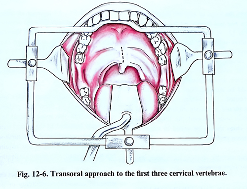
The patient is placed in a semi seated position on the operating table. A special mouth retractor is inserted, which keeps the access open. The blade of the retractor presses the tongue down and fixes the endotracheal tube (Fig. 12-6). The soft palate is incised along the midline except for the uvula, which goes to one side; both flaps are elevated with traction sutures or are fixed with stitches to the lateral walls of the mouth.
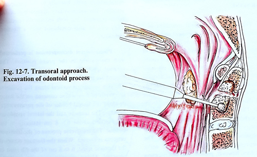
The anterior lamina of the atlas and the second and third cervical vertebra are palpated with a finger through the pharyngeal wall. The pharyngeal mucosa is cut along the midline (4 cm in length) after infiltration with local anaesthetics. The incision borders of the pharyngeal wall are retracted laterally with stitches, to expose the retropharyngeal space on both sides of the mid-line. After this the ligaments in the craniospinal region are stripped from the anterior surface of the atlas, using the surgical microscope.The anterior arch of the atlas is drilled out with the high speed power tool sufficiently to expose the odontoid. If it is a case of an odontoid process fracture, the surgeon penetrates into the fracture cleft and it is widened with the drill (Fig. 12-7).
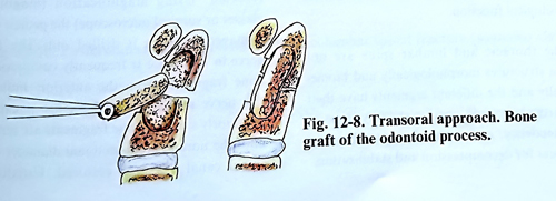
A bed is excavated for the bone graft, if grafting is intended. If required, the graft is taken from the iliac bone at the time of the drilling by a second operating team (Fig. 12-8). The traction is increased when the graft is inserted. The technique however should first resolve all needs for decompression by removal with the high speed drill and taking out of all dislocated fragments. A well preserved and aligned odontoid fragment can be fixed with a screw through the body of C2.
The pharyngeal wall is sutured with absorbable material. After completing the operation, a nasogastric tube is introduced. If instability persists or fixation has not been done, immobilisation is required until bone consolidation (at least 6 to 8 weeks) or fixation is performed on a separate session.
Posterior stabilisation in atlanto-axial fracture-dislocations. There are many different surgical techniques for stabilisation of atlanto-axial fracture-dislocations. The posterior stabilisation of the first and second cervical vertebrae is advisable in cases of anterior dislocation of the atlas with respect to the axis, with or without fracture of the base of the odontoid process. More complex cases of fractures, dislocation and remaining instability require the fixation to be extended to the occipital bone and C3. All techniques of stabilisation rely on the uncompromised integrity of the bony structures, used for fixation. There are many options with the use of metallic instrumentation and/or bone grafts.A combination of them is usually the most appropriate.
The approach to the bony structures is similar in all techniques, and it is via a midline incision, identical to that in upper cervical laminectomy. The most simple method of C1C2 fixation is by wiring, fixing the posterior lamina of the atlas with the base of the posterior apophysis of the axis.
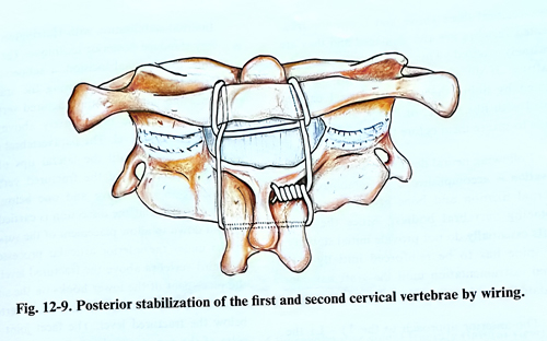
After a straight incision in the midline from the external occipital protuberance up to the level of the posterior apophysis of the IV - V cervical vertebrae, the penetration continues along the septum nuchae reaching the occipital bone and the posterior processes of the cervical vertebrae. Parts of the occipital bone near the foramen magnum and the laminae of the first, second and third vertebrae are stripped. A space is dissected under each lamina, lateral to the posterior apophysis for the introduction of a wire. The same can be done under the border of the foramen magnum, through a perforation at 1,5 - 2 cm from the midline if the occipital bone should be included in the fixation. In a C1-C2 fixation case, the loops of wire are passed under the laminae and tied crossed on the C2 spinous process (Fig. 12 - 9). Bone grafts are taken from the iliac crest sufficiently large to cover the distance over the fixed levels. They are tied with fine wires passed also under the laminae. The surface of the occipital bone and the vertebral laminae, which are in contact with the bone grafts should be stripped from the periosteum and decorticated. The contact surface of the bone grafts should be mainly on the surface of cancelous bone. Around the bone grafts the small remaining bone chips should be added to improve contact between graft and laminae.
Different metallic devices have been designed for the same fixation purposes.
OPERATIONS ON THE THORACOLUMBAR SPINE
The aim of the surgical treatment of a thoracolumbar fracture is alignment and stabilisation. The decision to operate depends on the stability of the fracture, radiological evidence of cord or cauda compression, neurological condition and the general condition with the associated injuries. It is not proven that surgical treatment can contribute to neurologic recovery. In that context, there is no indication that complete cord damage can benefit from surgical treatment. The evidence suggests that realignment and stabilisation decrease subsequent spinal deformity and pain, improving the background for physical treatment and rehabilitation. The decompression of the spinal cord and nerve roots does, however, prevent later deterioration in neurological function.
The thoracic and lumbar spine are not uniform structures morphologically and biomechanically and the different segments have their own particular conditions for stability, type of injury tendency, and required approaches and techniques for decompression and stabilization.
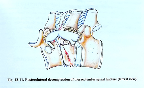
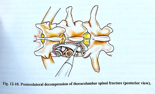
At the Tl – L1 segment of spine compression is almost always from the anterior. Decompression techniques can apply approaches from the anterior through a standard thoracotomy, from a posterolateral approach by a costo-transversectomy or via an extrapleural route, and posteriorly via the transpedicular approach (Fig. 12-10; 12-11).
The posterior transpedicular approach requires removal of facet and pedicle with the drill. Through the space created in this way, fragments indented to the neural structures are removed or repositioned. The method is useful when anterior grafting is not required, and it can be combined with posterior fixation.
The posterolateral approaches can be via a standard costotransversectomy or go through extracavitary path, laterally to the paraspinal muscles. The extracavitary approach provides better exposure, but both require rib resection. They can be also combined with a fixation technique.
Posterolateral decompression is performed with the patient in the prone position. The surgical level and the fracture to be decompressed are first identified by X-ray fluoroscopy.
The skin incision is vertical along the posterior apophysis of about 5 vertebrae, centered on the lesion or along the lateral border of the paravertebral muscles. The laminae and the lateral apophyses are exposed about 6 - 8 cm laterally to the midline. The transverse process of the fractured vertebra is excised. When the fracture occurs in the thoracic spine, 6 to 8 cm of the rib must be resected. Using magnification (magnifying glasses or surgical microscope) the pedicle of the damaged vertebra is drilled out. The spinal nerve at that level is frequently compressed by bone fragments from the anterior. Following this nerve medially, the dural sac is exposed. Anteriorly situated bone fragments are removed until the normal cross-sectional diameter of the spinal canal has been restored. Usually, the intervertebral discs above and below the fragmented vertebra are also damaged and they are routinely excised. This is a precondition for grafting. If dural tears are present, they are treated by lining the site with fascial patches sealed with fibrin glue, or better, an attempt is made to suture them before that.
Following the neural decompression, spinal stabilisation is accomplished by a combination of internal fixation and bone grafts (a strut graft replacing vertebral bodies). Since bone grafts essentially do not provide initial stability, the spine has to be reinforced initially with spinal instrumentation until the graft has consolidated enough to provide sufficient strength.
The anterior approach to the T1 - L1 spine segment is through a thoracotomy performed in a standard way; after retraction of the lung the fracture is reached extrapleurally. This provides a broad space for manipulation. The fracture is treated in a similar way as in the extracavitary approach and anterior fixation completes the task with an appropriate graft to be added. When T12 - Ll is operated on, this requires mobilization of the diaphragm. The uppermost thoracic level requires a special anterior approach. Anterior surgery does not, however, appear to be in way more efficient regarding neurological recovery.
Posterior instrumentation and fusion, in spite of the multiple designs, contain principally rods contoured to the curvature of the spinal segment. These rods are fixed with screws, wires, clamps or hooks. Wires tend to slip with additional deformity, and therefore are no longer the tool of choice. Arthrodesis with decortication and grafting must be added to these techniques, as it gives better outcome for long-term stability. Anterior instrumentation and fusion also uses plates to fix and incorporate grafts, interbody devices (cages), and some special rods, but it should be used only in association with anterior decompression.
Internal stabilisation with Harrington rods is a very standard posterior technique. Through a midline longitudinal incision, a subperiosteal dissection is performed exposing the spinous processes and laminae of the fractured vertebra and three vertebrae immediately above and below the injured level. The paravertebral muscles are retracted until the lateral tips of the transvers processes of the fractured vertebra together with one above and one below are exposed. After that the dissection is carried out up and down to allow placement of the superior hooks under the inferior articular processes of the third vertebra above the fractured level and the placement of the lower hooks on the superior edge of the lamina of the third vertebra below the fractured level. The facet joint capsules of the superior and inferior articular processes of the fractured vertebra are excised as well on both sides including the articular cartilage. The exposed bone is decorticated at three levels (the fractured vertebra, one above and one below out to the tips of the transverse processes. After placement of the hooks, the rods are selected such that the ratchet-rod junction is as close to the upper hook as possible, thereby minimising the stress at this critical part of the rod. The rods may be contoured if required. Bone grafts are harvested from the iliac crest through a separate incision and placed posterior to the transverse processes, facet joints, and laminae at the level of the fracture plus one vertebra above and below on decorticated areas.
Postoperatively patients are immediately placed in regular hospital beds. Mobilisation either independently or with a wheelchair is begun after three to seven days. The Harrington rods are removed approximately 6 months after their implantation, when spinal exercises are started.
The L2 - L4 segment is approached through either a transpedicular or anterior extraperitoneal approach. This last technique uses an incision in the abdomen, and dissection retroperitoneally to the psoas muscle; it requires sacrifice of the lumbar segmental vessels. The approach provides access to the vertebra even for corporectomy, grafting and anterior fixation. For this segment, however, posterior transpedicular fixation is much more to be preferred.
The LS vertebra is most appropriately exposed anteriorly after lower midline laparotomy or through a transpedicular approach. However, at this level the posterior transpedicular fixation can be also the technique of choice because of its lower morbidity.