ANTERIOR DECOMPRESSION AND STABILISATION
Anterior decompression followed by stabilisation is absolutely indicated in cases of anterior compression and for preservation of neural function below the level of the injury. It is also required when the anterior spinal column (applying the “three column” concept) must be reinforced to avoid kyphosis.
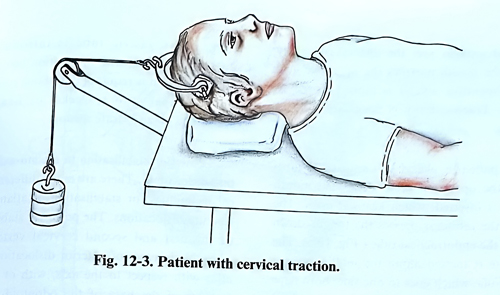
The patient is placed in a supine position on the operating table, with the neck elevated with a small support roll cushion, so that during the operation the lateral X-ray monitoring cannot be obstructed. Direct skeletal traction is applied with an initial weight of 2,5 kg. A horizontal or slightly oblique incision is performed on the anterior cervical skin along the skin creases, preferably on the left side and at the fracture level. The platysma and the neck fascia are divided along the same plane of the incision, after which is penetrated into the space between the sternocleidomastoid muscle and the great vessels of the neck laterally, and oesophagus, thyroid and pharynx medially.
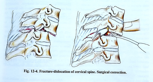
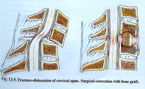
We recommend the penetration should begin in the angle between the sternocleidomastoid and the superior belly of the omohyoid. The anterior surface of the spine is exposed, covered by the deep cervical fascia of the neck and the prevertebral muscles. The fascia is incised and the muscles are separated on both sides of the mid-line. During these manipulations, a deformity usually can be observed or palpated on the anterior surface of the spine. A tip of a long needle is introduced into the disc where the vertebral dislocation is assumed to be, and a lateral X-ray control is made. The weight of the traction is increased to improve alignment and the disc with the adjacent parts of the neighbouring vertebrae are removed from the dislocation level until the posterior longitudinal ligament and the spinal canal are exposed (Fig 12-4). All fragments of disc, vertebrae and other compressing material are removed from the epidural space and the defect is filled with a bone graft (Fig. 12- 5). The latter is taken from the iliac bone crest by a second operating team at the time of reaching the vertebral bodies. The traction is released and the graft remains fixed. To avoid anterior displacement of the graft and to reduce the period of long-lasting and inconvenient immobilization, especially if the intervertebral joints and flavum ligaments are destroyed, stabilisation with a metallic plate and screws is mandatory. Screws are inserted in the adjacent intact vertebral bodies and the plate firmly covers the graft, preventing its migration. The operative wound is drained for 24 hours. The neck is immobilised with an orthopaedic collar.
There are no specific recommendations in the postoperative period. The patients must be carefully observed over the first 24 hours, particularly for respiratory disturbances, which are the most serious complications. If oedema of the larynx or pharynx produces obstruction of the airways, a tracheostomy may be required. X-ray control is needed immediately after surgery and at the time of mobilising the patient.
Different surgical approaches are introduced in practice for the first three cervical vertebrae. Most frequently, the transoral or extrapharyngeal anterolateral approach is applied.
The transoral approach has the advantage over the anterolateral in being direct and faster. The disadvantages are: the anaesthesia given through the mouth narrows the operative field and the operative wound is more exposed to infection. Tracheostomy is necessary only in rare cases.
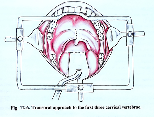
The patient is placed in a semi seated position on the operating table. A special mouth retractor is inserted, which keeps the access open. The blade of the retractor presses the tongue down and fixes the endotracheal tube (Fig. 12-6). The soft palate is incised along the midline except for the uvula, which goes to one side; both flaps are elevated with traction sutures or are fixed with stitches to the lateral walls of the mouth.
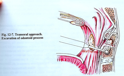
The anterior lamina of the atlas and the second and third cervical vertebra are palpated with a finger through the pharyngeal wall. The pharyngeal mucosa is cut along the midline (4 cm in length) after infiltration with local anaesthetics. The incision borders of the pharyngeal wall are retracted laterally with stitches, to expose the retropharyngeal space on both sides of the mid-line. After this the ligaments in the craniospinal region are stripped from the anterior surface of the atlas, using the surgical microscope.The anterior arch of the atlas is drilled out with the high speed power tool sufficiently to expose the odontoid. If it is a case of an odontoid process fracture, the surgeon penetrates into the fracture cleft and it is widened with the drill (Fig. 12-7).
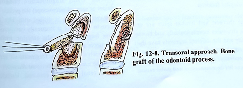
A bed is excavated for the bone graft, if grafting is intended. If required, the graft is taken from the iliac bone at the time of the drilling by a second operating team (Fig. 12-8). The traction is increased when the graft is inserted. The technique however should first resolve all needs for decompression by removal with the high speed drill and taking out of all dislocated fragments. A well preserved and aligned odontoid fragment can be fixed with a screw through the body of C2.
The pharyngeal wall is sutured with absorbable material. After completing the operation, a nasogastric tube is introduced. If instability persists or fixation has not been done, immobilisation is required until bone consolidation (at least 6 to 8 weeks) or fixation is performed on a separate session.
Posterior stabilisation in atlanto-axial fracture-dislocations. There are many different surgical techniques for stabilisation of atlanto-axial fracture-dislocations. The posterior stabilisation of the first and second cervical vertebrae is advisable in cases of anterior dislocation of the atlas with respect to the axis, with or without fracture of the base of the odontoid process. More complex cases of fractures, dislocation and remaining instability require the fixation to be extended to the occipital bone and C3. All techniques of stabilisation rely on the uncompromised integrity of the bony structures, used for fixation. There are many options with the use of metallic instrumentation and/or bone grafts.A combination of them is usually the most appropriate.
The approach to the bony structures is similar in all techniques, and it is via a midline incision, identical to that in upper cervical laminectomy. The most simple method of C1C2 fixation is by wiring, fixing the posterior lamina of the atlas with the base of the posterior apophysis of the axis.
After a straight incision in the midline from the external occipital protuberance up to the level of the posterior apophysis of the IV - V cervical vertebrae, the penetration continues along the septum nuchae reaching the occipital bone and the posterior processes of the cervical vertebrae. Parts of the occipital bone near the foramen magnum and the laminae of the first, second and third vertebrae are stripped. A space is dissected under each lamina, lateral to the posterior apophysis for the introduction of a wire. The same can be done under the border of the foramen magnum, through a perforation at 1,5 - 2 cm from the midline if the occipital bone should be included in the fixation. In a C1-C2 fixation case, the loops of wire are passed under the laminae and tied crossed on the C2 spinous process (Fig. 12 - 9). Bone grafts are taken from the iliac crest sufficiently large to cover the distance over the fixed levels. They are tied with fine wires passed also under the laminae. The surface of the occipital bone and the vertebral laminae, which are in contact with the bone grafts should be stripped from the periosteum and decorticated. The contact surface of the bone grafts should be mainly on the surface of cancelous bone. Around the bone grafts the small remaining bone chips should be added to improve contact between graft and laminae.
Different metallic devices have been designed for the same fixation purposes.