OPERATIONS ON THE THORACOLUMBAR SPINE
The aim of the surgical treatment of a thoracolumbar fracture is alignment and stabilisation. The decision to operate depends on the stability of the fracture, radiological evidence of cord or cauda compression, neurological condition and the general condition with the associated injuries. It is not proven that surgical treatment can contribute to neurologic recovery. In that context, there is no indication that complete cord damage can benefit from surgical treatment. The evidence suggests that realignment and stabilisation decrease subsequent spinal deformity and pain, improving the background for physical treatment and rehabilitation. The decompression of the spinal cord and nerve roots does, however, prevent later deterioration in neurological function.
The thoracic and lumbar spine are not uniform structures morphologically and biomechanically and the different segments have their own particular conditions for stability, type of injury tendency, and required approaches and techniques for decompression and stabilization.
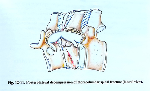
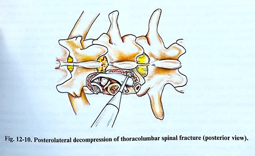
At the Tl – L1 segment of spine compression is almost always from the anterior. Decompression techniques can apply approaches from the anterior through a standard thoracotomy, from a posterolateral approach by a costo-transversectomy or via an extrapleural route, and posteriorly via the transpedicular approach (Fig. 12-10; 12-11).
The posterior transpedicular approach requires removal of facet and pedicle with the drill. Through the space created in this way, fragments indented to the neural structures are removed or repositioned. The method is useful when anterior grafting is not required, and it can be combined with posterior fixation.
The posterolateral approaches can be via a standard costotransversectomy or go through extracavitary path, laterally to the paraspinal muscles. The extracavitary approach provides better exposure, but both require rib resection. They can be also combined with a fixation technique.
Posterolateral decompression is performed with the patient in the prone position. The surgical level and the fracture to be decompressed are first identified by X-ray fluoroscopy.
The skin incision is vertical along the posterior apophysis of about 5 vertebrae, centered on the lesion or along the lateral border of the paravertebral muscles. The laminae and the lateral apophyses are exposed about 6 - 8 cm laterally to the midline. The transverse process of the fractured vertebra is excised. When the fracture occurs in the thoracic spine, 6 to 8 cm of the rib must be resected. Using magnification (magnifying glasses or surgical microscope) the pedicle of the damaged vertebra is drilled out. The spinal nerve at that level is frequently compressed by bone fragments from the anterior. Following this nerve medially, the dural sac is exposed. Anteriorly situated bone fragments are removed until the normal cross-sectional diameter of the spinal canal has been restored. Usually, the intervertebral discs above and below the fragmented vertebra are also damaged and they are routinely excised. This is a precondition for grafting. If dural tears are present, they are treated by lining the site with fascial patches sealed with fibrin glue, or better, an attempt is made to suture them before that.
Following the neural decompression, spinal stabilisation is accomplished by a combination of internal fixation and bone grafts (a strut graft replacing vertebral bodies). Since bone grafts essentially do not provide initial stability, the spine has to be reinforced initially with spinal instrumentation until the graft has consolidated enough to provide sufficient strength.
The anterior approach to the T1 - L1 spine segment is through a thoracotomy performed in a standard way; after retraction of the lung the fracture is reached extrapleurally. This provides a broad space for manipulation. The fracture is treated in a similar way as in the extracavitary approach and anterior fixation completes the task with an appropriate graft to be added. When T12 - Ll is operated on, this requires mobilization of the diaphragm. The uppermost thoracic level requires a special anterior approach. Anterior surgery does not, however, appear to be in way more efficient regarding neurological recovery.
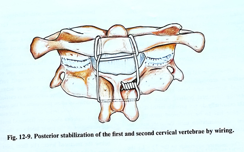
Posterior instrumentation and fusion, in spite of the multiple designs, contain principally rods contoured to the curvature of the spinal segment. These rods are fixed with screws, wires, clamps or hooks. Wires tend to slip with additional deformity, and therefore are no longer the tool of choice. Arthrodesis with decortication and grafting must be added to these techniques, as it gives better outcome for long-term stability. Anterior instrumentation and fusion also uses plates to fix and incorporate grafts, interbody devices (cages), and some special rods, but it should be used only in association with anterior decompression.
Internal stabilisation with Harrington rods is a very standard posterior technique. Through a midline longitudinal incision, a subperiosteal dissection is performed exposing the spinous processes and laminae of the fractured vertebra and three vertebrae immediately above and below the injured level. The paravertebral muscles are retracted until the lateral tips of the transvers processes of the fractured vertebra together with one above and one below are exposed. After that the dissection is carried out up and down to allow placement of the superior hooks under the inferior articular processes of the third vertebra above the fractured level and the placement of the lower hooks on the superior edge of the lamina of the third vertebra below the fractured level. The facet joint capsules of the superior and inferior articular processes of the fractured vertebra are excised as well on both sides including the articular cartilage. The exposed bone is decorticated at three levels (the fractured vertebra, one above and one below out to the tips of the transverse processes. After placement of the hooks, the rods are selected such that the ratchet-rod junction is as close to the upper hook as possible, thereby minimising the stress at this critical part of the rod. The rods may be contoured if required. Bone grafts are harvested from the iliac crest through a separate incision and placed posterior to the transverse processes, facet joints, and laminae at the level of the fracture plus one vertebra above and below on decorticated areas.
Postoperatively patients are immediately placed in regular hospital beds. Mobilisation either independently or with a wheelchair is begun after three to seven days. The Harrington rods are removed approximately 6 months after their implantation, when spinal exercises are started.
The L2 - L4 segment is approached through either a transpedicular or anterior extraperitoneal approach. This last technique uses an incision in the abdomen, and dissection retroperitoneally to the psoas muscle; it requires sacrifice of the lumbar segmental vessels. The approach provides access to the vertebra even for corporectomy, grafting and anterior fixation. For this segment, however, posterior transpedicular fixation is much more to be preferred.
The LS vertebra is most appropriately exposed anteriorly after lower midline laparotomy or through a transpedicular approach. However, at this level the posterior transpedicular fixation can be also the technique of choice because of its lower morbidity.