CERVICAL TRACTION OF THE SPINE
The application of external skeletal traction and immobilization is used in all injuries inducing axial deformation of the cervical spine, irrespective of whether they are fractures, fracture-dislocations or pure dislocations in the cervical region. This method corrects spinal deformity, and at times relieves the existing compression on the spinal cord and roots.
The patient's neck on admission is usually already immobilised and cervical traction is applied as early as possible to replace it. In some delayed cases this can be done within one week after the injury. Cervical traction is preferably followed by long-term immobilization or fixa-tion, which will permit early mobilisation and rehabilitation of the patient.
Traction devices have been designed for several decades; all follow the principle of applying a traction force to an instrument fixed to the skull with pins through the outer table of the calvaria. The last two decades have allowed a gradual replacement of these by the halo. This metal ring fixed with 4 pins can be used for traction and after repositioning, for immobilization connecting it with rigid bars to a vest. This permits the patient to be ambulatory without compromise of his immobilisation. The instrument of Crutchfield is one of the first to have been employed and is still occasionally in use.
Many other instruments, those of Gardner, Winke, Barton, Ramandier, etc, can also be used; they all have similar modes of application.
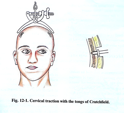
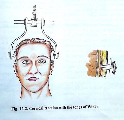
The midline of the cranium is marked. The points of fixation of the tongs are chosen in the plane of transverse processes of the cervical vertebrae symmetrically to the midline. The supposed pin penetration areas are infiltrated with local anaesthetic. Small skin perforations are made that reach and also incise the periosteum. The outer table may or may not be perforated, according to the design of the pins, and the instrument is applied (Fig. 12-1). The instrument of Winke specifically has the advantage of being firmly fixable to the cranium; it cannot be pulled out easily. At the end of the hook there is a special eccentric disc, which after perforation of the outer table is well fixed in the diploe (Fig. 12-2). Traction begins with small weights from 2 - 4 kg, which over a week are gradually increased and reach up to 10 - 12 kg if required for alignment. Daily follow-up of the neurological symptoms can be done and X-ray control if needed can follow. After the dislocation is corrected, the weight is gradually decreased to a level which maintains the spine in the correct position and at the same time allows the patient to feel comfortable; alternatively, the patient is immobilized in a halo vest. The traction may remain for long time if the patient is in a special bed, permitting position changes without removing the traction (Fig. 12-3).
The halo has four pins which screw through the ring. Insertion points are at the level below the frontal tubera and the external occipital protuberance and the screws are adjusted to the prescribed force of resistance in a crosswise order. This latter manoeuvre avoids leaving any screw loose. After alignment has been achieved, or the patient must be mobilized and rehabilitated, the halo is attached to the vest to continue neck immobilization. All pins fixed to the skull become loose in their beds over time and need to be tightened in the next few days after their initial insertion.