Legacy: "Atlast of Neurosurgery" / L.Karaguiosov, A. Ramadan, K.Karaguiosov / Kiwait/ 1998
PREV > 12. Surgical Treatment of Spinal Injury
13. TUMOURS OF THE SPINE AND SPINAL CORD
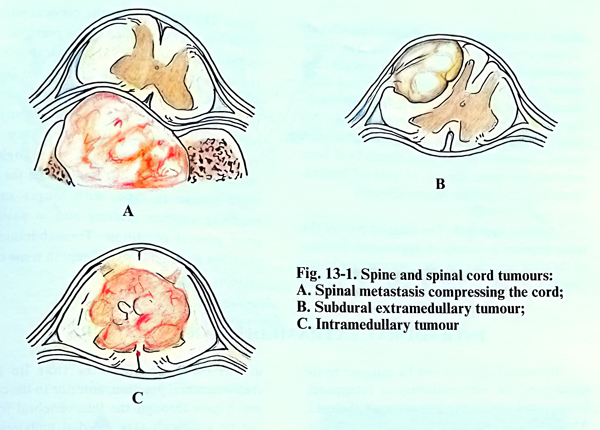
Tumours affecting the spinal canal, comprise only 10 - 15% of all tumours of the central nervous system. They can be intrathecal or secondarily invading spinal lesions. They occur with equal frequency in men and women, and are most common between the ages of 20 and 60 years. The most common tumour affecting the spinal cord is metastatic carcinoma. Primary benign tumours are rare and can be found more frequently in the first two decades of life. Of the primary intrathecal tumours about 30% are neurinomas, 26% meningiomas and 15% gliomas or ependymomas, and 29% others (Fig. 13-1).
EPIDURAL TUMOURS COMPRESSING THE SPINAL CORD
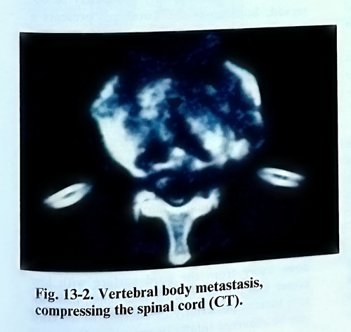
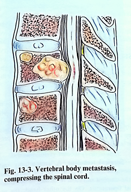
These are metastatic tumours of the spine, less often epidural space neoplastic growths that affect the content of the spinal canal. Four primary malignant tumours alone account for more than two-thirds of all cases of neoplastic cord compression - breast, lung, haematopoietic system, and prostate. The compression of the spinal cord results from extension of a focus in the vertebral body (Figs. 13-2; 13-3). Other possible mechanisms include direct invasion of the spine from a paraspinal tumour, as well as extension through the intervertebral foramina along the perineurium or its lymphatics without bone involvement.
Indications for surgical treatment. Primary benign tumours of the spine, affecting the spinal cord are usually indicated for surgical removal. To the contrary, an attempt to define the indications in cases of malignant spinal cord compression is affected by the fact that treatment has to be individualised in each patient.
Five major goals of the surgery of spinal tumours can be defined:
1.Treatment of the neoplasm by removal in cases of benign and malignant tumours.
2.Stabilisation of the spine.
3.Neurological recovery, stabilisation or prevention of rapid deterioration
4.Tissue diagnosis of the tumour.
5.Pain relief.
Advance knowledge of important prognostic factors is helpful in arriving at a decision for surgical treatment. The site of the primary tumour, its histological type, and the pre-treatment of neurological deficits are considered the most important factors. For optimal postoperative results, the patient should be ambulatory before surgery. Patients who have rapidly deteriorating neurological deficits will have a poor outcome regardless of therapy, especially if the deficit progresses in spite of a high dose of steroid therapy. The stage of the cancer patient's illness in which the treatment is undertaken is important. Patients with advanced disseminated disease and those who have failed previous treatment are much less likely to respond to surgery. In patients with lymphoma and other round cell malignancies such as neuroblastoma, Ewing's sarcoma, etc., the surgical treatment can be planned after a lack of response to radiotherapy. Patients with breast and prostate cancer with minimal neurological deficit can be treated initially with radiotherapy and other conservative measures.
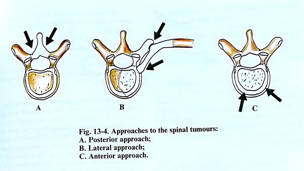
Operative approaches. There are three basic surgical approaches to the spine: posterior approach by laminectomy; lateral approach by transverse osteotomy, and anterior approach by vertebral body resection (Fig. 13-4). In addition, complete spondylectomy can be performed by a combination of an anterior and posterior approach. The choice of a surgical approach should be based on the extent of the tumour, and avoiding any additional destabilisation of the spinal segment. The patient's condition to tolerate the proposed operation should be based on the clinical and laboratory criteria, and the desired goal of therapy. The surgical approach shoud provide adequate exposure for stabilisation with instrumentation.
Laminectomy. Posterior decompression through laminectomy is indicated when the tumour is in a posterior or posterolateral location. It can be also indicated as a second stage of operation where a considerable residual tumour is left beyond the dural sac at the end of an anterior approach. More recently these indications have made the performance of a laminectomy relatively infrequent.
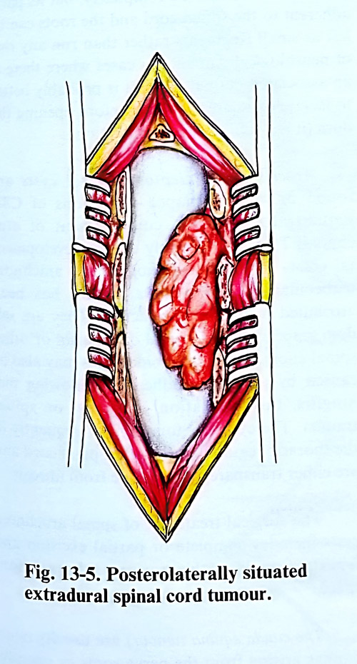
The patient is placed in a prone or lateral position on the operating table. The skin incision is straight in the midline over the spinous processes and sufficiently long to approach the affected vertebra. In the presence of atrophic changes provoked by previous irradiation, the incision should be placed laterally or in the shape of an arch in the territory of the healthy tissue. After separating the spinous processes and the laminae, an intraoperative X-ray control is mandatory, since it determines the exact extent at which laminectomy is carried out and spares unnecessary bone removal. It starts at the lower border of the lesion and minimises the removal of normal laminae (Fig. 13-5).
The neoplasm is most conveniently approached from its distal margin. In many occasions the laminae are infiltrated by the tumour tissue, which should be removed completely. If present, the epidural fatty tissue is split to reach the normal dura. A dissection plane is sought, gently elevating the tumour edges from the dura. The dura is not usually infiltrated and there is no serious problem in freeing the dural surface from the tumour tissue. Some of the malignant tumours are flexible or friable, easy detachable. The ultrasonic aspirator can be used successfully for tumour removal. Some other tumours are of firm consistency, and their excision is piecemeal with sharp instruments. Caution should be exercised not to compress or damage the dura when the tumour is scraped of its surface. In a case of very vascular lesions infiltrating the laminae, the dura must be exposed at both ends (up and down) of the lesion before starting the excision, as with more extensive bleeding, dissection will become more difficult and the dura can be damaged more easily. Neurological deterioration as a direct result of laminectomy in cancer patients is possible. Hence, every effort must be made to minimise iatrogenic damage. At times, intraoperative bleeding can be significant. It is controlled by application of bone wax or by local haemostatic agents such as oxidised cellulose, gelfoam or bipolar coagulation.
When the removal of the tumour is complete or optimal for the specific case, stabilisation is done at the same stage. The spine can be considered unstable if the intervertebral joints are destroyed or resected bilaterally, and there is vertebral body involvement, or if a pathological fracture dislocation is present. The three-column concept of spinal stability must be applied. In such cases the operation continues with fixation. It can be done with Harrington rods or other methods mentioned above according to the preferred surgical technique.
Posterolateral approach. The spine is approached paravertebrally by excision of the transverse process of the affected vertebra, as has been described in traumatic lesions of the spine. The technique has the advantage of not causing additional destabilisation of the spine. However, this permits the surgery to be concluded with fixation. It provides an sufficient approach to the vertebral body, without the complications and the postoperative morbidity of the anterior transthoracic and transabdominal approaches. That makes it very suitable for patients with malignancies and impaired general condition.
The skin incision can be curved or in a T- shape to the midline. The paraspinal muscles are divided over the transverse process. The corresponding spinal nerve is carefully dissected. The spinal resection should include a partial laminectomy, facetectomy, and pediculectomy, reaching the spinal canal and the vertebral body. No additional reconstruction is required after such lateral osteotomy procedures, especially if bone removal has been limited to part of lamina, facets joint and pedicle only on one side.
Anterior approach. The anterior part of the spine requires a variety of approaches because of the complex soft tissues anatomy anterior to the vertebral bodies in the different parts of the spine. The technique in the cervical part of the spine has been described in the previous chapter in traumatic lesions of the cervical spine. It is the method of choice for all affected bodies of the cervical vertebrae. The surgical approach to the thoracic vertebrae is by thoracotomy at the level of the tumour. It gives a view of the vertebral bodies through a wide angle after retraction of the lung. It is adequate for lesions more benign in type, with longer survival, requiring anterior grafting and in patients of good general condition. Transabdominal approaches are rarely used, except in some cases of vertebrectomies.
INTRADURAL EXTRAMEDULLARY TUMOURS
Intradural tumours can be extrinsic to the spinal cord, i.e. extramedullary or intramedullary, arising from and within the substance of the cord. About 70% of intradural tumours are extramedullary. They are almost always benign and the great majority are meningiomas or neurinomas. The rest are dermoid and epidermoid tumours, cysts, lipomas, and other rare lesions. At present this surgery is not associated with mortality, except under unique circumstances appearing accidentally. The very good outcome is the result of improved preoperative imaging, the application of steroids, and the much-improved intraoperative technique and monitoring. This last technological improvement - microsurgery, SSEP, MEP, the surgical laser and cavitron aspiration, spinal instrumentation - are highlights in the improvement of neurosurgical technique in all areas of the field.
The removal of those tumours at the level of the craniovertebral junction, anterior to the cord, or which pass through an intervertebral foramen can be a difficult task. Medial unilateral facetectomy is required for access to the ventral part of the canal for anteriorly and anterolaterally located tumours. All manoeuvres are intended to minimise cord manipulations.
After a laminectomy is made, the epidural space seems narrowed in a limited region, and a decrease in epidural fatty tissue may be observed. Sometimes the dural sac presents a bulge, which is harder on gentle palpation. The opening of the dura is best started cranially towards the tumour. The arachnoid should be preserved if possible at this stage.
The goal of surgery in these tumours is complete excision. Cases of neurofibromatosis and multiple tumours can be followed up, with only the symptomatic ones being excised when they are found. The standard approach is limited, but precise precise laminectomy.
Surgical technique. A laminectomy and removal of a small accessible tumour can be one of the simplest and most rewarding of neurosurgical operations.
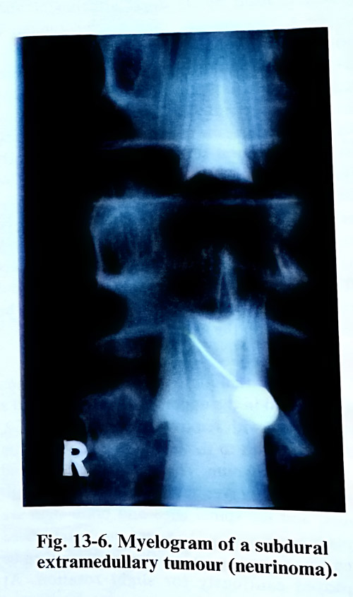
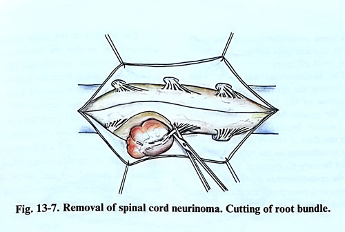
Neurinomas situated posteriorly or postero-laterally to the spinal cord are easily removed (Fig. 13-6). After the dura is opened, the arachnoid is divided over the tumour together with surrounding adhesions. A large part of the tumour surface is exposed with a fine forceps or dissector. In dissecting the tumour it may very occasionally be necessary to coagulate vessels. Such coagulation should be done away from the cord surface with low power and flushing with saline. Small neurinomas located posteriorly or laterally to the cord can be removed totally in one piece. In case of big tumours situated laterally or anteriorly, it is safer to debulk the tumour, thus allowing withdrawal of the capsule and the remaining fragments from the spinal cord without applying any pressure. Root fibers attached to the tumour capsule can be dissected from the tumour, but some of them may need to be divided (Fig. 13-7).
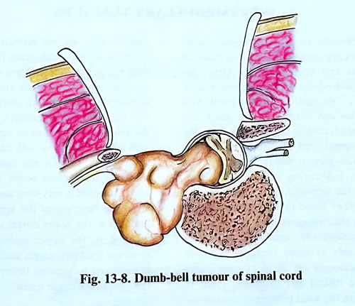
Large neurinomas (schwannomas) with extravertebral extension cause difficulty due to their fixation along the course of the nerve root of origin. Unroofing the intervertebral foramen allows removal of the foraminal component and if the extraspinal portion of a dumbbell tumour is large, laminectomy is combined with an extra-spinal approach (Fig. 13-8).
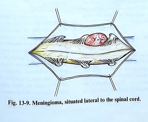
The removal of meningiomas differs from that of schwannomas in some aspects. A meningioma is fixed by its dural attachment and, unlike a schwannoma, even a small tumour is not initially mobile (Fig. 13-9). It may be possible to remove a small posterior tumour in one piece together with its dural attachment. In general terms it is preferable to exenterate the tumour before dissecting the capsule from the cord, and dividing the dural origin. In case of anteriorly situated menigiomas, a lateral enlargement of the laminectomy by facetectomy and, rarely, removal of the pedicle are needed. Dura at the attachment is not initially completely divided, and only the accessible parts and attachment are resected. In any dural defect after tumour attachment removal grafting will be necessary, and this can be difficult on the anterior side of the canal.
Anterior tumours - close to the foramen magnum and in the upper cervical region need a laminectomy with removal of the pedicle of C2, part of the atlas to the lateral mass and the rim of foramen magnum. The vertebral arteries and lower cranial nerves mainly straddle these tumours, and the spinal cord and brain stem are displaced posteriorly. The cord can be mobilised by cutting a dentate ligaments accessible, which can allow cautiously slight rotation. At times, broad-based meningiomas and recurrent tumours in this region require a transcondylar approach.
An anterior approach using a transoral or transcervical route involves removal of the vertebral bodies to allow direct access to the tumour without retraction of the spinal cord. However, the exposure of the lateral margin of the tumour may be inadequate and there is a serious risk of a CSF fistula and meningitis; therefore an anterior approach is nowadays rarely applied.
Dermoid and epidermoid cysts are congenital and in many cases are associated with dysraphism, (spina bifida, dermal sinus, myelomeningocele, diastematomyelia) and syringomyelitic cavities. Radical removal is desirable but not always possible or even required if they are intrinsic to the cord. The content of the cyst should be evacuated, taking care not to spread the fluid into the subarachnoid space. The capsule is removed completely, but its parts adherent to the spinal cord and the roots can be left as small fragments rather than run any risk of neurological damage. In cases where there is an associated dermal sinus, it is probably better to isolate or excise the sinus before opening the dura (it is a source of contamination).
Arachnoidal or leptomeningeal cysts are round or sausage-shaped collections of CSF sequestrated within a compartment of arachnoid. They are probably due to developmental errors in the distribution of the arachnoid trabeculae, and their enlargement has been attributed to hydrostatic factors. They are seldom associated with spinal dysraphism or other spinal anomalies. Arachnoidal cysts may also be caused by arachnoid adhesions following meningitis, the intrathecal instillation of drugs, or spinal trauma. The cysts are found most frequently in the thoracic region dorsal to the spinal cord and are either transparent or opaque from fibrosis. The surgical treatment of spinal arachnoid cysts includes complete or partial excision and fenestration into the normal subarachnoid space.
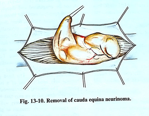
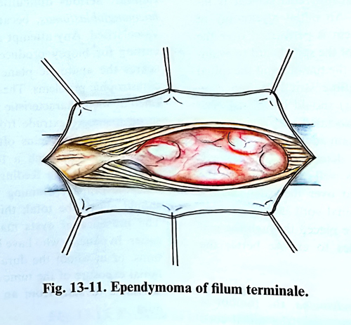
The cauda equina tumours are usually neurinomas arising from the nerve roots or ependymomas of filum terminale. The removal of these tumours has some particularities, as they grow rather large in size and may compress the nerve roots at the level of more than two lumbar vertebrae. The laminectomy has to be wide enough and during the operation of the spinal canal a thinning of the spinal laminae and widening of the spaces between neighbouring laminae may be seen. In cases of large tumours of cauda equina the dura is very often thinned and sometimes is so adherent to the tumour surface that it will not be easy to separate. Some of the filum terminale ependymomas comprise all cauda equina roots and even penetrate the intervertebral foramina (Figs. 13-10; 13-11).
The opening of the spinal canal should be wide enough, so that the superior and inferior ends of the tumour are reached. The nerve roots are separated gently from the tumour surface without injuring them. After that the debulking of the tumour will facilitate the dissection of the roots from the tumour capsule. If the tumours have soft consistency they can be removed with the ultrasound aspirator or usual suction tip. In case of ependymoma, filum terminale should be divided up and below the tumour, which facilitates the mobilisation of the tumour and its separation from the roots.
INTRAMEDULLARY TUMOURS
Approximately 15% of all primary intradural tumours are intramedullary, and most are astrocytomas and ependymomas. Other tumours as oligodendroglioma, haemangioblastoma, lipoma, dermoid, epidermoid and even metastasis, but they are much less common. Ependymomas are distinct from the surrounding spinal cord tissue, are firm in consistency and reddish-grey or yellow. Astrocytomas, however, are infiltrating and can be distinguished from the surrounding cord by their grey appearance. They blend without margin with the spinal cord. They are usually of low grade histologically, appearing in children, but about two-thirds of these tumours occur in adults. Astrocytomas are frequently associated with extensive rostral and or caudal cysts. In such a situation the solid portion of the tumour can occupy a variable length of the cord, but the cysts may become huge and extend to the conus or even into the brain stem.
SURGICAL INDICATIONS
The diagnosis of an intramedullary tumour does not necessarily mandate surgical removal. The decision to operate on patients with far-advanced neurological deficits must be made with realistic expectations. These include a desire to preserve residual sensory function in patients who are not ambulatory and to preserve of residual sphincter function. Patients who are unable to stand are unlikely to regain enough motor function to ambulate as a result of tumour resection. An operation may maintain the quality of a patient's life by preserving the ability to transfer or to turn in bed. Patients with total paraplegia will not regain function with surgery, and are not operative candidates.
In astrocytoma cases below the middle dorsal area, extension is very slow and usually does not affect the function of the upper limbs. Similar tumours in the upper dorsal area can affect the function of the upper limbs, and despite the resultant total paraplegia such a tumour should be removed to prevent future neurological disfunction in the upper limbs. The patients with progressive neurological deficits who are still able to walk represent the ideal candidates. In patients with a cervical intramedullary tumours with neck pain and mild motor and sensory deficit, the tumour removal is related to a risk of inducing neurological deficit. In similar patients the reasonable policy is to follow the patient closely for the appearance of additional symptoms. Once patients realise that their progressive deterioration is developing beyond what they can tolerate, they are more prepared psychologically to face the risks of operative treatment as it is justified regardless these risks.
Surgical technique. The prone position is generally used for intramedullary tumours at all levels of the spinal cord. The monitoring of motor evoked and somatosensory evoked potentials can be recommended peroperatively. The full extent of the tumour must be known before the surgery. This is usually demonstrated on the MRI study. In a case of suspected haemangioblastoma, the blood supply should be demonstrated by angiography. A wide laminectomy is then performed over the entire extent of the tumour. The dura must be opened carefully since the pia-arachnoid must be opened carefully since the pia-arachnoid may be attached to the inner surface of the dura. Any bleeding should be avoided, because it will hide the vessels on the spinal cord surface and compromise the surgical effort.
Extensive peritumoural cysts above and below the solid part of tumour can be reached and exposed easily.
The spinal cord shows various pathological changes: there is considerable increase in its volume with pathologically enlarged vessels in some cases, and change of colour - bright, yellowish or greyish, etc. The surface of the cord at the area of the greatest enlargement is inspected (Fig. 14-12). An initial myelotomy of approximately 1 - 2 cm is performed over the greatest enlargement of the spinal cord to evaluate the plane between the tumour and the spinal cord tissue. The midline strictly (often even shifted by the tumour) should be incised. The presence of a cyst associated with the tumour provides a chance to gain additional room by its removal. Once it is determined that the neoplasm is cystic or has well-defined planes, the myelotomy is enlarged over the extent of the tumour. Following the spinal cord incision, 6-0 or 7-0 traction sutures are placed through the pial margins on both sides to expose better the interior of the cord.
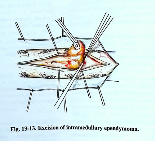
In a case of ependymoma, the tumour is visible a few millimetres under the spinal cord surface. In most instances there is a clear border between the tumour and the spinal cord tissue. Using a microsurgical technique, a sharp plane of dissection is developed around the tumour, taking care to retract the tumour and not the spinal cord. The removal starts from any of the ends of the tumour. If possible, the pole of the tumour is lifted, and gentle traction with a tumour forceps facilitates its removal from the interior of the cord (Fig. 13-13). All fine vessels should be coagulated and sharply divided. In cases of ependymomas, the blood supply comes from the ventral surface. Blunt dissection of the tumour is not advisable - especially where vessels connect the tumour to the spinal cord. It is important to keep the operative field dry so that the plane between the tumour and the spinal cord can be clearly visualised.
For those tumours that are infiltrative (usually astrocytomas), the removal of the tumour is easier with ultrasonic aspiration. The hard part of the tumour may need the use of a laser for this purpose. The removal of intramedullary epidermoids and dermoids is an easy task because they are usually avascular and well delineated. The tumour capsule remains in place if it is adherent to the spinal cord tissue. With teratomas, the stalk between the cord and the dura should be removed as an integral part of the tumour.
Serious difficulties are present with haemangioblastomas, because they are highly vascularised. Any attempt at decompression or cutting for biopsy produces bleeding that obscures the anatomic plane and may result in catastrophic problems. These tumours are identified by their characteristic red appearance, and in all instances extrude from the pial surface. Large arterialised veins often surround them. The removal of such a tumour starts with interruption of the feeding arteries and ends with the primary draining veins. The removal should always be total as this lesion is curable. The presence of cysts makes their removal easier. In patients who have had previous operations, or in whom the dura was left open, the initial exposure of the tumour is difficult and is advisable to start from an intact part of the dura.
Lipomas of the spinal cord occur as isolated tumours in the thoracic and cervical spinal cord. Histologically identical to normal fat, these tumours are located on the dorsal surface of the spinal cord covered by little or no neural tissue. The most effective operative strategy consists of subtotal removal leaving a layer of tumour tissue on the surface of the cord. The laser is ideal to remove lipomas. On the other hand, the ultrasonic aspirator is not effective due to the fibrous stroma of the lesion.
Postoperative management. Steroids are routinely used pre- and postoperatively in high doses for limited periods. Prophylactic antibiotics can be used intraoperatively and up to three days postoperatively. If the surgery has involved the upper cervical area and motor impairment is expected, the endotracheal tube is left in place for at least 24 hours, regardless of the patient's condition at the end of the surgery and an evaluation afterwards.
Radiotherapy is considered postoperatively for patients with malignant spinal tumours and intramedullary astrocytomas, where tumour removal has been incomplete. Careful follow-up of spinal stability is needed. Bracing might be necessary for children because of the possible development of spinal deformity following the tumour removal.