
INTRADURAL EXTRAMEDULLARY TUMOURS
Intradural tumours can be extrinsic to the spinal cord, i.e. extramedullary or intramedullary, arising from and within the substance of the cord. About 70% of intradural tumours are extramedullary. They are almost always benign and the great majority are meningiomas or neurinomas. The rest are dermoid and epidermoid tumours, cysts, lipomas, and other rare lesions. At present this surgery is not associated with mortality, except under unique circumstances appearing accidentally. The very good outcome is the result of improved preoperative imaging, the application of steroids, and the much-improved intraoperative technique and monitoring. This last technological improvement - microsurgery, SSEP, MEP, the surgical laser and cavitron aspiration, spinal instrumentation - are highlights in the improvement of neurosurgical technique in all areas of the field.
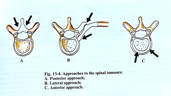
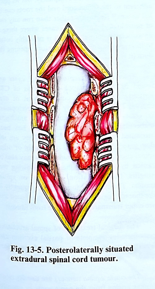
The removal of those tumours at the level of the craniovertebral junction, anterior to the cord, or which pass through an intervertebral foramen can be a difficult task. Medial unilateral facetectomy is required for access to the ventral part of the canal for anteriorly and anterolaterally located tumours. All manoeuvres are intended to minimise cord manipulations.
After a laminectomy is made, the epidural space seems narrowed in a limited region, and a decrease in epidural fatty tissue may be observed. Sometimes the dural sac presents a bulge, which is harder on gentle palpation. The opening of the dura is best started cranially towards the tumour. The arachnoid should be preserved if possible at this stage.
The goal of surgery in these tumours is complete excision. Cases of neurofibromatosis and multiple tumours can be followed up, with only the symptomatic ones being excised when they are found. The standard approach is limited, but precise precise laminectomy.
Surgical technique. A laminectomy and removal of a small accessible tumour can be one of the simplest and most rewarding of neurosurgical operations.
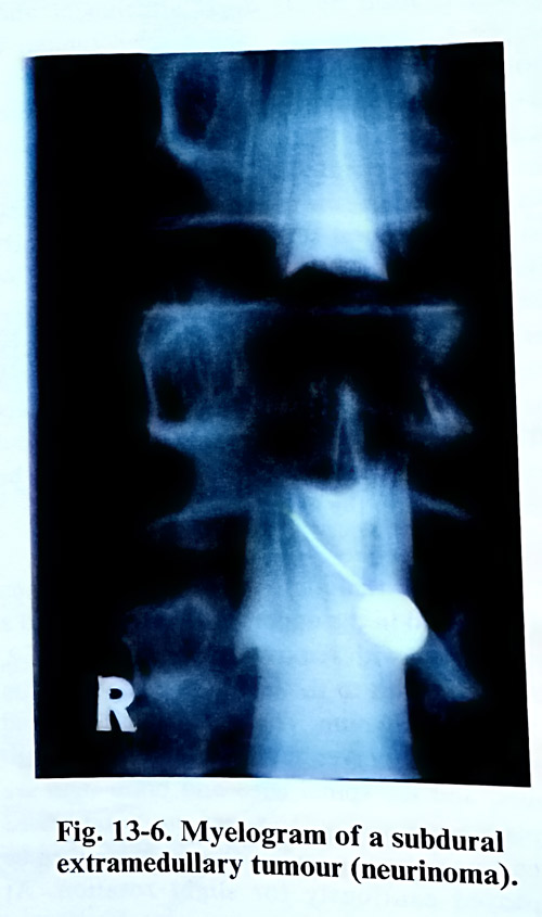
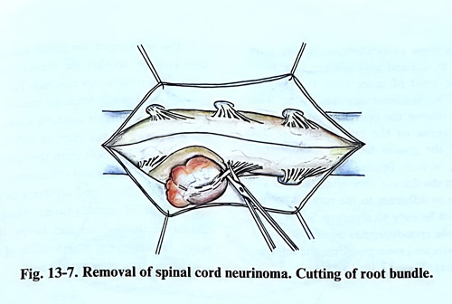
Neurinomas situated posteriorly or postero-laterally to the spinal cord are easily removed (Fig. 13-6). After the dura is opened, the arachnoid is divided over the tumour together with surrounding adhesions. A large part of the tumour surface is exposed with a fine forceps or dissector. In dissecting the tumour it may very occasionally be necessary to coagulate vessels. Such coagulation should be done away from the cord surface with low power and flushing with saline. Small neurinomas located posteriorly or laterally to the cord can be removed totally in one piece. In case of big tumours situated laterally or anteriorly, it is safer to debulk the tumour, thus allowing withdrawal of the capsule and the remaining fragments from the spinal cord without applying any pressure. Root fibers attached to the tumour capsule can be dissected from the tumour, but some of them may need to be divided (Fig. 13-7).
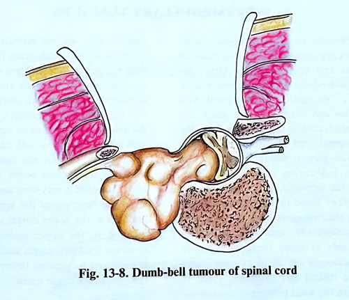
Large neurinomas (schwannomas) with extravertebral extension cause difficulty due to their fixation along the course of the nerve root of origin. Unroofing the intervertebral foramen allows removal of the foraminal component and if the extraspinal portion of a dumbbell tumour is large, laminectomy is combined with an extra-spinal approach (Fig. 13-8).
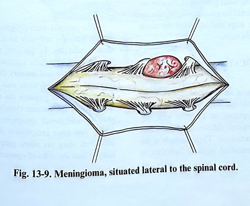
The removal of meningiomas differs from that of schwannomas in some aspects. A meningioma is fixed by its dural attachment and, unlike a schwannoma, even a small tumour is not initially mobile (Fig. 13-9). It may be possible to remove a small posterior tumour in one piece together with its dural attachment. In general terms it is preferable to exenterate the tumour before dissecting the capsule from the cord, and dividing the dural origin. In case of anteriorly situated menigiomas, a lateral enlargement of the laminectomy by facetectomy and, rarely, removal of the pedicle are needed. Dura at the attachment is not initially completely divided, and only the accessible parts and attachment are resected. In any dural defect after tumour attachment removal grafting will be necessary, and this can be difficult on the anterior side of the canal.
Anterior tumours - close to the foramen magnum and in the upper cervical region need a laminectomy with removal of the pedicle of C2, part of the atlas to the lateral mass and the rim of foramen magnum. The vertebral arteries and lower cranial nerves mainly straddle these tumours, and the spinal cord and brain stem are displaced posteriorly. The cord can be mobilised by cutting a dentate ligaments accessible, which can allow cautiously slight rotation. At times, broad-based meningiomas and recurrent tumours in this region require a transcondylar approach.
An anterior approach using a transoral or transcervical route involves removal of the vertebral bodies to allow direct access to the tumour without retraction of the spinal cord. However, the exposure of the lateral margin of the tumour may be inadequate and there is a serious risk of a CSF fistula and meningitis; therefore an anterior approach is nowadays rarely applied.
Dermoid and epidermoid cysts are congenital and in many cases are associated with dysraphism, (spina bifida, dermal sinus, myelomeningocele, diastematomyelia) and syringomyelitic cavities. Radical removal is desirable but not always possible or even required if they are intrinsic to the cord. The content of the cyst should be evacuated, taking care not to spread the fluid into the subarachnoid space. The capsule is removed completely, but its parts adherent to the spinal cord and the roots can be left as small fragments rather than run any risk of neurological damage. In cases where there is an associated dermal sinus, it is probably better to isolate or excise the sinus before opening the dura (it is a source of contamination).
Arachnoidal or leptomeningeal cysts are round or sausage-shaped collections of CSF sequestrated within a compartment of arachnoid. They are probably due to developmental errors in the distribution of the arachnoid trabeculae, and their enlargement has been attributed to hydrostatic factors. They are seldom associated with spinal dysraphism or other spinal anomalies. Arachnoidal cysts may also be caused by arachnoid adhesions following meningitis, the intrathecal instillation of drugs, or spinal trauma. The cysts are found most frequently in the thoracic region dorsal to the spinal cord and are either transparent or opaque from fibrosis. The surgical treatment of spinal arachnoid cysts includes complete or partial excision and fenestration into the normal subarachnoid space.
The cauda equina tumours are usually neurinomas arising from the nerve roots or ependymomas of filum terminale. The removal of these tumours has some particularities, as they grow rather large in size and may compress the nerve roots at the level of more than two lumbar vertebrae. The laminectomy has to be wide enough and during the operation of the spinal canal a thinning of the spinal laminae and widening of the spaces between neighbouring laminae may be seen. In cases of large tumours of cauda equina the dura is very often thinned and sometimes is so adherent to the tumour surface that it will not be easy to separate. Some of the filum terminale ependymomas comprise all cauda equina roots and even penetrate the intervertebral foramina (Figs. 13-10; 13-11).
The opening of the spinal canal should be wide enough, so that the superior and inferior ends of the tumour are reached. The nerve roots are separated gently from the tumour surface without injuring them. After that the debulking of the tumour will facilitate the dissection of the roots from the tumour capsule. If the tumours have soft consistency they can be removed with the ultrasound aspirator or usual suction tip. In case of ependymoma, filum terminale should be divided up and below the tumour, which facilitates the mobilisation of the tumour and its separation from the roots.