
EPIDURAL TUMOURS COMPRESSING THE SPINAL CORD
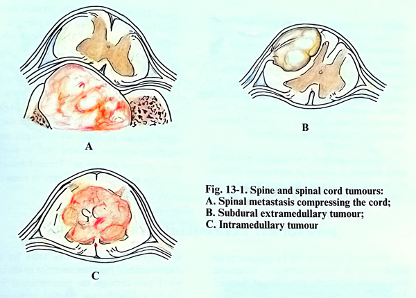
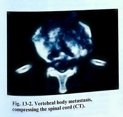
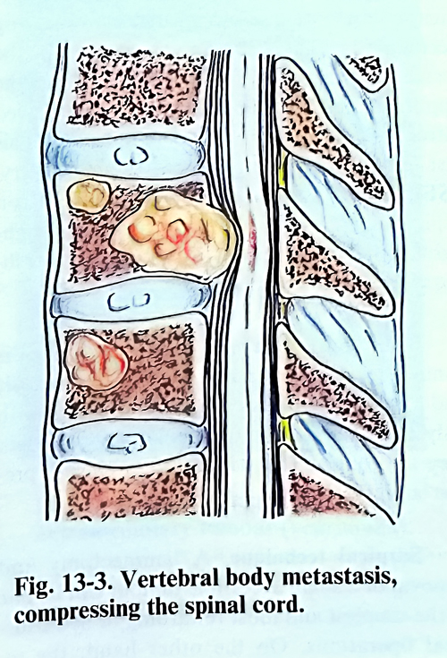
These are metastatic tumours of the spine, less often epidural space neoplastic growths that affect the content of the spinal canal. Four primary malignant tumours alone account for more than two-thirds of all cases of neoplastic cord compression - breast, lung, haematopoietic system, and prostate. The compression of the spinal cord results from extension of a focus in the vertebral body (Figs. 13-2; 13-3). Other possible mechanisms include direct invasion of the spine from a paraspinal tumour, as well as extension through the intervertebral foramina along the perineurium or its lymphatics without bone involvement.
Indications for surgical treatment. Primary benign tumours of the spine, affecting the spinal cord are usually indicated for surgical removal. To the contrary, an attempt to define the indications in cases of malignant spinal cord compression is affected by the fact that treatment has to be individualised in each patient.
Five major goals of the surgery of spinal tumours can be defined:
1.Treatment of the neoplasm by removal in cases of benign and malignant tumours.
2.Stabilisation of the spine.
3.Neurological recovery, stabilisation or prevention of rapid deterioration
4.Tissue diagnosis of the tumour.
5.Pain relief.
Advance knowledge of important prognostic factors is helpful in arriving at a decision for surgical treatment. The site of the primary tumour, its histological type, and the pre-treatment of neurological deficits are considered the most important factors. For optimal postoperative results, the patient should be ambulatory before surgery. Patients who have rapidly deteriorating neurological deficits will have a poor outcome regardless of therapy, especially if the deficit progresses in spite of a high dose of steroid therapy. The stage of the cancer patient's illness in which the treatment is undertaken is important. Patients with advanced disseminated disease and those who have failed previous treatment are much less likely to respond to surgery. In patients with lymphoma and other round cell malignancies such as neuroblastoma, Ewing's sarcoma, etc., the surgical treatment can be planned after a lack of response to radiotherapy. Patients with breast and prostate cancer with minimal neurological deficit can be treated initially with radiotherapy and other conservative measures.
Operative approaches. There are three basic surgical approaches to the spine: posterior approach by laminectomy; lateral approach by transverse osteotomy, and anterior approach by vertebral body resection (Fig. 13-4). In addition, complete spondylectomy can be performed by a combination of an anterior and posterior approach. The choice of a surgical approach should be based on the extent of the tumour, and avoiding any additional destabilisation of the spinal segment. The patient's condition to tolerate the proposed operation should be based on the clinical and laboratory criteria, and the desired goal of therapy. The surgical approach shoud provide adequate exposure for stabilisation with instrumentation.
Laminectomy. Posterior decompression through laminectomy is indicated when the tumour is in a posterior or posterolateral location. It can be also indicated as a second stage of operation where a considerable residual tumour is left beyond the dural sac at the end of an anterior approach. More recently these indications have made the performance of a laminectomy relatively infrequent.
The patient is placed in a prone or lateral position on the operating table. The skin incision is straight in the midline over the spinous processes and sufficiently long to approach the affected vertebra. In the presence of atrophic changes provoked by previous irradiation, the incision should be placed laterally or in the shape of an arch in the territory of the healthy tissue. After separating the spinous processes and the laminae, an intraoperative X-ray control is mandatory, since it determines the exact extent at which laminectomy is carried out and spares unnecessary bone removal. It starts at the lower border of the lesion and minimises the removal of normal laminae (Fig. 13-5).
The neoplasm is most conveniently approached from its distal margin. In many occasions the laminae are infiltrated by the tumour tissue, which should be removed completely. If present, the epidural fatty tissue is split to reach the normal dura. A dissection plane is sought, gently elevating the tumour edges from the dura. The dura is not usually infiltrated and there is no serious problem in freeing the dural surface from the tumour tissue. Some of the malignant tumours are flexible or friable, easy detachable. The ultrasonic aspirator can be used successfully for tumour removal. Some other tumours are of firm consistency, and their excision is piecemeal with sharp instruments. Caution should be exercised not to compress or damage the dura when the tumour is scraped of its surface. In a case of very vascular lesions infiltrating the laminae, the dura must be exposed at both ends (up and down) of the lesion before starting the excision, as with more extensive bleeding, dissection will become more difficult and the dura can be damaged more easily. Neurological deterioration as a direct result of laminectomy in cancer patients is possible. Hence, every effort must be made to minimise iatrogenic damage. At times, intraoperative bleeding can be significant. It is controlled by application of bone wax or by local haemostatic agents such as oxidised cellulose, gelfoam or bipolar coagulation.
When the removal of the tumour is complete or optimal for the specific case, stabilisation is done at the same stage. The spine can be considered unstable if the intervertebral joints are destroyed or resected bilaterally, and there is vertebral body involvement, or if a pathological fracture dislocation is present. The three-column concept of spinal stability must be applied. In such cases the operation continues with fixation. It can be done with Harrington rods or other methods mentioned above according to the preferred surgical technique.
Posterolateral approach. The spine is approached paravertebrally by excision of the transverse process of the affected vertebra, as has been described in traumatic lesions of the spine. The technique has the advantage of not causing additional destabilisation of the spine. However, this permits the surgery to be concluded with fixation. It provides an sufficient approach to the vertebral body, without the complications and the postoperative morbidity of the anterior transthoracic and transabdominal approaches. That makes it very suitable for patients with malignancies and impaired general condition.
The skin incision can be curved or in a T- shape to the midline. The paraspinal muscles are divided over the transverse process. The corresponding spinal nerve is carefully dissected. The spinal resection should include a partial laminectomy, facetectomy, and pediculectomy, reaching the spinal canal and the vertebral body. No additional reconstruction is required after such lateral osteotomy procedures, especially if bone removal has been limited to part of lamina, facets joint and pedicle only on one side.
Anterior approach. The anterior part of the spine requires a variety of approaches because of the complex soft tissues anatomy anterior to the vertebral bodies in the different parts of the spine. The technique in the cervical part of the spine has been described in the previous chapter in traumatic lesions of the cervical spine. It is the method of choice for all affected bodies of the cervical vertebrae. The surgical approach to the thoracic vertebrae is by thoracotomy at the level of the tumour. It gives a view of the vertebral bodies through a wide angle after retraction of the lung. It is adequate for lesions more benign in type, with longer survival, requiring anterior grafting and in patients of good general condition. Transabdominal approaches are rarely used, except in some cases of vertebrectomies.