INTRAMEDULLARY TUMOURS
Approximately 15% of all primary intradural tumours are intramedullary, and most are astrocytomas and ependymomas. Other tumours as oligodendroglioma, haemangioblastoma, lipoma, dermoid, epidermoid and even metastasis, but they are much less common. Ependymomas are distinct from the surrounding spinal cord tissue, are firm in consistency and reddish-grey or yellow. Astrocytomas, however, are infiltrating and can be distinguished from the surrounding cord by their grey appearance. They blend without margin with the spinal cord. They are usually of low grade histologically, appearing in children, but about two-thirds of these tumours occur in adults. Astrocytomas are frequently associated with extensive rostral and or caudal cysts. In such a situation the solid portion of the tumour can occupy a variable length of the cord, but the cysts may become huge and extend to the conus or even into the brain stem.
SURGICAL INDICATIONS
The diagnosis of an intramedullary tumour does not necessarily mandate surgical removal. The decision to operate on patients with far-advanced neurological deficits must be made with realistic expectations. These include a desire to preserve residual sensory function in patients who are not ambulatory and to preserve of residual sphincter function. Patients who are unable to stand are unlikely to regain enough motor function to ambulate as a result of tumour resection. An operation may maintain the quality of a patient's life by preserving the ability to transfer or to turn in bed. Patients with total paraplegia will not regain function with surgery, and are not operative candidates.
In astrocytoma cases below the middle dorsal area, extension is very slow and usually does not affect the function of the upper limbs. Similar tumours in the upper dorsal area can affect the function of the upper limbs, and despite the resultant total paraplegia such a tumour should be removed to prevent future neurological disfunction in the upper limbs. The patients with progressive neurological deficits who are still able to walk represent the ideal candidates. In patients with a cervical intramedullary tumours with neck pain and mild motor and sensory deficit, the tumour removal is related to a risk of inducing neurological deficit. In similar patients the reasonable policy is to follow the patient closely for the appearance of additional symptoms. Once patients realise that their progressive deterioration is developing beyond what they can tolerate, they are more prepared psychologically to face the risks of operative treatment as it is justified regardless these risks.
Surgical technique. The prone position is generally used for intramedullary tumours at all levels of the spinal cord. The monitoring of motor evoked and somatosensory evoked potentials can be recommended peroperatively. The full extent of the tumour must be known before the surgery. This is usually demonstrated on the MRI study. In a case of suspected haemangioblastoma, the blood supply should be demonstrated by angiography. A wide laminectomy is then performed over the entire extent of the tumour. The dura must be opened carefully since the pia-arachnoid must be opened carefully since the pia-arachnoid may be attached to the inner surface of the dura. Any bleeding should be avoided, because it will hide the vessels on the spinal cord surface and compromise the surgical effort.
Extensive peritumoural cysts above and below the solid part of tumour can be reached and exposed easily.
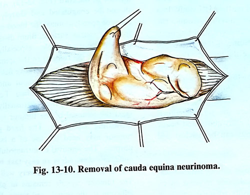
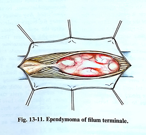
The spinal cord shows various pathological changes: there is considerable increase in its volume with pathologically enlarged vessels in some cases, and change of colour - bright, yellowish or greyish, etc. The surface of the cord at the area of the greatest enlargement is inspected (Fig. 14-12). An initial myelotomy of approximately 1 - 2 cm is performed over the greatest enlargement of the spinal cord to evaluate the plane between the tumour and the spinal cord tissue. The midline strictly (often even shifted by the tumour) should be incised. The presence of a cyst associated with the tumour provides a chance to gain additional room by its removal. Once it is determined that the neoplasm is cystic or has well-defined planes, the myelotomy is enlarged over the extent of the tumour. Following the spinal cord incision, 6-0 or 7-0 traction sutures are placed through the pial margins on both sides to expose better the interior of the cord.
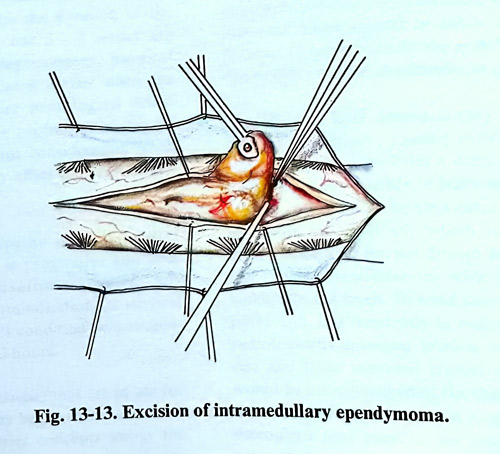
In a case of ependymoma, the tumour is visible a few millimetres under the spinal cord surface. In most instances there is a clear border between the tumour and the spinal cord tissue. Using a microsurgical technique, a sharp plane of dissection is developed around the tumour, taking care to retract the tumour and not the spinal cord. The removal starts from any of the ends of the tumour. If possible, the pole of the tumour is lifted, and gentle traction with a tumour forceps facilitates its removal from the interior of the cord (Fig. 13-13). All fine vessels should be coagulated and sharply divided. In cases of ependymomas, the blood supply comes from the ventral surface. Blunt dissection of the tumour is not advisable - especially where vessels connect the tumour to the spinal cord. It is important to keep the operative field dry so that the plane between the tumour and the spinal cord can be clearly visualised.
For those tumours that are infiltrative (usually astrocytomas), the removal of the tumour is easier with ultrasonic aspiration. The hard part of the tumour may need the use of a laser for this purpose. The removal of intramedullary epidermoids and dermoids is an easy task because they are usually avascular and well delineated. The tumour capsule remains in place if it is adherent to the spinal cord tissue. With teratomas, the stalk between the cord and the dura should be removed as an integral part of the tumour.
Serious difficulties are present with haemangioblastomas, because they are highly vascularised. Any attempt at decompression or cutting for biopsy produces bleeding that obscures the anatomic plane and may result in catastrophic problems. These tumours are identified by their characteristic red appearance, and in all instances extrude from the pial surface. Large arterialised veins often surround them. The removal of such a tumour starts with interruption of the feeding arteries and ends with the primary draining veins. The removal should always be total as this lesion is curable. The presence of cysts makes their removal easier. In patients who have had previous operations, or in whom the dura was left open, the initial exposure of the tumour is difficult and is advisable to start from an intact part of the dura.
Lipomas of the spinal cord occur as isolated tumours in the thoracic and cervical spinal cord. Histologically identical to normal fat, these tumours are located on the dorsal surface of the spinal cord covered by little or no neural tissue. The most effective operative strategy consists of subtotal removal leaving a layer of tumour tissue on the surface of the cord. The laser is ideal to remove lipomas. On the other hand, the ultrasonic aspirator is not effective due to the fibrous stroma of the lesion.
Postoperative management. Steroids are routinely used pre- and postoperatively in high doses for limited periods. Prophylactic antibiotics can be used intraoperatively and up to three days postoperatively. If the surgery has involved the upper cervical area and motor impairment is expected, the endotracheal tube is left in place for at least 24 hours, regardless of the patient's condition at the end of the surgery and an evaluation afterwards.
Radiotherapy is considered postoperatively for patients with malignant spinal tumours and intramedullary astrocytomas, where tumour removal has been incomplete. Careful follow-up of spinal stability is needed. Bracing might be necessary for children because of the possible development of spinal deformity following the tumour removal.