Legacy: "Atlast of Neurosurgery" / L.Karaguiosov, A. Ramadan, K.Karaguiosov / Kiwait/ 1998
PREV>14. Operations on Intervertebral Discs
NEXT< 16. Arteriovenous Malformations of the Spinal Cord
15. MALFORMATIONS OF THE SPINE AND SPINAL CORD
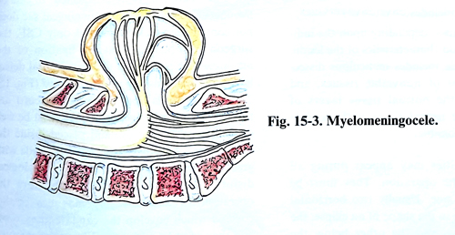
There is a wide variety of congenital pathologic conditions of the spine and spinal cord, of which the most frequent of them are open neural tube defects (meningocele, myelomeningocele, myeloschisis, tethered spinal cord, split-cord malformations, syringomyelia, dermal sinus with or without tumour. The common denominator of this variety of condition is a congenital defect in the posterior elements of one or more vertebral segments. In myelomeningocele there is a characteristic dorsal protrusion of more or less severely malformed spinal cord elements and their coverings between the bifid bony structure on the surface of the back.
Neurological deficits in the lower extremities are often severe, with an associated loss of bladder and bowel control. These deficits are present at birth and usually do not alter. The Chiari malformation and progressive hydrocephalus are common.
The group of malformations leading to a tethered cord have fewer external manifestations, but sometimes produce progressive neurological deficits.
Syringomyelia has a special place among congenital malformations. It is caused by an enlarging central canal (hydromyelia) at paramedian location with accumulation of fluid within the spinal cord. This malformation is frequently related to the Chiari malformation.
The surgical treatment in general cannot eradicate the malformation, but in many cases, it can stop the progression of the neurological deficit, prevent infection and in some cases it may bring about improvement of a recently developed neurological deficit. In many cases, the operation has a cosmetic purpose.
OPEN NEURAL TUBE DEFECTS
Meningoceles and myelomeningoceles are the most common of the congenital dysraphic states, occurring in approximately 1 per two thousand live births with the tendency to decrease in the last decade. They are a result of failure of normal midline fusion of the neural tube. Dorsal fusion defects are much more common than are their ventral counterparts.
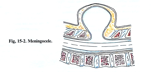
Meningocele and myelomeningocele are characterised by an epithelium-covered sac filled with
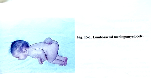
CSF that communicates with the spinal subarachnoid space, or the sac also includes nerve roots, and spinal cord incorporated into the sac (Figs. 15-1; 15-2; 15-3).
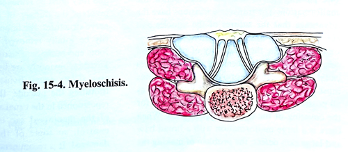
Myeloschisis represents a severe dysraphic state in which a large placode is present at the site of the defect without meningeal encasement. This defect involves multiple levels and presents severe deficits in neurological function (Fig. 15-4).
Measuring alpha fetal protein and confirming it with amniocentesis, monitoring fetal movements, and performing imaging using ultrasonography can allow prenatal diagnosis. This allows abortion to be carried out in those women who desire to terminate their pregnancy.
Dysraphic lesions are generally affecting the normal CSF flow pattern resulting in hydrocephalus and hydromyelia. Hydrocephalus is not always evident at birth, but in about 80% of the cases may soon develop post-partum.
The aim of surgical treatment of meningoceles and myelomeningoceles is to release all structures of the nervous system inside the meningeal sac and to reposition them into the spinal canal again, to close the meningeal sac to its normal proportions and to perform plastic repair the defect of the spinal canal, the soft tissues, including the skin. These makes it possible to ensure a free flow of CSF around the neural structures, which is their natural environment and to close the spinal canal as it should be in normal conditions.
Eighty percent of open neural tube defects are found in the lubo-sacral area, 4% in the cervical area, and 16% in the thoracic area. Many children with myelomeningoceles and myeloschisis also have an associated Chiari malformation, accompanied with polymicrogyria, hydromyelia, and abnormalities of the skull base. The initial surgical problem is closing the skin defect. This is followed closely by treatment of the urological problems when the sacral nerves are involved, and of associated orthopedic problems when the extremities and the spine become deformed as a result of imbalanced motor and sensory denervations.
The preoperative studies include CT and/or and ultrasonic examination of the head to assess the ventricular size and any other malformations. A spinal X-rays helps to demonstrate the degree of kyphosis. More recently MRI studies give the greatest details of the malformations.
TIMING AND SELECTION OF PATIENTS FOR SURGICAL TREATMENT
In meningoceles covered with nomal skin, irrespective of their size and provided there is no neurological deficit, the of neurological function. operative treatment has only cosmetic goals and can be performed after some time. In cases of meningomyelocele and myeloschisis, recent research suggests tha the earlier the defect is closed, the lower the incidence of infection, and that closure within the first 48 hours of life offers optimal preservation.
SURGICAL TECHNIQUE
The operation is performed under general anaesthesia. The patient is in a prone position, the malformation area being a little higher than the ventricular system level so that intracranial hypotension will not occur when CSF flows out during the operation. Many details of the operative technique depend on the size of the malformation, its characteristics, the condition of the skin, which covers the meningeal sac and its content. The operation must solve the following problems:
1. Reliese of all neural structures in the meningeal sac and replacing them into the spinal canal.
2. Complete closure of the neck of the meningeal sac.
3. Restoration with plastic surgical techniques of the spinal canal defect, fascia and skin.
The different types of defects require different procedures and techniques, depending upon the individual anatomy and characteristics of the lesion.
The basic principle includes meticulous dissection, debridement of nonviable tissues, and preservation of the normal tissue layers and potentially viable neural tissues with as little manipulation as possible.
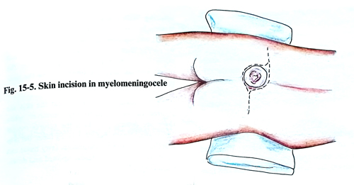
Some difficulties may appear during the different stages of the operation. They must be resolved individually. Usually, two horizontal incisions are made in the shape of an ellipse, the one passing above and the other below the malformation. The transverse incisions have the advantage that the sutures of the muscles and the aponeurosis are perpendicular to one another. With this, the possibility of provoking a CSF fistula decreases, and the spinal canal wall becomes more resilient. The incisions must pass along the limits of the intact skin, so all possible and available skin can be used for the covering of the defect. When there is a large malformation with a broad base and large skin defect, the problem of making the skin incisions is more important and on its solution depends whether the skin will be safely closed. In such cases it is very convenient to make two incisions in the form of the letter S, perfomed laterally on each side, and that will provide the possibility during closure to mobilise the skin edges not only in a vertical direction, but also transversally (Fig. 15-5). For the same purpose the incisions may be made longer in the transverse direction and by a cuneiform resection of one of the edges to obtain a greater mobilisation of the flap. In a small herniation a vertical incision around the malformation can be made, if it is far from the anus. The help of a plastic surgeon is necessary with very large skin defects.
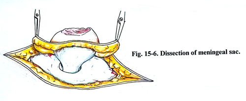
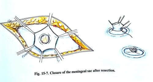
After this, the meningeal sac is separated carefully with blunt instruments until its neck and the spinal canal defect edges are dissected (Fig.15-6). The meningeal sac is opened on the midline and its content is well inspected. In case of meningoceles, i.e. when the meningeal sac does not contain nerve elements, but only CSF, the surgical problem is less complex. A ligation of the meningeal sac is made at the spinal canal defect level and the remaining part is resected (Fig. 15-7). If nerve roots and/or the spinal cord are revealed in the meningeal sac, they must be well dissected without injury, and placed inside the spinal canal. This dissection must be made with great care so as not to worsen the neurological deficit, which usually exists before the operation. In some cases lipomas may be discovered which envelop the cauda equina roots and they must be removed. When their removal involves the danger of injuring some of the nerve structures, the lipoma should not necessarily be totally removed. If the cauda equina roots end in the scar tissue in the fundus of the sac, they obviously are without function. However, if any neural tissue is suspicious of only transiting the placode, the attachment area is repositioned in the canal as a whole. The closing of the meningeal sac is made under visual control to avoid root damage. It is recommended that the meningeal sac is watertight closed, so that a CSF fistula may not occur in the postoperative period.
Very rarely, the anterior wall of the spinal canal is protruding on the surface near the skin level and in order to reposition the spinal cord and the cauda equina roots, it is necessary to excavate an additional spinal canal space within the vertebral bodies.
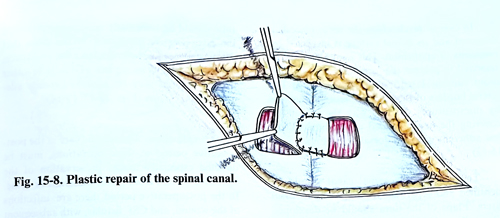
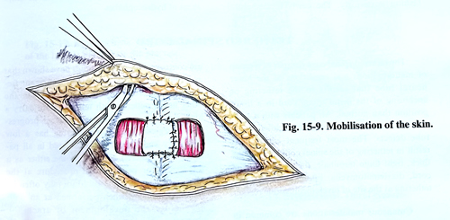
The last stage of the operation is the plastic closure of the spinal canal defect. In round defects and when they are small, their edges can be mobilised, by excising on both sides and then suturing them. With a larger defect, the plastic closure is done by two flaps, wide enough that they can be left and sutured one over the other (Figs. 15- 8; 15-9). With much larger defects it is not possible for the flaps to be put one over the other; instead, they are sutured along the midline.
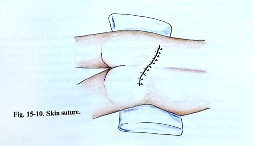
The subcutaneous tissue and the skin are sutured in the ordinary way. However, when there are defects, in which the sutures remain under great distension, before suturing them, the skin together with the subcutaneous fatty tissue can be separated from the fascia upward and downward (Fig. 15-10).
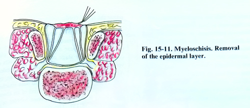
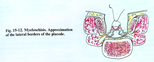
In case of myeloschisis the nerve placode is dissected and its dorsal surface is inspected for remnants of epithelium. This remnants must be removed to prevent the formation of an epidermoid tumour later in the patient's life. The lateral borders of the placode are approximated by microsutures. In this way the intact pia covering the newly created neural tube will be less adherent with the dorsal dura (Figs. 15-11; 15-12).
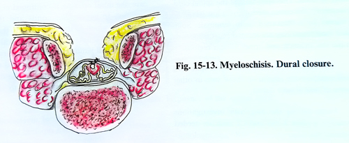
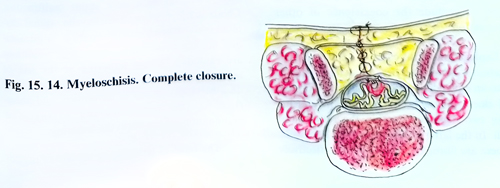
The dura of the spinal canal should be identified and separated from the lumbodorsal fascia. Flaps of the dura should be developed bilaterally and closed over the placode in a stable fashion posteriorly. The newly formed dural tube should be sufficiently capacious to minimize the postoperative adhesions. In the case when the closure does not provide adequate space, grafting materials as a piece of fascia or other plastic material may be used to provide adequate closure (figs. 15-13; 15-14).
Complications. The specific care in the postoperative period in these operations must be directed specifically to prevent contaminations of the operative wound from faecal materials or urine. In the postoperative period can be observed infections of the wound and CSF fistulae, with subsequent development of meningitis or acute development of hydrocephalus.
TETHERED SPINAL CORD
The tethered spinal cord consists of a group of malformations and conditions of the lower neural tube that result in the tethering or anchoring of the distal spinal cord in a lower position than normal. All patients with a tethered cord usually have evidence of closed spinal tube defects. The most frequent lesions that result in tethering of the spinal cord include the thick, tight filum terminale, the attached spinal cord, diastematomyelia, lipomeningocele, and tethering at the site of myelomeningocele repair.
Cutaneous manifestations are common in patients with a tethered spinal cord. These skin manifestations may consist of a hair patch, a dimple, a haemangioma, a lipoma, or an area of atrophic skin. Patients with lipomeningocele will always have a fatty mass visible in the lumbosacral region, which can vary from a very large expansive fatty mass to a small lipoma. X-rays, CT and MRI are essential studies for confirming the malformation and the tethered cord.
Surgical indications. In case of evidence of neurological deterioration, there is widespread agreement that the cord should be untethered as soon as possible. In cases of stable neurological manifestations, opinions on the role of surgery are controversial. Many studies have shown that an improvement can occur with significant frequency after releasing the tethering. On the other hand, patients with tethered cord carry a high risk of subsequent neurological progression. Surgical release of tethering has a low morbidity and should be considered in all patients with tethered cord syndrome either as a therapeutic or prophylactic procedure at the time of diagnosis. The procedure may often be technically simple when performed at an early age before the development of arachnoid adhesions.
SURGICAL TECHNIQUE
Thick filum terminale. Patients with a thick filum terminale need a single-level laminectomy, usually at the level of the fifth lumbar and first sacral vertebrae. It is very easy to differentiate the thickened filum terminale from the surrounding nerve roots, because of its size, its tautness, and its richness of blood vessels. The filum terminale is divided after coagulation. It is not necessary to put a metallic marker in its cut ends for monitoring of cord ascent, because MRI can be used for a follow-up study.
Attached spinal cord. The surgical technique in patients with an attached spinal cord is similar to that with a thick filum terminale. The laminectomy is performed in accordance with the MRI data, usually in the sacral area. The cord is detached from the terminal dural sac and allowed to ride free. Frequently, the attached spinal cord is thinned out and stretched because of the tethering, and can be differentiated from the tethered filum terminale by the nerve roots, which can be seen leaving the spinal cord and ascending to reach their exit foramina.
Split-cord malformations. These malformations are of two types: Diastematomyelia, in which the spinal cord is split into two hemicords and each has a single set of dorsal and ventral nerve roots and is contained within its own dural sheath. The second type is diplomyelia where the two spinal cord segments are completely duplicated within a single dural sleeve.
When the two dural sleeves are present (diastematomyelia), a bony or fibrocartilagenous spur exists at the caudal end of the cleft and may be attached to the surrounding bone dorsally, ventrally or both. This spur tethers the spinal cord.
Frequently the conus medullaris is low lying and tethered by a thickened filum terminale. Split-cord malformations are located most frequently in the low thoracic and upper lumbar regions.
The clinical manifestations of the split-cord malformation are similar to those seen in other forms of neural tube defect. Among the diagnostic investigations available, a plain X-ray and CT scan are needed to detect the bony changes. MRI and myelography are needed too. If a split-cord malformation is present, the entire spinal cord should be imaged to exclude the coexistence of other malformation.
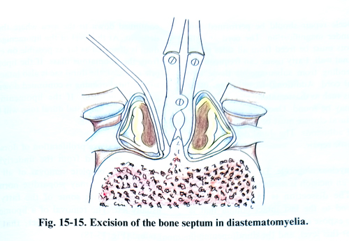
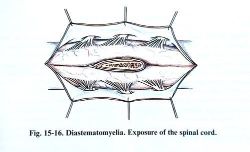
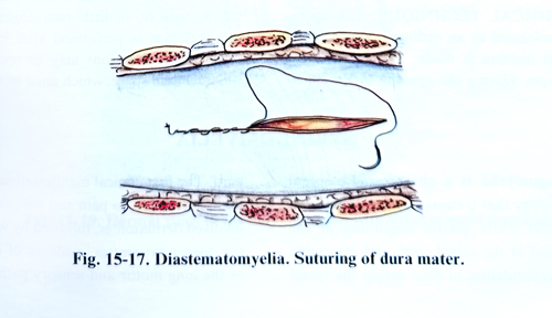
Exploration of all cases of split-cord malformation is advocated because deterioration is seen to an equal degree in patients with a double dural sleeve plus a bony spur and in patients having two hemicords within the same dural sheath. In the latter group there is no bony spur but there are fibroglial adhesions that effectively tether the spinal cord. The spinal canal is opened surgically in the usual way, by laminectomy of one or two vertebrae at the place of the split-cord. During this stage of operation defects of vertebral laminae may frequently be revealed. After the dissection of the split dural sac, the bony spur, (that ends in the broad base attached to two vertebral bodies at the level of an absent disc space) is excised with a bone nibbler or is drilled (Fig. 15-15). The dura is opened with a vertical incision in one of the single dural tubes, and in the area of splitting dural tube it passes near to the midline, extended in the midline on the other single dural tube (Fig. 15-16). The split spinal cord is exposed and, if arachnoid adhesions are fixing it, they need to be divided. The anterior dura is then left either open or is closed, if possible with some single sutures. The posterior dura is reconstructed, either directly or with a dural graft (Fig. 15-17). If the diastematomyelia is in the lumbosacral area, it is usually associated with a thick filum terminale. In such a case, the dural opening is continued in a caudal direction to expose the conus and the filum terminale, which is then divided. If the diastematomyelia is not in the lumbosacral area, a separate small laminectomy is necessary at L5 - S1. In case of diplomyelia the surgical task is a little easier. After the opening of the dura, the adhesions fixing the spinal cord to the dura should be released.
Tethering at the site of myelomeningocele repair. The patient with a repaired myelomeningocele who shows evidence of secondary deterioration probably has some condition other than tethering such as a shunt malfunction, Chiari malformation, hydromyelia, spinal dermoid cysts, split-cord malformation, or intracranial midline cysts. Only when these conditions are excluded, tethering at the site of repair should be considered as a cause of the deterioration. MRI, and a lack of movement and pulsation of the conus during ultrasound examination may confirm the tethering.
The relief of tethering at the site of myelo-meningocele repair should be performed cautiously under magnification. The cord and the nerve roots must be freed from all sides of the spinal canal wall. Fatty tissue can frequently be seen extending from the subcutaneous layer into the spinal cord. Additional to tethering, other conditions such as diastematomyelia or dermoid tumour may be found. Once the spinal cord is free, the dural closure must be secure to prevent arachnoid adhesions. The dural defects usually need a plastic repair.
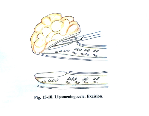
Lipomyelomeningocele. To repair a lipomeningocele, an skin incision is made in the midline. The dissection is caried out to the cranial end to the last intact lamina that is removed to expose an intact dural sac. The dura is opened at this level, and the dural opening is continued down to the area where the dura is deficient. At the level of the lipomenigocele, the dura is dissected as far as possible on either side from the lipomatous mass. If the lipomeningocele is dorsal, the dural sac is also intact distally, so the dural opening is continued down into the normal anatomical structures. If the lipomeningocele is lumbosacral, the distal dural tube will be abnormal or non-existing. The bulk of dorsally located fat can now be removed from the underlying nerve structures. Complete removal of all the fatty tissue, however, may jeopardise normal conus function, so that some of the fatty tissue is usually left. The removal of a lipomeningocele always leaves a dural defect, that must be grafted (Fig. 15-18).
SPINAL DERMAL SINUS
The spinal dermal sinus should always be treated surgically, as it may be the path of local infection that may penetrate into the spinal canal and cause serious disturbances, such as meningitis or abscess. The appearance of a meningeal syndrome in a patient with dermal sinus of the spine, locally infected, represents an immediate indication for surgery. The presence of neurological disturbances is also an indication for surgical treatment that must be performed as early as possible, if these disturbances show progressive deterioration.
SURGICAL TECHNIQUE
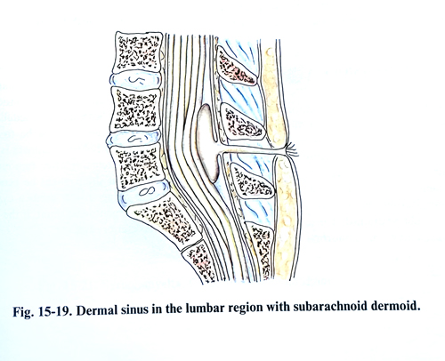
The operation is performed as an ordinary laminectomy. A vertical incision is made, that includes the skin changes. During the opening of the spinal canal, a dissection is made of the fibrous tract, which may enter the epidural space and widen into a dermoid or epidermoid (Fig. 16-19). The entire tract, together with the tumour must be removed in one piece. If there are adhesions to the dura or penetration into the dural sac, the latter must be opened and its content well inspected for adhesions between the tract and the meninges, or penetration of the tract itself into the spinal cord, where it widens into a dermoid or epidermoid. These types of tumours are clearly delimited from the spinal cord tissue and are totally removed under optical magnification, with no or little neurological deficit. If the operation is performed after infection has taken place, adhesions may be revealed in the subarachnoid space, which must be divided.
SYRINGOMYELIA
Syringomyelia is a characteristic spinal cord pathology that is caused by the destruction of grey and white matter beginning close to the central canal of the spinal cord, and associated with an accumulation of fluid within the spinal cord. The first clinical manifestation is usually a loss of sensation to pain and temperature in the involved dermatomes, followed by weakness. As the disease progresses, evidence of involvement of the long motor and sensory pathways in the spinal cord is often observed as well. The typical spinal syndrome is frequently accompanied by a dysfunction of the low brain stem due to the same cystic development - syringobulbia. Most frequently the enlarged central canal of the spinal cord communicates with the fourth ventricle and Chiari malformation invariably accompanies this condition.
The second most common cystic spinal lesion that can produce a syringomyelitic-type cord syndrome is an intramedullary tumour. The cysts do not communicate with the central canal and are not associated with a hindbrain malformation. Cystic cavities can develop within the spinal cord after traumatic injury too.
From all investigations available of the brain and spinal cord, the following are useful: a plain X-ray, CT, myelography, and an MRI provide all the information needed to plan a surgical approach to this condition. The extent of the descent of the cerebellar tonsils and fourth ventricle into the foramen magnum, the transverse diameter and caudal extent of the hydromyelitic cavity, and the size of the ventricle system can be seen very well. These investigations can also demonstrate the disappearance of the hydromyelitic cavity with successful therapy.
SURGICAL TECHNIQUE
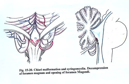
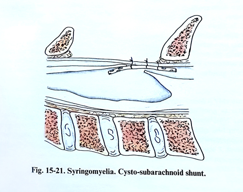
Patients with Chiari malformations and hydromyelia should be treated with a suboccipital craniectomy and upper cervical laminectomy to decompress the malformation at the foramen magnum. If the fourth ventricle is blocked, free communication should be established using a microsurgical technique, and the hydromyelitic cavity should be drained. The elongated cerebellar tonsils have an abnormal appearance in colour and consistency. The caudal loop of the posterior inferior cerebellar artery often descends to the level of C2. The next step is the penetration between the two tonsils toward the foramen Magendi. This penetration should be very cautious; if there are adhesions their division should be non traumatic. This procedure should be done under a microscope. If the foramen Magendie is blocked, its lower part should be opened in the midline (Fig. 15-20). A myelotomy is performed through the laminectomy of one vertebra at the level of the inferior part of the hydromyelitic cyst. The cord should be incised longitudinally in the dorsal root entry zone between lateral and posterior columns because this is usually the thinnest area. A needle should be introduced into the cavity through the thinnest area of its wall and fluid collected for investigation before the cord is incised. Than a vertical incision is made of at least 1 cm in length along the dorsal entry zone. To prevent closure a silastic fine catheter may be introduced into the cystic cavity and fixed to the dura (Fig. 15-21). The dura in the posterior fossa and in the laminectomy is closed with a graft allowing some expansion.
In a patient with hydrocephalus associated with syringomyelia, a ventriculoperitoneal shunt is the procedure of first choice, especially if increased CSF pressure is present. If spinal cord symptoms have not improved, shunting of the intramedullary cyst should be considered. For a cyst associated with an intramedullary tumour, the goal of surgery should be to remove the tumour; usually further treatment is not needed.
If the cavity remains after tumour removal a cystoperitoneal shunt might be beneficial. Post-traumatic syringomyelia needs a cysto-subarachnoid or cystoperitoneal shunt, if the neurological deficit is continuing to deteriorate.