
TIMING AND SELECTION OF PATIENTS FOR SURGICAL TREATMENT
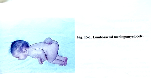
In meningoceles covered with nomal skin, irrespective of their size and provided there is no neurological deficit, the of neurological function. operative treatment has only cosmetic goals and can be performed after some time. In cases of meningomyelocele and myeloschisis, recent research suggests tha the earlier the defect is closed, the lower the incidence of infection, and that closure within the first 48 hours of life offers optimal preservation.
SURGICAL TECHNIQUE
The operation is performed under general anaesthesia. The patient is in a prone position, the malformation area being a little higher than the ventricular system level so that intracranial hypotension will not occur when CSF flows out during the operation. Many details of the operative technique depend on the size of the malformation, its characteristics, the condition of the skin, which covers the meningeal sac and its content. The operation must solve the following problems:
1. Reliese of all neural structures in the meningeal sac and replacing them into the spinal canal.
2. Complete closure of the neck of the meningeal sac.
3. Restoration with plastic surgical techniques of the spinal canal defect, fascia and skin.
The different types of defects require different procedures and techniques, depending upon the individual anatomy and characteristics of the lesion.
The basic principle includes meticulous dissection, debridement of nonviable tissues, and preservation of the normal tissue layers and potentially viable neural tissues with as little manipulation as possible.
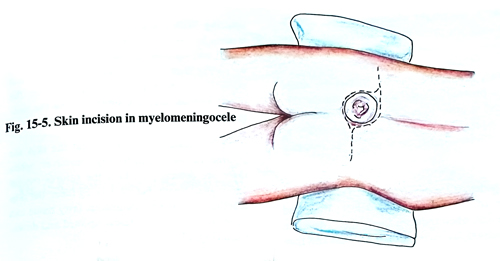
Some difficulties may appear during the different stages of the operation. They must be resolved individually. Usually, two horizontal incisions are made in the shape of an ellipse, the one passing above and the other below the malformation. The transverse incisions have the advantage that the sutures of the muscles and the aponeurosis are perpendicular to one another. With this, the possibility of provoking a CSF fistula decreases, and the spinal canal wall becomes more resilient. The incisions must pass along the limits of the intact skin, so all possible and available skin can be used for the covering of the defect. When there is a large malformation with a broad base and large skin defect, the problem of making the skin incisions is more important and on its solution depends whether the skin will be safely closed. In such cases it is very convenient to make two incisions in the form of the letter S, perfomed laterally on each side, and that will provide the possibility during closure to mobilise the skin edges not only in a vertical direction, but also transversally (Fig. 15-5). For the same purpose the incisions may be made longer in the transverse direction and by a cuneiform resection of one of the edges to obtain a greater mobilisation of the flap. In a small herniation a vertical incision around the malformation can be made, if it is far from the anus. The help of a plastic surgeon is necessary with very large skin defects.
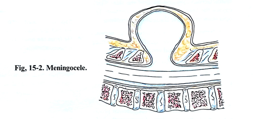
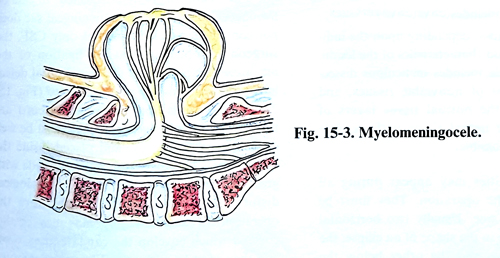
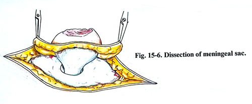
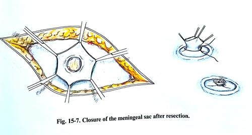
After this, the meningeal sac is separated carefully with blunt instruments until its neck and the spinal canal defect edges are dissected (Fig.15-6). The meningeal sac is opened on the midline and its content is well inspected. In case of meningoceles, i.e. when the meningeal sac does not contain nerve elements, but only CSF, the surgical problem is less complex. A ligation of the meningeal sac is made at the spinal canal defect level and the remaining part is resected (Fig. 15-7). If nerve roots and/or the spinal cord are revealed in the meningeal sac, they must be well dissected without injury, and placed inside the spinal canal. This dissection must be made with great care so as not to worsen the neurological deficit, which usually exists before the operation. In some cases lipomas may be discovered which envelop the cauda equina roots and they must be removed. When their removal involves the danger of injuring some of the nerve structures, the lipoma should not necessarily be totally removed. If the cauda equina roots end in the scar tissue in the fundus of the sac, they obviously are without function. However, if any neural tissue is suspicious of only transiting the placode, the attachment area is repositioned in the canal as a whole. The closing of the meningeal sac is made under visual control to avoid root damage. It is recommended that the meningeal sac is watertight closed, so that a CSF fistula may not occur in the postoperative period.
Very rarely, the anterior wall of the spinal canal is protruding on the surface near the skin level and in order to reposition the spinal cord and the cauda equina roots, it is necessary to excavate an additional spinal canal space within the vertebral bodies.
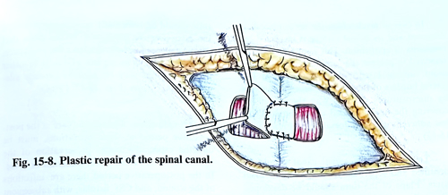
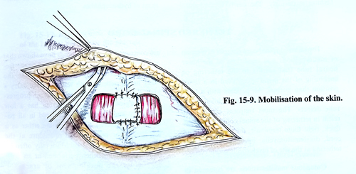
The last stage of the operation is the plastic closure of the spinal canal defect. In round defects and when they are small, their edges can be mobilised, by excising on both sides and then suturing them. With a larger defect, the plastic closure is done by two flaps, wide enough that they can be left and sutured one over the other (Figs. 15- 8; 15-9). With much larger defects it is not possible for the flaps to be put one over the other; instead, they are sutured along the midline.
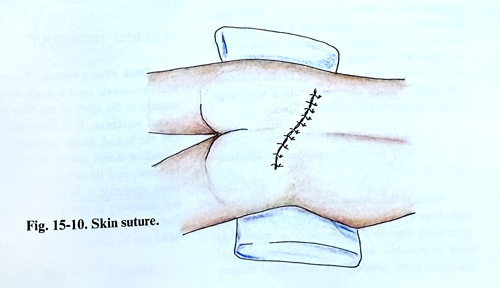
The subcutaneous tissue and the skin are sutured in the ordinary way. However, when there are defects, in which the sutures remain under great distension, before suturing them, the skin together with the subcutaneous fatty tissue can be separated from the fascia upward and downward (Fig. 15-10).
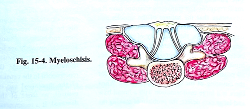
In case of myeloschisis the nerve placode is dissected and its dorsal surface is inspected for remnants of epithelium. This remnants must be removed to prevent the formation of an epidermoid tumour later in the patient's life. The lateral borders of the placode are approximated by microsutures. In this way the intact pia covering the newly created neural tube will be less adherent with the dorsal dura (Figs. 15-11; 15-12).
The dura of the spinal canal should be identified and separated from the lumbodorsal fascia. Flaps of the dura should be developed bilaterally and closed over the placode in a stable fashion posteriorly. The newly formed dural tube should be sufficiently capacious to minimize the postoperative adhesions. In the case when the closure does not provide adequate space, grafting materials as a piece of fascia or other plastic material may be used to provide adequate closure (figs. 15-13; 15-14).
Complications. The specific care in the postoperative period in these operations must be directed specifically to prevent contaminations of the operative wound from faecal materials or urine. In the postoperative period can be observed infections of the wound and CSF fistulae, with subsequent development of meningitis or acute development of hydrocephalus.