SYRINGOMYELIA
Syringomyelia is a characteristic spinal cord pathology that is caused by the destruction of grey and white matter beginning close to the central canal of the spinal cord, and associated with an accumulation of fluid within the spinal cord. The first clinical manifestation is usually a loss of sensation to pain and temperature in the involved dermatomes, followed by weakness. As the disease progresses, evidence of involvement of the long motor and sensory pathways in the spinal cord is often observed as well. The typical spinal syndrome is frequently accompanied by a dysfunction of the low brain stem due to the same cystic development - syringobulbia. Most frequently the enlarged central canal of the spinal cord communicates with the fourth ventricle and Chiari malformation invariably accompanies this condition.
The second most common cystic spinal lesion that can produce a syringomyelitic-type cord syndrome is an intramedullary tumour. The cysts do not communicate with the central canal and are not associated with a hindbrain malformation. Cystic cavities can develop within the spinal cord after traumatic injury too.
From all investigations available of the brain and spinal cord, the following are useful: a plain X-ray, CT, myelography, and an MRI provide all the information needed to plan a surgical approach to this condition. The extent of the descent of the cerebellar tonsils and fourth ventricle into the foramen magnum, the transverse diameter and caudal extent of the hydromyelitic cavity, and the size of the ventricle system can be seen very well. These investigations can also demonstrate the disappearance of the hydromyelitic cavity with successful therapy.
SURGICAL TECHNIQUE
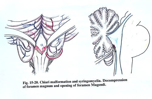
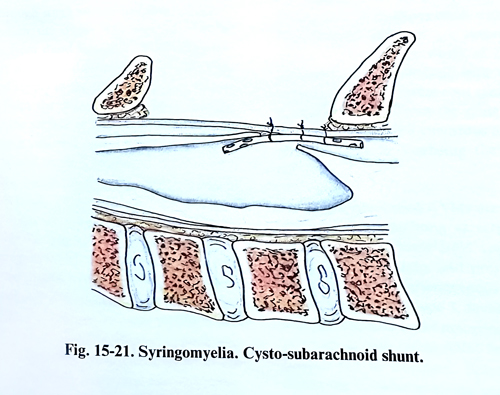
Patients with Chiari malformations and hydromyelia should be treated with a suboccipital craniectomy and upper cervical laminectomy to decompress the malformation at the foramen magnum. If the fourth ventricle is blocked, free communication should be established using a microsurgical technique, and the hydromyelitic cavity should be drained. The elongated cerebellar tonsils have an abnormal appearance in colour and consistency. The caudal loop of the posterior inferior cerebellar artery often descends to the level of C2. The next step is the penetration between the two tonsils toward the foramen Magendi. This penetration should be very cautious; if there are adhesions their division should be non traumatic. This procedure should be done under a microscope. If the foramen Magendie is blocked, its lower part should be opened in the midline (Fig. 15-20). A myelotomy is performed through the laminectomy of one vertebra at the level of the inferior part of the hydromyelitic cyst. The cord should be incised longitudinally in the dorsal root entry zone between lateral and posterior columns because this is usually the thinnest area. A needle should be introduced into the cavity through the thinnest area of its wall and fluid collected for investigation before the cord is incised. Than a vertical incision is made of at least 1 cm in length along the dorsal entry zone. To prevent closure a silastic fine catheter may be introduced into the cystic cavity and fixed to the dura (Fig. 15-21). The dura in the posterior fossa and in the laminectomy is closed with a graft allowing some expansion.
In a patient with hydrocephalus associated with syringomyelia, a ventriculoperitoneal shunt is the procedure of first choice, especially if increased CSF pressure is present. If spinal cord symptoms have not improved, shunting of the intramedullary cyst should be considered. For a cyst associated with an intramedullary tumour, the goal of surgery should be to remove the tumour; usually further treatment is not needed.
If the cavity remains after tumour removal a cystoperitoneal shunt might be beneficial. Post-traumatic syringomyelia needs a cysto-subarachnoid or cystoperitoneal shunt, if the neurological deficit is continuing to deteriorate.