TETHERED SPINAL CORD
The tethered spinal cord consists of a group of malformations and conditions of the lower neural tube that result in the tethering or anchoring of the distal spinal cord in a lower position than normal. All patients with a tethered cord usually have evidence of closed spinal tube defects. The most frequent lesions that result in tethering of the spinal cord include the thick, tight filum terminale, the attached spinal cord, diastematomyelia, lipomeningocele, and tethering at the site of myelomeningocele repair.
Cutaneous manifestations are common in patients with a tethered spinal cord. These skin manifestations may consist of a hair patch, a dimple, a haemangioma, a lipoma, or an area of atrophic skin. Patients with lipomeningocele will always have a fatty mass visible in the lumbosacral region, which can vary from a very large expansive fatty mass to a small lipoma. X-rays, CT and MRI are essential studies for confirming the malformation and the tethered cord.
Surgical indications. In case of evidence of neurological deterioration, there is widespread agreement that the cord should be untethered as soon as possible. In cases of stable neurological manifestations, opinions on the role of surgery are controversial. Many studies have shown that an improvement can occur with significant frequency after releasing the tethering. On the other hand, patients with tethered cord carry a high risk of subsequent neurological progression. Surgical release of tethering has a low morbidity and should be considered in all patients with tethered cord syndrome either as a therapeutic or prophylactic procedure at the time of diagnosis. The procedure may often be technically simple when performed at an early age before the development of arachnoid adhesions.
SURGICAL TECHNIQUE
Thick filum terminale. Patients with a thick filum terminale need a single-level laminectomy, usually at the level of the fifth lumbar and first sacral vertebrae. It is very easy to differentiate the thickened filum terminale from the surrounding nerve roots, because of its size, its tautness, and its richness of blood vessels. The filum terminale is divided after coagulation. It is not necessary to put a metallic marker in its cut ends for monitoring of cord ascent, because MRI can be used for a follow-up study.
Attached spinal cord. The surgical technique in patients with an attached spinal cord is similar to that with a thick filum terminale. The laminectomy is performed in accordance with the MRI data, usually in the sacral area. The cord is detached from the terminal dural sac and allowed to ride free. Frequently, the attached spinal cord is thinned out and stretched because of the tethering, and can be differentiated from the tethered filum terminale by the nerve roots, which can be seen leaving the spinal cord and ascending to reach their exit foramina.
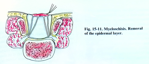
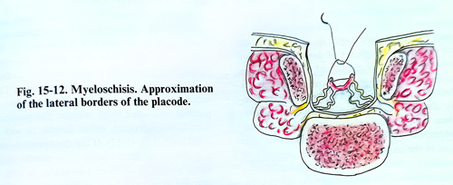
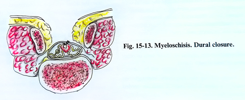
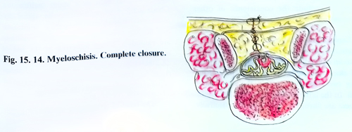
Split-cord malformations. These malformations are of two types: Diastematomyelia, in which the spinal cord is split into two hemicords and each has a single set of dorsal and ventral nerve roots and is contained within its own dural sheath. The second type is diplomyelia where the two spinal cord segments are completely duplicated within a single dural sleeve.
When the two dural sleeves are present (diastematomyelia), a bony or fibrocartilagenous spur exists at the caudal end of the cleft and may be attached to the surrounding bone dorsally, ventrally or both. This spur tethers the spinal cord.
Frequently the conus medullaris is low lying and tethered by a thickened filum terminale. Split-cord malformations are located most frequently in the low thoracic and upper lumbar regions.
The clinical manifestations of the split-cord malformation are similar to those seen in other forms of neural tube defect. Among the diagnostic investigations available, a plain X-ray and CT scan are needed to detect the bony changes. MRI and myelography are needed too. If a split-cord malformation is present, the entire spinal cord should be imaged to exclude the coexistence of other malformation.
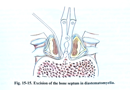
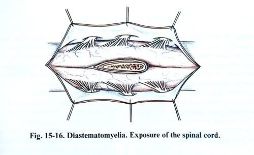
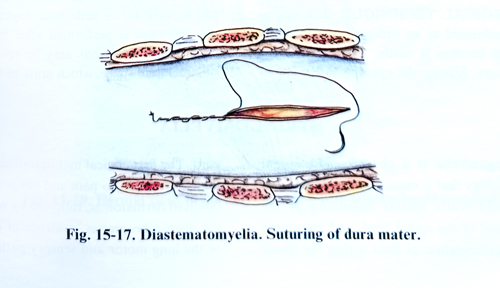
Exploration of all cases of split-cord malformation is advocated because deterioration is seen to an equal degree in patients with a double dural sleeve plus a bony spur and in patients having two hemicords within the same dural sheath. In the latter group there is no bony spur but there are fibroglial adhesions that effectively tether the spinal cord. The spinal canal is opened surgically in the usual way, by laminectomy of one or two vertebrae at the place of the split-cord. During this stage of operation defects of vertebral laminae may frequently be revealed. After the dissection of the split dural sac, the bony spur, (that ends in the broad base attached to two vertebral bodies at the level of an absent disc space) is excised with a bone nibbler or is drilled (Fig. 15-15). The dura is opened with a vertical incision in one of the single dural tubes, and in the area of splitting dural tube it passes near to the midline, extended in the midline on the other single dural tube (Fig. 15-16). The split spinal cord is exposed and, if arachnoid adhesions are fixing it, they need to be divided. The anterior dura is then left either open or is closed, if possible with some single sutures. The posterior dura is reconstructed, either directly or with a dural graft (Fig. 15-17). If the diastematomyelia is in the lumbosacral area, it is usually associated with a thick filum terminale. In such a case, the dural opening is continued in a caudal direction to expose the conus and the filum terminale, which is then divided. If the diastematomyelia is not in the lumbosacral area, a separate small laminectomy is necessary at L5 - S1. In case of diplomyelia the surgical task is a little easier. After the opening of the dura, the adhesions fixing the spinal cord to the dura should be released.
Tethering at the site of myelomeningocele repair. The patient with a repaired myelomeningocele who shows evidence of secondary deterioration probably has some condition other than tethering such as a shunt malfunction, Chiari malformation, hydromyelia, spinal dermoid cysts, split-cord malformation, or intracranial midline cysts. Only when these conditions are excluded, tethering at the site of repair should be considered as a cause of the deterioration. MRI, and a lack of movement and pulsation of the conus during ultrasound examination may confirm the tethering.
The relief of tethering at the site of myelo-meningocele repair should be performed cautiously under magnification. The cord and the nerve roots must be freed from all sides of the spinal canal wall. Fatty tissue can frequently be seen extending from the subcutaneous layer into the spinal cord. Additional to tethering, other conditions such as diastematomyelia or dermoid tumour may be found. Once the spinal cord is free, the dural closure must be secure to prevent arachnoid adhesions. The dural defects usually need a plastic repair.
Lipomyelomeningocele. To repair a lipomeningocele, an skin incision is made in the midline. The dissection is caried out to the cranial end to the last intact lamina that is removed to expose an intact dural sac. The dura is opened at this level, and the dural opening is continued down to the area where the dura is deficient. At the level of the lipomenigocele, the dura is dissected as far as possible on either side from the lipomatous mass. If the lipomeningocele is dorsal, the dural sac is also intact distally, so the dural opening is continued down into the normal anatomical structures. If the lipomeningocele is lumbosacral, the distal dural tube will be abnormal or non-existing. The bulk of dorsally located fat can now be removed from the underlying nerve structures. Complete removal of all the fatty tissue, however, may jeopardise normal conus function, so that some of the fatty tissue is usually left. The removal of a lipomeningocele always leaves a dural defect, that must be grafted (Fig. 15-18).