
Legacy: "Atlast of Neurosurgery" / L.Karaguiosov, A. Ramadan, K.Karaguiosov / Kiwait/ 1998
PREV > 18. Functional Surgery of the Spinal Cord
PART THREE
PERIPHERAL NERVE SURGERY
19. GENERAL PRINCIPLES OF PERIPHERAL NERVE REPAIR
STRUCTURE OF THE PERIPHERAL NERVES. DEGENERATION AND REGENERATION
Every peripheral nerve consists of a large number of nerve fibres (axons) that are extended processes of nerve cell bodies situated in the spinal cord, dorsal root ganglia, and sympathetic ganglia. The ratio between the size of a cell body and its axon is considerable, as axon may extend to many thousands of times the cell body diameter. In this special cellular structure of the neuron, the axon contains more than 90% of the total cytoplasmic volume of the nerve cell. As a consequence of this, any localised lesion of an axon implies serious structural and functional problems affecting the survival of the whole neuron. The fibres of the peripheral nerves are divided into three nerve groups: 1. Motor or efferent fibres, which transmit impulses from the central nervous, system toward the muscles; 2. Sensory or afferent fibres, which transmit sensation from muscles, joints, ten-dons, skin, etc.; 3. Autonomic fibres, which innervate the smooth muscles and glands.
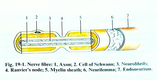
Nerve fibers are either myelinated or non-myelinated. A chain of Schwann cells arranged end to end surrounds both myelinated and nonmyelinated axons. The specific relationship between the Schwann cell and axon differs. The myelinated nerve fiber consists of one axon, which is associated with only one Schwann cell at any one level. The membrane of the Schwann cell, the myelin sheath, is wrapped spirally around the axon, creating a multilaminate sleeve of lipids and protein. The Schwann cells, arranged in a longitudinal sequence, approach each other at the nodes of Ranvier, where finger like cellular processes interdigitate (Fig. 19- 1). In nonmyelinated fibers, Schwann cells accommodate a large number of axons, which are located in internal troughs.
The nerve fibers are divided into three groups according to the relationship between the form of the compound axon potential and the fibre size. Group A fibers are the largest and have the fastest conduction velocities. These fibres are myelinated somatic afferents and effer-ents. Group A fibres are subdivided according to fibre size into A-alpha, A-ß, and A-gamma. An efferent motor fibre, which has a large diameter, belongs to the A-alpha fibre group. A-o fibres transmit the sensation of sharp, pricking pain and temperature, while the A-ß fibres are associated with touch sensation. Myelinated autonomic and preganglionic fibres belong to group B. Group C fibres are thin, slowly conducting nonmyelinated visceral and somatic afferent fibres. Postganglionic, autonomic efferent fibres are also in group C. Nonmyelinated C fibres transfer the sensation of deep burning pain.
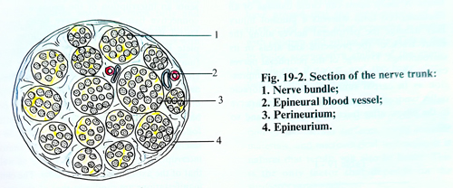
With the exception of the cutaneous nerves, the nerve trunks of the limbs, which are most often subject to surgical operation, are mixed, i. e. they have sensory, motor and autonomic fibres. The nerve fibres are connected in the form of fascicles whose cross-sections are round. A membrane of connective tissue or perineurium envelops each fascicle; every fascicle has a stroma of connective tissue called endoneurium.
The nerve fascicles covered by their perineurium, are separated from each other by connective tissue of a loose character that connects them into a nerve trunk. The same tissue forms another external sheath of the nerve trunk called epineurium (Fig. 19-2).
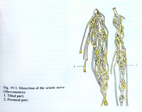
Enveloped in perineurium, the nerve fascicles are not isolated from one another along the nerve trunk, i.e, they are not in the form of a cable. The nerve trunk has a plexiform internal structure and the nerve bundles exchange fibres among themselves. Because of this the number and disposition of the nerve fascicles vary in a series of cross-sections of a given nerve trunk, (Fig. 19 - 3).
The nerve trunks are supplied by the blood vessels in their vicinity. Peripheral nerves are well vascularised structures with separate but extensive interconected microvascular systems in the epineurium, perineurium, and endoneurium. Along the course of the nerve, this vascular system is reinforced by segmental regional vessels of extrinsic origin. These regional vessels travel in an adventítia of connective tissue that surrounds the nerve, allowing it to move longitudinally in its bed with movements of the extremity.
After continuity interruption of a peripheral nerve, the peripheral segment of the interrupted nerve loses its transmission capability in 3 - 4 days. The axons and myelin sheaths suffer from desintegration and are object of fagocytosis by macrophages and Schwann cells. These changes are called Valerian degeneration. The proliferation of Schwann cells and endoneural fibrocytes lead to an increase in the volume of the distal segment of the interrupted nerve, but with time the cell populations diminishes, the endoneural tubes shrink, and the diameter of all distal segments diminishes. Similar changes occur also in the proximal segment of the interrupted nerve without diminishing of the nerve volume.
When an axon is interrupted, chromatolysis in the body of the corresponding nerve cell develops. This is considered more a manifestation of regeneration than of degeneration. After the fourth day of the injury, the amount as well as the metabolism of the ribonucleic acid increase. The increase in the amount and activity of the ribonucleic acid remain until the regeneration and maturation of the axon. The nearer the injury to the spinal cord the more numerous are the degenerative changes of the neurons, whereas in a distal direction, these changes become less expressed. The axoplasm of the axon goes out into the chaotic growth of Schwann cells and the proximal end of the axon increases in volume.
The advance of axons over the zone of injury is a critical process that may determine the success or failure of functional regeneration.
The growing axon of the central interrupted part may penetrate into the extension of the Schwann cells made canal in its peripheral part, i. e. in order that regeneration may be possible, certain conditions are necessary. One of the most important conditions is that the distance between the two ends of the interrupted nerve should be as small as possible. The axons grow through this space in about two weeks, before a thick cicatrix is formed. In injury of the peripheral nerves, no doubt, the ends of the interrupted nerve are separated from one another at a comparatively large distance because of their great elasticity.
The muscle traction and sometimes the traumatic agent may destroy part of the nerve trunk. The presence of haematomas, destruction of muscle fibres, bone fragments and foreign bodies obstructs additionally the regeneration. In cases of a long duration of wound infection, the regeneration may be obstructed because of the presence of neuritis, which in the end causes sclerosis of the nerve fibres. When the axons growing from the proximal end do not penetrate into the peripheral end of the interrupted nerve, a thickening in the shape of a bulb is formed, which is the result of the grouping of a large number of chaotically growing axons; this is called a neuroma. The lack of a neuroma at the proximal end always signifies bad regeneration.
MECHANISMS AND CLASSIFICATION OF NERVE INJURY
The lesions of the peripheral nerves may be provoked by different agents and in different mechanisms (Tab. 19-1). The lesion can affect some or all structures of the nerve in different proportions and in different extents. For example, a cutting injury affects a small segment of the nerve trunk, whereas traction injuries provoke lesions of large extent and without clear limits. A sharp agent can cause damage of an circumscribed form whereas a gunshot injury may damage the peripheral nerves along the entire track of the projectile and areas in its surroundings. Damage of the peripheral nerves can also be chronic, provoked by long compression as in carpal tunnel syndrome, thoracic outlet syndrome, compression of the ulnar nerve in the elbow after fracture in this area and so on.
Table 19 - 1
MECHANISM OF INJURY
Laceration and contusion
Traction
Compression
Chemical lesion - injection
Thermal lesion
Electrical lesion
The peripheral nerve fibres that are composed of thousands of axons with varied dimensions, each of which reacts in a different way to the forces that may cause an injury. J. Seddon has introduced three degrees of axon injury. The temporary physiological blocking of the axon without interruption is neuropraxia. Histological changes if present, will be those of segmental demyelination without axonal injury. The motor and sensory functions rapidly return usually within minutes to hours. However, a patient with more severe neuropraxic injury may require as long as 6 weeks to recover. A common example of neuropraxic injury is partial peroneal palsy, which can result from a prolonged cross-leg position, or "Saturday night palsy" involving the posterior cord of the brachial plexus, or the radial nerve. Axonotmesis represents an interruption of the axons without the connective tissue of the nerve being touched.
Distal Wallerian degeneration follows. Complete loss of motor, sensory, and autonomic function usually occurs distal to the lesion.
However, because the nerve is still in continuity, distal growth of axonal sprouts from the point of injury is facilitated, and reinnervation is possible. This type of injury can be encountered following traction, compression, contusion and laceration wounds. Neurotmesis shows anatomic interruption of the connective tissue, as well as that of the axons with their sheaths. The clinical manifestations are similar to that of axonotmesis: complete loss of motor, sensory, and autonomic function distal to the lesion. A neurotmetic lesion requires either direct repair or grafting if nerve function is to be restored. In an injury of the nerves over a large segment, the fibres suffer to a varied extent from all three stages of injury.
According to the classification of Sunder-land, there are five grades of peripheral nerve injuries. Sunderland's grade I injury corresponds to neuropraxia. Grade Il injuries involve loss of axon continuity but preservation of the endoneurium and corresponds to the axonotmesis of Seddon's classification. The more severe nerve injuries (neurotmesis according to Seddon) are subdivided. Sunderland IIIrd degree represents loss of axonal continuity as well as of continuity of endoneural tubes. Grade IV injury comprises of the loss of axons, endoneurium, and the fascicular structure, but continuity is maintained by the epineurium. A grade V injury is a transected nerve.
FACTORS INFLUENCING THE REGENERATION AFTER NERVE SUTURE
Many are the factors, on which the regeneration of an interrupted and sutured peripheral nerve depend. The most important of them to be taken into consideration in determining the operative indications and evaluating the outcome are the following:
1. Characteristics of the injury. The smooth cutting injuries are those most fully and quickly re, all othercovered conditions being equal, while the lacerated injuries with segmental damage have a poorer outcome.
2. Time elapsed after injury. Outcome deteriorates with increasing delay between the time of injury and repair, but determining a specific time interval in which to repair the nerve is difficult. In general the suture made later than 3 months after the injury is less favourable to the regeneration and it is even more so, if this time is longer.
3. Level of injury. The nearer the injury is to the spinal column the more the regeneration process is not complete. This is due to anterograde degeneration, which is much more expressed the nearer the injury is to the spinal column. The interruption of the nerve is a real separation of a part of the protoplasm of the ganglion cells. The ganglion cells perish, when a greater part of the protoplasm is torn off, ie. injury is proximal.
4. The distance from the injury to the terminal ends of the injured nerve is also an important factor. The regenerating axons grow at a speed of 1 - 2 mm a day, and it is natural that the longer the period of penetration of the regenerating axons toward the peripheral receptors and muscle fibres, the lesser the possibility for their regeneration. Numerous clinical studies have demonstrated that distal injuries have a better prognosis.
5. The type of the interrupted nerve, i.e. whether it is primary motor, sensory or mixed, is of great significance for the outcome. In primary sensory or primary motor nerves the possibility of the regenerating axons of the proximal segment penetrating into their respective motor or sensory Schwann cells tubes of the distal nerve segment is greater.
6. The age of the patient. Clinical observations have proved that the regeneration of an interrupted nerve after repairing is better with children and young people. This is due to better regenerative ability of the axons, the shorter distance, and the better functional adaptation in children.
7. Quality of the nerve suture. When the suturing of the nerve is made with good adaptation of the section surfaces, using suitable materials, and microsurgical technique, it is natural that results will also be better. This is the only factor that depends on the surgeon's experience.
PRINCIPLES OF SURGICAL MANAGEMENT OF PERIPHERAL NERVE INJURY
Not all peripheral nerve injuries need surgical treatment, but in some cases inappropriate delay of surgery or considering it unnecessary can be a cause of permanent neurological deficit and disability. The selection of patients and the proper timing for an operation in some aspects are controversial.
With proximal brachial plexus injuries, early myelography, or CT and MRI may confirm evidence of nerve root avulsion. In such a case nonoperative management is indicated or an early neurotization procedure may be considered.
For penetrating injuries of the laceration type, early exploration is appropriate. In grossly contaminated injuries, and in nerve transection with ragged ends, the nerve ends should be tagged before wound closure. A delayed repair (2 - 6 weeks) should be planned (end-to-end or nerve graft). If the transection of the nerve is sharp, immediate repair is indicated. Interrupted nerves in cutting wounds need an additional incision to find the two segments of the interrupted nerve. Primary suture is indicated in clean wounds, with a good general condition of the patient permitting complete investigation of motor power and sensation before surgery. For gunshot and other missile injuries, early surgery often is performed to correct the vascular damage. Through the same surgical procedure, the nerve should be exposed and evaluated, eventually repaired. Otherwise surgical exploration in unaffected continuity will be required after 4 - weeks when the manifestations of the neuropraxia are over, and if there are no electrophysiological or clinical signs of improvement.
In closed injury of peripheral nerves, when it appears that the nerve continuity has not been compromised, a period of observation of some weeks will be necessary. If an EMG or clinical evidence of regeneration or recovery is evident over a 12-week period, surgical intervention is most likely not indicated. Partial injury of a nerve trunk is an exception. If, at the time of surgery, the nerve is observed to be transected, an end-to-end of graft repair is performed. If a neuroma or scar tissue distorts or squeezes the nerve, dissection to free the nerve of its confinement (external neurolysis) should be performed. An intraoperative Nerve Action Potential [NAP] recording across the injury site should be recorded before and after the neurolysis. An improvement may be observed immediately. If so, this indicates that at least a moderate chronic neuropraxic component is present.
If the injury is at about 20 cm or more distant from the end organ (muscle), the surgeon should consider follow-up of the patient for another 1 - 2 months. This delay may allow spontaneous recovery to manifest itself and eliminate the need for surgery. In all fractures accompanied by peripheral nerve injuries, an operation is indicated comparatively rarely as, in a majority of cases, a spontaneous recovery occurs.
OPERATIVE APPROACHES TO THE NERVES
@@@Various kinds of anaesthesia are applied in operations on the peripheral nerves: general, local and lumbar. Concerning the choice of anaesthesia there is no unified opinion. The long duration of the operation on the peripheral nerves is a reason to prefer general anaesthesia in the majority of cases. Because a nerve graft is necessary in some cases, this provides another argument in favour of the frequent use of general anaesthesia. Muscle relaxants disturb diagnostic nerve stimulation and should be avoided. In a large number of operations on peripheral nerves, local anaesthesia with general sedation can be used.
The patient's position on the operative table differs according to the different nerve trunks that are being approached and the level of the lesion. One of the two legs is prepared as an operative field to be ready if it is necessary to take a sural nerve as a graft. The limbs should be free for observation of the results of intraoperative electrodiagnostic tests and to evaluate the different muscles and tendons.
The operative exposure of the peripheral nerves should aim not only to reach them very quickly and easily but also to establish their sufficiently broad exposure so that all anatomic details related to the injury be established. Skin incisions are usually made along the limbs. In open injuries of the nerves, when there is a skin scar, it is excised if this is possible, at least in those parts, toward the deep tissues and where there is no subcutaneous fatty tissue. With transverse disposition of the skin scars, two curved incisions are sufficient as a continuation superior and inferior to the skin scar so that a common incision is formed in the shape of letter S. It is very important to comply with the rules - to have the incisions of the skin, subcutaneous tissue and fascia made a little away, i.e. outside the projection of the nerves, so that the scar acquired from the suturing be far away from the nerve trunks. The incisions outside the nerve projection are suitable in operations of the medial surface of the arm, in the lower third of the forearm, and around the capitulum fibulae. In some other regions, where the nerves are situated among powerful muscles, as for instance the sciatic nerve in the superior and middle third of the thigh, the incisions outside the nerve projection have no advantage over the approaches along it. It is important also to do the skin incisions along the skin creases in the flexion surface of the elbow and the knee. The incisions crossing the skin creases perpendicularly frequently leave a badly disposed scar, which stretches when the respective joint is extended. Incisions applied in the approaches to the nerves in the neck and infra-clavicular region should coincide with skin creases. They leave better scars than do incisions along the nerve projections.
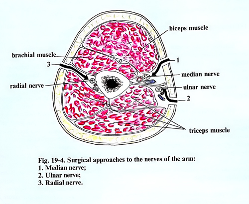
In reaching the nerve trunks under the fascia, it should be penetrated carefully between the muscles and the muscle groups. No incision of muscles near or above the nerve trunks should be performed. If cutting of muscles is necessary, this is done outside the projection of the nerve near their insertion (Fig. 20-4).
The operative wounds should be broad and convenient for work, so that the surgeon may carry out all manipulations around the nerve trunk. The skin scar, if it is found above the injured nerve, is cleaned from the surrounding muscles. The scar in the depth of the wound is left temporarily and the nerve trunk is revealed within normal tissues, above and below the place of injury. After the separation the nerve trunks above and below the place of the lesion, and as necessary, various interventions are made upon them, such as neurolysis (external and internal), resection of a neuroma, nerve sutures, etc.
NEUROLYSIS
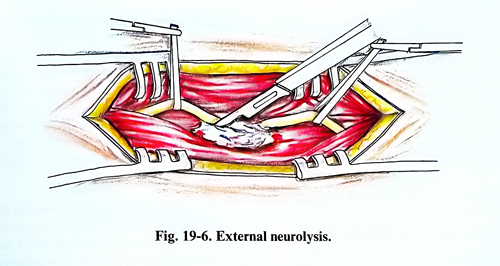
The external neurolysis consists of removing the scar changes, situated around the nerves, when they exert certain compression. The excision of the scar tissue is made with a sharp scalpel, always dividing tissues along the nerve axis. The operation should be done under optic magnification. The scar changes must be removed around the whole nerve up to normal tissue. Injecting local anaesthetic into the scars ensures their easier separation from the nerve.
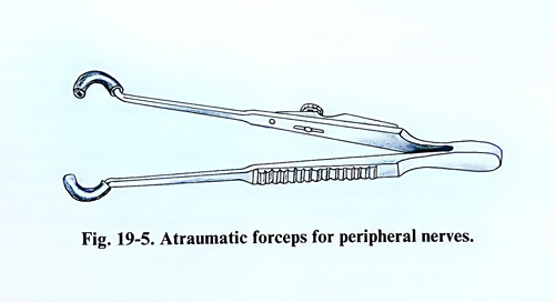
During the dissection, the nerve should be held with special forceps (Fig. 19-5), or with a rubber band, which is pulled lightly. In carrying out neurolysis it is necessary to have the epineurium intact. The nerve fascicles seem thinner on the spot, where they have been enveloped by scar tissue (Fig. 19-6).
In neurolysis, special attention should be paid to haemostasis. The haemorrhage from the small vessels near the nerve stops easily, when oxidised cellulose is put under light pressure. The vessels that continue to bleed even after this tamponade are coagulated. In applying coagulation, there should be no direct afection on the nerve surface and in its immediate neighbourhood.
During the carrying out of the neurolysis, one may notice that the tendons in the proximity of the nerve are enveloped by fibrous tissue. The latter should also be well dissected. In the reverse case, a good restoration of mobility of the limb could not be expected. Special attention should be paid to the cicatricial tissue, which envelops both the vessels and nerve. This cicatricial tissue is removed very carefully, not only from the nerves, but also from the arteries and veins, as these cause severely painful syndromes. In the presence of causalgia an excision of the sympathetic fibres that envelop the arteries could be made.
In combined lesions of nerves and bone fractures, the neurolysis has some characteristics. The nerves may be enveloped and compressed by a bone callus. In such cases fine drilling and bone instrumentation is used until the nerve is freed. This should be done very carefully, so that the nerve fascicles are not be damaged. During the mobilization of the nerve out from the bone callus a high-speed diamond drill can be used. This should not be used in the immediate vicinity of the nerve, because the high temperature can damage nerve fibres.
External neurolysis can be a separate operation, but very often it is only a stage and always precedes the resection of a neuroma, suturing the nerve, etc. The favourable influence of neurolysis for nerve regeneration is not proven, except in cases where the nerve is evidently compressed by scar tissue.
Internal neurolysis is made, when scar changes are found in the nerve itself among the nervous bundles. It should be avoided because there is a risk of a lesion of the nerve fascicles.
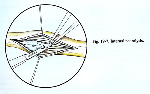
The careful longitudinal incision of the thick epineurium can ensure some decompression, especially if the nerve conduction is partially affected. Technically, internal neurolysis is performed in the following way: after the nerve is well isolated from the surrounding scar changes, 1 - 2% local anaesthetic is injected under the epineurium. This is done with a very fine needle along the whole nerve length, where scar changes are noticed in it. After this is made a longitudinal incision of the epineurium.
The epineurium be slightly retracted and the scar tissue between the nerve fascicles is removed with a small sharp scalpel (Fig. 19-7). This is done very slowly and carefully under a surgical microscope, taking cares not to injure nerve fascicles. The hemostasis on the nerve must be made by slight compression with cottonoids, soaked in saline or oxidised cellulose. Electrocoagulation should not be applied for this purpose. After removing the fibrous tissue, the epineurium is sutured with a few interrupted stitches, if that is possible. Resection and suture of the nerve are made, when there are clinical data of complete functional interruption, the scar tissue has massively penetrated between the nerve bundles, and when the preoperative investigations confirm this interruption. In the presence of a painful syndrome, especially of the causalgia type, it is better to make a resection of the nerve, and not internal neurolysis.
TECHNIQUE OF NERVE REPAIR
Suturing of the peripheral nerves is one of the most frequent and most responsible surgical techniques, when operations are performed on them. The result of the operation depends on how the suture of an interrupted nerve is made, i.e. whether certain degree of regeneration will appear or not.
The operation includes the following stages: dissection of the nerve, inspection, additional evaluation for final selection of the method of intervention, mobilisation of the nerve segments, preparation of the bed, where the injured and sutured nerve will remain, resection of the nerve ends, the performance of the suture, closing the operative wound, and immobilisation of the limb.
In open injuries of peripheral nerves, a primary suture is made when the surgical repair of the wound is performed in the first hours or days after injury. The exploration of the injured peripheral nerves and their dissection for the primary surgical repair of the wounds do not presents difficulties. In many occasions one or both ends of the interrupted nerve are found only after the retraction of the wound. In other cases should be penetrated into the respective intermuscular or intertendinous spaces to expose the nerve. Sometimes the position of the wound and its form are convenient for the easy exposure of the injured nerve, while in other cases it is necessary to make additional incisions. In lacerated and contaminated wounds, especially in a gunshot injury, the performance of additional incisions and the penetration between the muscles must be considered inapropriate, and is to be avoided. In such cases it is better to give up the search of the nerve ends and the suture of the nerve is left to be made later (secondary suture).
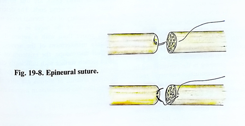
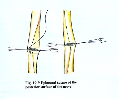
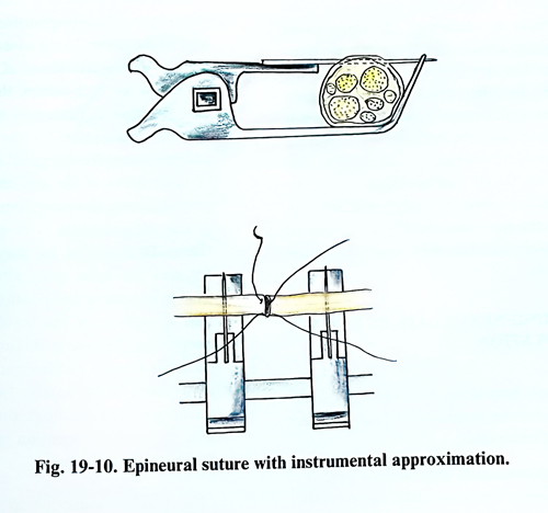
Epineural suture has been the traditional method of repair, using magnification, either with magnifying loupes or the surgical microscope. The goal of epineural repair is to establish continuity without tension and proper rotational alignment. The nerve is inspected for longitudinal blood vessels that can be aligned in the epineurium. Also, the fascicular arrangement is noted and the fascicles from both stumps are matched. This ensures appropriate rotational alignment. The sutures with the affixed needle are passed through the epineurium of the central and peripheral ends of the interrupted nerve from both opposite section surfaces (at 1800). The perforation of the epineurium is made along the axis of the nerve (Fig. 19-8). The number of sutures used is the minimal number required ensuring approximation of the nerve stumps. If the nerve cannot be approximated with 8-0 nylon suture, tension can be considered excessive. Either further mobilisation or a nerve graft is then performed. No fascicles should protrude between the suture line. An 8-0 monofilament suture is recommended for large nerves and 10-0 for small nerves, such as digital nerves. The sutures of both opposite sides of the nerve section are tightened until the nerve surfaces are approximated, but compression or folding of the nerve fascicles should not be provoked. After these two main sutures are made, the threads are caught with fine haemostatic forcepces a few centimetres from the adjoined section surface and are used to immobilize them. The hemostats are slightly pulled and sutures are applied on the accessible nerve surface. After that the direction of the pulling is changed in the opposite way and sutures are placed on the other side of the nerve (Fig. 19-9). For the same purpose a nerve approximator with needles for fixation and approximation of the nerve stumps may be used. (Fig. 19-10).
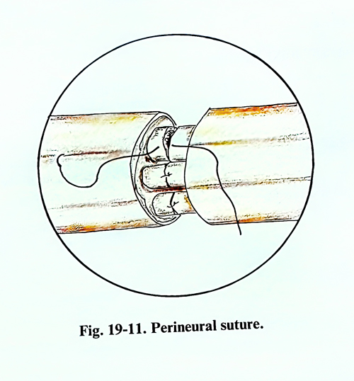
A potentially more accurate technique is the group fascicular repair. The nerve ends are matched by resecting damaged tissue, followed by a careful analysis of the anatomical cross-sectional appearance of the nerve stumps. Fascicular groups are drawn out and dissected and the groups of fascicles are connected by interfascicular perineural suture (Fig. 19-11). In case of a small nerve or nerve branches, individual fascicle repair is recommended.
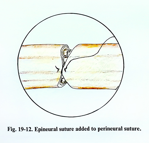
A combination of the two types of nerve repair (epineural and fascicular suture) can be used on a specific area within an extremity (Fig.19-12).
In delayed nerve repair and complete anatomical interruption, the nerve ends are separated from one another, and a neuroma has been formed in the proximal part. In such cases the problems are the extent of the resection, realising the mobilisation necessary to overcome the gap between the nerve ends, and performing the suture without excessive tension. Ordinary inspection and palpation are not enough to choose the operative method on the nerve trunk. In such cases it is inappropriate to simply reconstruct the nerve without some consideration to the motor and sensory alignment of the proximal and distal nerve stumps. The techniques that can be used to identify motor and sensory fascicles in the proximal stump include the use of anatomical clues (surgical identification of fascicles going to specific muscles or cutaneous territories), topography maps, awake stimulation, and enzyme staining. Distally it is not possible to use awake stimulation and histochemical staining, and the surgeon will need to extend the surgical dissection in order to identify motor and sensory groups anatomically. Awake stimulation is a useful technique in determining motor and sensory fascicular grouping in the proximal end in secondary nerve reconstruction and nerve grafting. The co-operation of the patient and support by the anaesthetist are important in this technique. The initial dissection of the nerve must be carried out with the patient under local anaesthesia. The surgeon will stimulate the proximal surface of the nerve. Gradually the stimulus will be increased until the patient perceives it. The patient will interpret the stimulation of the sensory fascicles as a sharp or painful response in a specific cutaneous distribution. By contrast, stimulation of the motor fibres (which will contain afferent sensory fibres) will be interpreted by the patient as dull, non-specific stimulus that usually localises to the midportion of the muscle belly of the corresponding motor nerve. Chemical staining can also be used in the proximal stump to identify motor (acetylcholinesterase or choline acetyltransferase ) and sensory (carbo-anhydrase) fibres. This technique needs at least one hour's time and depends upon a pathologist or a technician.
The nerve ends are mobilised at the minimal distance that is necessary for approximation of both ends of the nerve without affecting its blood supply. If the mobilisation of the nerve is necessary at a greater distance and, during the separation, blood vessels going to the nerve are discovered, they are preserved. The rough mobilisation of the nerve with interruption of its vessels leads to unfavourable post-operative results.
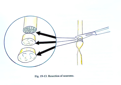
The resection of the injured parts on both ends of the nerve is one of the most responsible manipulations in making late sutures and requires great attention. Poor results after a suture in many cases are due to insufficient resection of the nerve ends. The determination of the boundary of the normal nerve tissue in a cross-section of the nerve in a late suture is much easier than with a suture during the primary management of the nerves. Using a surgical microscope, the existence of a granulated section surface with sufficiently clear fascicular pattern, associated with slight bleeding, and the absence of fibrosis are sure signs that the section has been made in normal tissue (Fig. 19-13).
Neuroma in continuity. Neuroma-in-continuity is the most challenging surgical problem of peripheral nerve injury reconstruction. The damaged components of the nerve must be reconstructed, and, at the same time, the normal fascicles or those with a potential for recovery must not be affected. Internal neurolysis is essential in management of the neuroma-in-continuity in which fascicular groups are separated carefully from one another to facilitate the reconstruction. Intraoperative electrodiagnostic testing is utilised when necessary to assess functional continuity across the neuroma-in-continuity. A simple disposable nerve stimulator can be used to identify the motor fascicles proximal and distal to the level of injury. These motor fascicules are protected, and the electrically silent sensory fascicles are divided proximal and distal to the neuroma-in-continuity and reconstructed with a nerve graft.
OVERCOMING DEFECTS OF PERIPHERAL NERVES
In case of late suture of interrupted peripheral nerves, after resecting the neuroma and the scar of the peripheral nerve segment, a gap is usually obtained. Overcoming such a gap ordinarily presents no difficulty. In some cases bigger gaps are found, usually when the traumatic agent has destroyed a segment of the nerve trunk. In other cases large nerve gaps remain when a malignant tumour of a nerve is excised and it must be resected to unaffected regions of the nerve proximally and distally. The defects that are found after the dissection of the nerve stumps are called "primary", and after the resection of the neuroma and the scar tissue in the peripheral segment they bear the name "final" defects.
OVERCOMING NERVE GAPS BY END-TO-END COAPTATION
There are three main techniques for overcoming gaps to achieve direct coaptation of nerve ends:
1. Nerve mobilisation in situ. With a long distance between the nerve ends, the mobilisation of the proximal and distal stumps helps overcome the gap. The nerves have reserve elasticity and their function begins to suffer from ischaemia after an 8% to 10% elongation. With some patients it is necessary to pay greater attention to the mobilisation of the proximal segment, while with other cases a better effect is obtained by the mobilisation of the peripheral segment. Mobilisation of the nerves in the forearm is of greater significance in interruption of the nerves in proximity with the wrist. Mobilisation of the nerves of the hand contributes little to the overcoming of gaps. In high injuries of the brachial plexus, overcoming of the gaps is achieved primary by mobilisation of the distal segments of the interrupted nerve. The rough manipulation on the nerve as well as the rough pulling may lead to intraneural injuries, which obstruct proper regeneration.
2. Fixing the limb in a convenient position. Flexion of a joint leads to relaxation of the nerves, if they are situated anteriorly to that joint. When the nerve is located behind the joint, extension is responsible for the relaxation of the nerve. In the case of injury of the median and radial nerves of the arm about the elbow for both nerves, the most favourable position is flexion in the elbow with maximum supination for the median nerve and pronation for the radial nerve. In injury of ulnar nerve of the arm and forearm, the reverse is true, the most favourable position of the upper limb is to be extended at the elbow. In injury of the same nerve of the forearm the wrist must also be put in a flexion and adduction position. It is obvious that in injuries of the brachial plexus the nerves will be under minimal traction in elevating the arm and inclination of the head to the side of the injury. In suture of the sciatic nerve of the thigh the most favour-able position is extension of the hip and flexion of the knee joint.
The greatest reduction in tension at the repair site occurs with positioning the joints closest to the site of the injury. The shortening of the gaps are shown in table 19-2.
Table 19 – 2
SHORTENING OF NERVE GAPS BY JOINT POSITION
Shoulder abduction to 90° 2 cm
Elbow 90° flexion 4 cm
Wrist 40° palmar flexion 2 cm
Knee 90° flexion 5 cm
Ankle 10° dorsal or plantar flexion 2 cm
3. Transposition of the nerves. Some of the peripheral nerves, such as the ulnar and radial can be shifted to overcome nerve gaps. In transposing the ulnar nerve from the posterior surface of the elbow to the anterior, and putting the elbow joint in a flexed position, quite large gaps can be over-come. A similar technique is also applied for the shifting of the radial nerve from the dorsal to the anterior arm surface. The technique of transposition will be described with the operations of the individual nerves.
NERVE GRAFTING
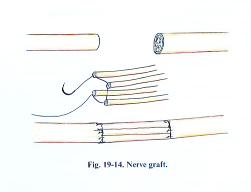
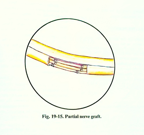
Of all the methods of free nerve grafting the most efficient is the autograft, taken intraoperatively (autotransplantation). Most often cutaneous nerves of little functional importance such as the sural nerve, or mixed nerves such as intercostal nerves or others are used as grafts. Ordinarily the graft is thinner than the recipient nerve. According to the classical method, some segments of the graft are united in the form of a cable and are sutured to the nerve gap by epineural sutures. On this account several pieces must be taken and placed side to side to one another, but so that the common diameter is the same. For instance, if it is necessary to make a transplantation of a 4-mm thick nerve, 4 pieces 2 mm thick must be taken. The cross-section nerve surfaces from which the graft is made are sutured one to another by an epineural suture, which remains in the middle. After that the nerve is sutured according to the general rules, applying microsurgical techniques (Figs. 19-14; 19-15). In delayed reconstruction when nerve grafting is performed, it is important to make motor and sensory alignment at the proximal and distal nerve stumps. The technique that can be used to identify motor and sensory fascicles in the proximal stump includes the use of anatomical clues (surgical identification of fascicles going to specific muscles or cutaneous territories), topography maps, awake stimulation and enzyme staining. It is not possible distally to use wake stimulation or enzyme staining and the surgeon must rely on anatomical dissection aided by anatomical maps to orient motor and sensory fascicular groups. In order to assess in advance which of the skin nerves are convenient for graft material in a given injury of the peripheral nerve, the approximate thickness of the more important nerves must be known beforehand.
According to T. Tarlov's data the average of the thickness of the more important peripheral nerves that need grafting is:
Median nerve at the beginning 4,1 mm
Median nerve at the elbow 3,5 mm
Radial nerve at the beginning 4,3 mm
Ulnar nerve at the beginning 3,8 mm
Ulnar nerve at the wrist 2,5 mm
Sciatic nerve at the middle of the thigh - 8-3 mm
Tibial nerve at popliteal fossa 4,7 mm
Tibial nerve at the ankle 3,5 mm
Peroneal nerve at popliteal fossa 3,6 mm
Femoral nerve at the inguinal region 4,8 mm
Most important nerves, used for grafts
have the following thickness and lengths:
Sural nerve – 25-40 cm length and 2-mm thickness.
Intercostal nerve – 15-20 cm length and 1-2 mm thickness.
Lateral cutaneous nerve of thigh – 15-20 cm length and 2 mm thickness.
Saphenous nerve – 40 cm length and 1 mm thickness.
NEUROTIZATION
Neurotization is the technique by which the proximal portion of one nerve is anastomosed to the distal end of another nerve. The proximal end of the donor nerve is sacrificed and is anastomosed to a nerve that mediates an indispensable function, but has sustained an irreparable injury. A great experience has been amassed of reinnervation of the interrupted facial nerve after anastomosis with accessory, hypoglossal, or phrenic nerve. In cases of brachial plexus avulsion, neurotization can be performed using superior intercostal nerves, the accessory nerve or another nerve from the neighbouring area. In case of long and irreparable gaps of the peroneal nerve in the popliteal fossa, a part of intact branches of the tibial nerve for the lateral parts of the gastrocnemius muscle can be sacrificed by being sutured to the peripheral branches of the peroneal nerve.
POSTOPERATIVE PERIOD
After applying the dressing, the limb is fixed with a splint in the position, in which the limb was at the time of suturing, and the injured nerve is under the weakest traction. The immobilisation continues for three weeks. Any earlier mobilisation may be followed by tearing of the suture, while immobilisation for too long is also undesirable, as it may favour the development of scar tissue and stiff joints. Physical treatment must begin for all parts of the limb, which did not need to be immobilised. In peripheral nerve operations the early patient getting out of bed, usually on the next day of surgery is recommended.
Very often in the postoperative period, pain appears in the operative wound or in the territory of the affected nerves. They are due to the irritation of both vessels and nerve trunks. The characteristics and the intensity of the pain depend on the structure of the nerves operated on. Thus, with nerves with a larger quantity of sensory and sympathetic fibres the pain is more frequent and more intense (for instance with operations on the sciatic and unar nerve). Pain in the postoperative period is more frequently associated with combined operations on vessels and nerves or nerves and bones. Routine analgesics and sedative drugs usually control this type of pain. It is necessary to avoid giving narcotic drugs. If the pain is not due to a septic complication, they usually subside within some days after the operation.
In the postoperative check-up of the operative wound great attention should be paid to the development of haematomas in the operative wound, which are manifested by tension and bluish coloration of the skin. These haematomas must always be evacuated because they lead to the formation of adhesions around the operated nerve. When there are indications of haematoma accumulation, several stitches are removed and the blood is evacuated. If the removal of the haematoma is not possible in this way, the wound must be opened widely, the haematoma cleaned, haemostasis made, and the wound sutured anew.
The penetration of the infection deep into the wound and consequent tissues septic affection in the postoperative period frequently leads to the development of neuritis and to the formation of serious scar changes between the muscle and the nerves. If there is a septic complication, particularly with pus accumulation, part of the stitches must be removed and the wound drained, as well as applying active treatment with antibiotics.
Physiotherapy should begin, if possible, early in the postoperative period. Of course, this kind of treatment is a prolongation of the overall general treatment, which has begun before the operation. In the great majority of cases physiotherapy in the postoperative period helps to obtain a softening of the cicatrices, protect the muscles from atrophy and the joints from ankylosis. In case of neurolysis and when a nerve suture has been made without tension, these treatments should also begin earlier.