STRUCTURE OF THE PERIPHERAL NERVES. DEGENERATION AND REGENERATION
Every peripheral nerve consists of a large number of nerve fibres (axons) that are extended processes of nerve cell bodies situated in the spinal cord, dorsal root ganglia, and sympathetic ganglia. The ratio between the size of a cell body and its axon is considerable, as axon may extend to many thousands of times the cell body diameter. In this special cellular structure of the neuron, the axon contains more than 90% of the total cytoplasmic volume of the nerve cell. As a consequence of this, any localised lesion of an axon implies serious structural and functional problems affecting the survival of the whole neuron. The fibres of the peripheral nerves are divided into three nerve groups: 1. Motor or efferent fibres, which transmit impulses from the central nervous, system toward the muscles; 2. Sensory or afferent fibres, which transmit sensation from muscles, joints, ten-dons, skin, etc.; 3. Autonomic fibres, which innervate the smooth muscles and glands.
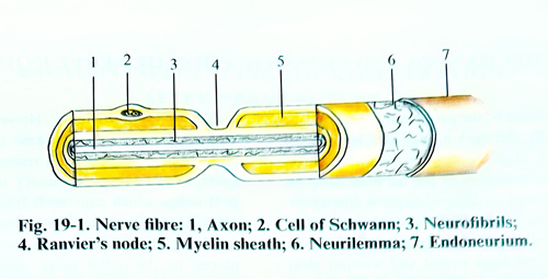
Nerve fibers are either myelinated or non-myelinated. A chain of Schwann cells arranged end to end surrounds both myelinated and nonmyelinated axons. The specific relationship between the Schwann cell and axon differs. The myelinated nerve fiber consists of one axon, which is associated with only one Schwann cell at any one level. The membrane of the Schwann cell, the myelin sheath, is wrapped spirally around the axon, creating a multilaminate sleeve of lipids and protein. The Schwann cells, arranged in a longitudinal sequence, approach each other at the nodes of Ranvier, where finger like cellular processes interdigitate (Fig. 19- 1). In nonmyelinated fibers, Schwann cells accommodate a large number of axons, which are located in internal troughs.
The nerve fibers are divided into three groups according to the relationship between the form of the compound axon potential and the fibre size. Group A fibers are the largest and have the fastest conduction velocities. These fibres are myelinated somatic afferents and effer-ents. Group A fibres are subdivided according to fibre size into A-alpha, A-ß, and A-gamma. An efferent motor fibre, which has a large diameter, belongs to the A-alpha fibre group. A-o fibres transmit the sensation of sharp, pricking pain and temperature, while the A-ß fibres are associated with touch sensation. Myelinated autonomic and preganglionic fibres belong to group B. Group C fibres are thin, slowly conducting nonmyelinated visceral and somatic afferent fibres. Postganglionic, autonomic efferent fibres are also in group C. Nonmyelinated C fibres transfer the sensation of deep burning pain.
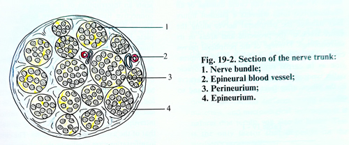
With the exception of the cutaneous nerves, the nerve trunks of the limbs, which are most often subject to surgical operation, are mixed, i. e. they have sensory, motor and autonomic fibres. The nerve fibres are connected in the form of fascicles whose cross-sections are round. A membrane of connective tissue or perineurium envelops each fascicle; every fascicle has a stroma of connective tissue called endoneurium.
The nerve fascicles covered by their perineurium, are separated from each other by connective tissue of a loose character that connects them into a nerve trunk. The same tissue forms another external sheath of the nerve trunk called epineurium (Fig. 19-2).
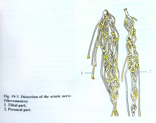
Enveloped in perineurium, the nerve fascicles are not isolated from one another along the nerve trunk, i.e, they are not in the form of a cable. The nerve trunk has a plexiform internal structure and the nerve bundles exchange fibres among themselves. Because of this the number and disposition of the nerve fascicles vary in a series of cross-sections of a given nerve trunk, (Fig. 19 - 3).
The nerve trunks are supplied by the blood vessels in their vicinity. Peripheral nerves are well vascularised structures with separate but extensive interconected microvascular systems in the epineurium, perineurium, and endoneurium. Along the course of the nerve, this vascular system is reinforced by segmental regional vessels of extrinsic origin. These regional vessels travel in an adventítia of connective tissue that surrounds the nerve, allowing it to move longitudinally in its bed with movements of the extremity.
After continuity interruption of a peripheral nerve, the peripheral segment of the interrupted nerve loses its transmission capability in 3 - 4 days. The axons and myelin sheaths suffer from desintegration and are object of fagocytosis by macrophages and Schwann cells. These changes are called Valerian degeneration. The proliferation of Schwann cells and endoneural fibrocytes lead to an increase in the volume of the distal segment of the interrupted nerve, but with time the cell populations diminishes, the endoneural tubes shrink, and the diameter of all distal segments diminishes. Similar changes occur also in the proximal segment of the interrupted nerve without diminishing of the nerve volume.
When an axon is interrupted, chromatolysis in the body of the corresponding nerve cell develops. This is considered more a manifestation of regeneration than of degeneration. After the fourth day of the injury, the amount as well as the metabolism of the ribonucleic acid increase. The increase in the amount and activity of the ribonucleic acid remain until the regeneration and maturation of the axon. The nearer the injury to the spinal cord the more numerous are the degenerative changes of the neurons, whereas in a distal direction, these changes become less expressed. The axoplasm of the axon goes out into the chaotic growth of Schwann cells and the proximal end of the axon increases in volume.
The advance of axons over the zone of injury is a critical process that may determine the success or failure of functional regeneration.
The growing axon of the central interrupted part may penetrate into the extension of the Schwann cells made canal in its peripheral part, i. e. in order that regeneration may be possible, certain conditions are necessary. One of the most important conditions is that the distance between the two ends of the interrupted nerve should be as small as possible. The axons grow through this space in about two weeks, before a thick cicatrix is formed. In injury of the peripheral nerves, no doubt, the ends of the interrupted nerve are separated from one another at a comparatively large distance because of their great elasticity.
The muscle traction and sometimes the traumatic agent may destroy part of the nerve trunk. The presence of haematomas, destruction of muscle fibres, bone fragments and foreign bodies obstructs additionally the regeneration. In cases of a long duration of wound infection, the regeneration may be obstructed because of the presence of neuritis, which in the end causes sclerosis of the nerve fibres. When the axons growing from the proximal end do not penetrate into the peripheral end of the interrupted nerve, a thickening in the shape of a bulb is formed, which is the result of the grouping of a large number of chaotically growing axons; this is called a neuroma. The lack of a neuroma at the proximal end always signifies bad regeneration.