TECHNIQUE OF NERVE REPAIR
Suturing of the peripheral nerves is one of the most frequent and most responsible surgical techniques, when operations are performed on them. The result of the operation depends on how the suture of an interrupted nerve is made, i.e. whether certain degree of regeneration will appear or not.
The operation includes the following stages: dissection of the nerve, inspection, additional evaluation for final selection of the method of intervention, mobilisation of the nerve segments, preparation of the bed, where the injured and sutured nerve will remain, resection of the nerve ends, the performance of the suture, closing the operative wound, and immobilisation of the limb.
In open injuries of peripheral nerves, a primary suture is made when the surgical repair of the wound is performed in the first hours or days after injury. The exploration of the injured peripheral nerves and their dissection for the primary surgical repair of the wounds do not presents difficulties. In many occasions one or both ends of the interrupted nerve are found only after the retraction of the wound. In other cases should be penetrated into the respective intermuscular or intertendinous spaces to expose the nerve. Sometimes the position of the wound and its form are convenient for the easy exposure of the injured nerve, while in other cases it is necessary to make additional incisions. In lacerated and contaminated wounds, especially in a gunshot injury, the performance of additional incisions and the penetration between the muscles must be considered inapropriate, and is to be avoided. In such cases it is better to give up the search of the nerve ends and the suture of the nerve is left to be made later (secondary suture).
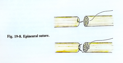
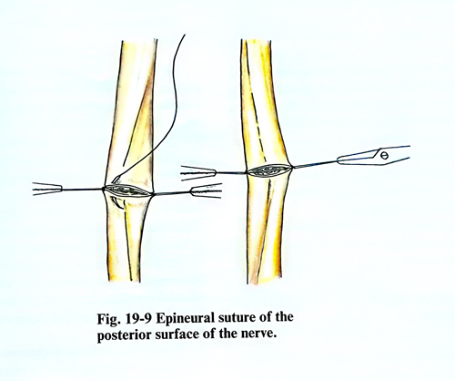
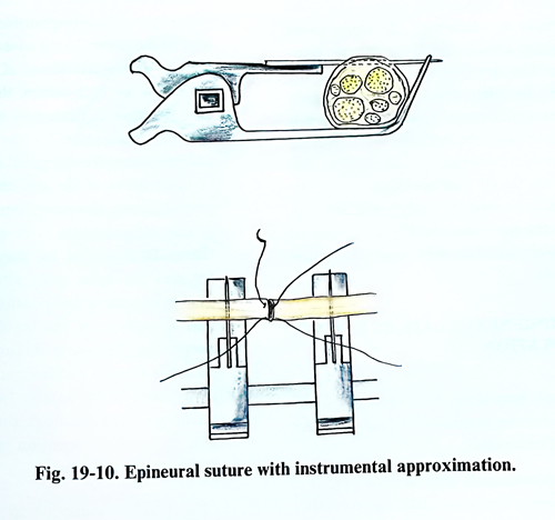
Epineural suture has been the traditional method of repair, using magnification, either with magnifying loupes or the surgical microscope. The goal of epineural repair is to establish continuity without tension and proper rotational alignment. The nerve is inspected for longitudinal blood vessels that can be aligned in the epineurium. Also, the fascicular arrangement is noted and the fascicles from both stumps are matched. This ensures appropriate rotational alignment. The sutures with the affixed needle are passed through the epineurium of the central and peripheral ends of the interrupted nerve from both opposite section surfaces (at 1800). The perforation of the epineurium is made along the axis of the nerve (Fig. 19-8). The number of sutures used is the minimal number required ensuring approximation of the nerve stumps. If the nerve cannot be approximated with 8-0 nylon suture, tension can be considered excessive. Either further mobilisation or a nerve graft is then performed. No fascicles should protrude between the suture line. An 8-0 monofilament suture is recommended for large nerves and 10-0 for small nerves, such as digital nerves. The sutures of both opposite sides of the nerve section are tightened until the nerve surfaces are approximated, but compression or folding of the nerve fascicles should not be provoked. After these two main sutures are made, the threads are caught with fine haemostatic forcepces a few centimetres from the adjoined section surface and are used to immobilize them. The hemostats are slightly pulled and sutures are applied on the accessible nerve surface. After that the direction of the pulling is changed in the opposite way and sutures are placed on the other side of the nerve (Fig. 19-9). For the same purpose a nerve approximator with needles for fixation and approximation of the nerve stumps may be used. (Fig. 19-10).
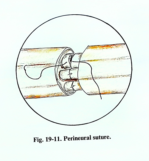
A potentially more accurate technique is the group fascicular repair. The nerve ends are matched by resecting damaged tissue, followed by a careful analysis of the anatomical cross-sectional appearance of the nerve stumps. Fascicular groups are drawn out and dissected and the groups of fascicles are connected by interfascicular perineural suture (Fig. 19-11). In case of a small nerve or nerve branches, individual fascicle repair is recommended.
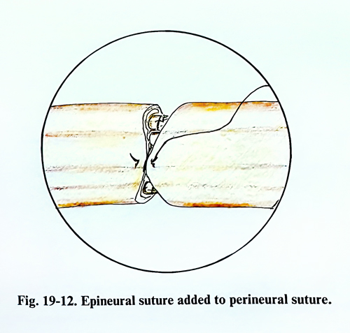
A combination of the two types of nerve repair (epineural and fascicular suture) can be used on a specific area within an extremity (Fig.19-12).
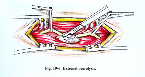
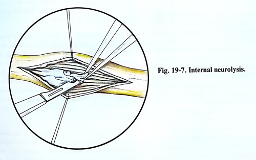
In delayed nerve repair and complete anatomical interruption, the nerve ends are separated from one another, and a neuroma has been formed in the proximal part. In such cases the problems are the extent of the resection, realising the mobilisation necessary to overcome the gap between the nerve ends, and performing the suture without excessive tension. Ordinary inspection and palpation are not enough to choose the operative method on the nerve trunk. In such cases it is inappropriate to simply reconstruct the nerve without some consideration to the motor and sensory alignment of the proximal and distal nerve stumps. The techniques that can be used to identify motor and sensory fascicles in the proximal stump include the use of anatomical clues (surgical identification of fascicles going to specific muscles or cutaneous territories), topography maps, awake stimulation, and enzyme staining. Distally it is not possible to use awake stimulation and histochemical staining, and the surgeon will need to extend the surgical dissection in order to identify motor and sensory groups anatomically. Awake stimulation is a useful technique in determining motor and sensory fascicular grouping in the proximal end in secondary nerve reconstruction and nerve grafting. The co-operation of the patient and support by the anaesthetist are important in this technique. The initial dissection of the nerve must be carried out with the patient under local anaesthesia. The surgeon will stimulate the proximal surface of the nerve. Gradually the stimulus will be increased until the patient perceives it. The patient will interpret the stimulation of the sensory fascicles as a sharp or painful response in a specific cutaneous distribution. By contrast, stimulation of the motor fibres (which will contain afferent sensory fibres) will be interpreted by the patient as dull, non-specific stimulus that usually localises to the midportion of the muscle belly of the corresponding motor nerve. Chemical staining can also be used in the proximal stump to identify motor (acetylcholinesterase or choline acetyltransferase ) and sensory (carbo-anhydrase) fibres. This technique needs at least one hour's time and depends upon a pathologist or a technician.
The nerve ends are mobilised at the minimal distance that is necessary for approximation of both ends of the nerve without affecting its blood supply. If the mobilisation of the nerve is necessary at a greater distance and, during the separation, blood vessels going to the nerve are discovered, they are preserved. The rough mobilisation of the nerve with interruption of its vessels leads to unfavourable post-operative results.
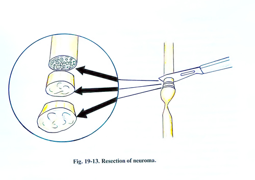
The resection of the injured parts on both ends of the nerve is one of the most responsible manipulations in making late sutures and requires great attention. Poor results after a suture in many cases are due to insufficient resection of the nerve ends. The determination of the boundary of the normal nerve tissue in a cross-section of the nerve in a late suture is much easier than with a suture during the primary management of the nerves. Using a surgical microscope, the existence of a granulated section surface with sufficiently clear fascicular pattern, associated with slight bleeding, and the absence of fibrosis are sure signs that the section has been made in normal tissue (Fig. 19-13).
Neuroma in continuity. Neuroma-in-continuity is the most challenging surgical problem of peripheral nerve injury reconstruction. The damaged components of the nerve must be reconstructed, and, at the same time, the normal fascicles or those with a potential for recovery must not be affected. Internal neurolysis is essential in management of the neuroma-in-continuity in which fascicular groups are separated carefully from one another to facilitate the reconstruction. Intraoperative electrodiagnostic testing is utilised when necessary to assess functional continuity across the neuroma-in-continuity. A simple disposable nerve stimulator can be used to identify the motor fascicles proximal and distal to the level of injury. These motor fascicules are protected, and the electrically silent sensory fascicles are divided proximal and distal to the neuroma-in-continuity and reconstructed with a nerve graft.
OVERCOMING DEFECTS OF PERIPHERAL NERVES
In case of late suture of interrupted peripheral nerves, after resecting the neuroma and the scar of the peripheral nerve segment, a gap is usually obtained. Overcoming such a gap ordinarily presents no difficulty. In some cases bigger gaps are found, usually when the traumatic agent has destroyed a segment of the nerve trunk. In other cases large nerve gaps remain when a malignant tumour of a nerve is excised and it must be resected to unaffected regions of the nerve proximally and distally. The defects that are found after the dissection of the nerve stumps are called "primary", and after the resection of the neuroma and the scar tissue in the peripheral segment they bear the name "final" defects.
OVERCOMING NERVE GAPS BY END-TO-END COAPTATION
There are three main techniques for overcoming gaps to achieve direct coaptation of nerve ends:
1. Nerve mobilisation in situ. With a long distance between the nerve ends, the mobilisation of the proximal and distal stumps helps overcome the gap. The nerves have reserve elasticity and their function begins to suffer from ischaemia after an 8% to 10% elongation. With some patients it is necessary to pay greater attention to the mobilisation of the proximal segment, while with other cases a better effect is obtained by the mobilisation of the peripheral segment. Mobilisation of the nerves in the forearm is of greater significance in interruption of the nerves in proximity with the wrist. Mobilisation of the nerves of the hand contributes little to the overcoming of gaps. In high injuries of the brachial plexus, overcoming of the gaps is achieved primary by mobilisation of the distal segments of the interrupted nerve. The rough manipulation on the nerve as well as the rough pulling may lead to intraneural injuries, which obstruct proper regeneration.
2. Fixing the limb in a convenient position. Flexion of a joint leads to relaxation of the nerves, if they are situated anteriorly to that joint. When the nerve is located behind the joint, extension is responsible for the relaxation of the nerve. In the case of injury of the median and radial nerves of the arm about the elbow for both nerves, the most favourable position is flexion in the elbow with maximum supination for the median nerve and pronation for the radial nerve. In injury of ulnar nerve of the arm and forearm, the reverse is true, the most favourable position of the upper limb is to be extended at the elbow. In injury of the same nerve of the forearm the wrist must also be put in a flexion and adduction position. It is obvious that in injuries of the brachial plexus the nerves will be under minimal traction in elevating the arm and inclination of the head to the side of the injury. In suture of the sciatic nerve of the thigh the most favour-able position is extension of the hip and flexion of the knee joint.
The greatest reduction in tension at the repair site occurs with positioning the joints closest to the site of the injury. The shortening of the gaps are shown in table 19-2.
Table 19 – 2
SHORTENING OF NERVE GAPS BY JOINT POSITION
Shoulder abduction to 90° 2 cm
Elbow 90° flexion 4 cm
Wrist 40° palmar flexion 2 cm
Knee 90° flexion 5 cm
Ankle 10° dorsal or plantar flexion 2 cm
3. Transposition of the nerves. Some of the peripheral nerves, such as the ulnar and radial can be shifted to overcome nerve gaps. In transposing the ulnar nerve from the posterior surface of the elbow to the anterior, and putting the elbow joint in a flexed position, quite large gaps can be over-come. A similar technique is also applied for the shifting of the radial nerve from the dorsal to the anterior arm surface. The technique of transposition will be described with the operations of the individual nerves.