Legacy: "Atlast of Neurosurgery" / L.Karaguiosov, A. Ramadan, K.Karaguiosov / Kiwait/ 1998
PREV> 7. Surgical treatment of intracranial infections
NEXT< 11. Principles of Spinal Cord Surgery
8. RECONSTRUCTIVE SURGERY OF THE SCALP
GENERAL PRINCIPLES OF SCALP RECONSTRUCTION
Plastic repair of scalp defects is required after injury, surgical removal of malignant tumors or defects caused by radiotherapy, burns, electric burns, etc. In case of a defect, comprising the scalp, the cranial bones, and intracranial structures, the scalp defect is restored first and later on, in a separate surgical session, 3 - 6 months later, plastic repair of the skull is carried out. The close attachment of the skin to galea aponeurotica makes it insufficiently elastic, and defects larger than 2 cm square need plastic repair technique application.
The scalp defect should be assessed regarding the size, local vascularity condition, the nature of the defect, and local tissue availability before taking any decision on which surgical method to apply. A precise measurement of the defect should be done before excision of the scalp lesion. In defects that are already present, light pulling of its edges may show the real size of the scalp deficit. If the blood supply to the defect edges is adequate, free grafting may suffice. If the local blood supply is inadequate because of scar tissue, radiation, or exposed bone, the graft must carry its own circulation. Exposed diploic bone and dura will easily support a free skin graft. The easiest reconstruction usually is accomplished with local tissue. If tissue can be mobilised by flap techniques, this will provide the most efficient closure.
SURGICAL TECHNIQUE
All reconstructive techniques applicable to soft tissues can be used in scalp defect reconstruction.
FLAP ROTATION TECHNIQUE
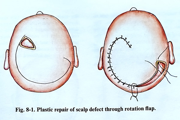
In the flap rotation technique the cover of one scalp defect produces a new defect with an extent which is less than the previous one; the new one must be also covered. For example a defect of 5 by 4 cm can be covered with a rotation flap and the new defect produced is either small enough for easy closure afterward or it does not need to be covered at all. That is achieved with a long curved marginal incision of the rotation flap and the production of a very narrow new defect that can be sutured without tension (Fig. 8-1).
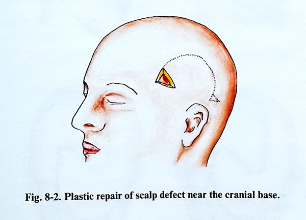
The flap consists of skin, subcutaneous tissue and galea from the neighbourhood of the defect, preserving the pedicle which provides blood supply to the flap. The length of the flap should be no longer than twice its base. The length of the flap, however can be longer in those regions supplied by the major scalp arteries. This applies to the temporal region, with the superficial temporal artery or the occipital artery, accompanied by veins, which provide an abundant circulation. As the course of the arteries is from skullbase to vertex, the flaps should have their base oriented to the origin of these vessels. If there is a scalp defect near to the cranial base, the pedicle of the flap must be lateral to the defect and must have a relatively broader base (Fig. 8-2). The lax fibrous tissue between the galea and periosteum permits easy dissection of the flap. To increase the length of the flap, the galea can be undercut on the internal surface of the flap, with incisions perpendicular to the direction of flap extension. These incisions should not affect the big vessels of the flap; remember that in their initial part they are situated on the surface of the galea when the flap is turned. An additional incision can be made on the base of the flap directed towards the defect. These incisions have a limited depth and should not affect the big arteries of the scalp. A triangular excision of the scalp at the end of the curved incision may be made for better edge adjustment.
FREE SKIN GRAFT
The skin graft is taken by the standard method, excising it with a scalpel or using special dermatomes. The donor place should be cosmetically acceptable as a location. The lower abdominal region, or groin regions are suitable for this purpose.
When the free skin graft is placed on tissue with a normal blood supply and maintained under slight compression over the course of five days, its blood supply begins to be restored from the vessels of the underlying tissue. It is very important during the first days of the postoperative period to maintain the compression without displacement in order to obtain good revascularization. When the skin graft is placed upon an intact periosteum, grafting is usually successful. If the periosteum is removed, the survival of the graft is in doubt. This method does not offer a good cosmetic effect, as the graft is much thinner than the normal scalp, and has no hair. The method is not applicable when there is a defect both of scalp and bone, as second stage bone grafting will not be possible. Therefore free skin grafts in scalp defect repair have a limited application.
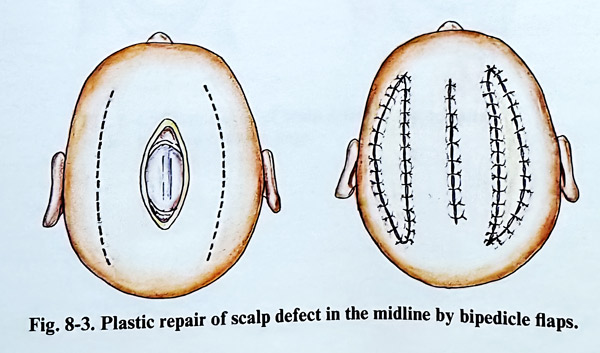
In defects along the midline of the hairy part of the head, which include scalp and bone, repair can be undertaken with bipedicle flaps. This is accomplished by two incisions in a sagittal direction, laterally from the defect, and they must be longer than the anteroposterior length of the defect itself (Fig. 8-3). The scalp and the bone defect situated laterally of the skull vault can be covered by one bipedicle flap. For this purpose an incision parallel to the defect is made in an anterior-posterior direction.
The new scalp defect, obtained after mobilisation of the flap, is covered with a free skin graft.
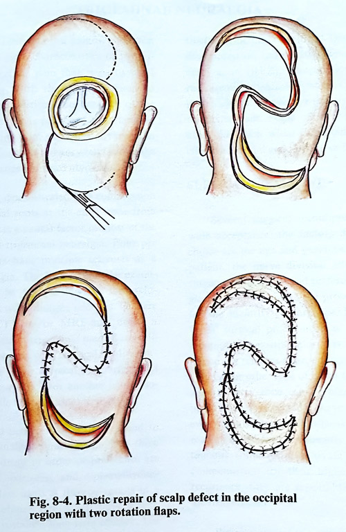
Two rotational flaps can be applied in large defects with a round shape. Two new defects are caused after flap rotation, which are covered with free skin grafts (Fig. 8- 4).
The reconstruction of more complicated defects require an individualised approach and application of more complex plastic surgical techniques.
REIMPLANTATION OF THE SCALP
With the introduction of microvascular anastomosis it became possible to make skin grafts with a pedicle containing artery and vein from the graft territory. For this purpose grafts are produced from skin and subcutaneous tis-sue, taken from the inguinal region and grafted onto scalp defects immediately after the injury and their harvesting.
Using microsurgical technique, it is possible to reimplant an avulsed scalp flap. This reimplantation should be done immediately after admission of the patient and stabilisation of the general condition. The avulsed scalp is preserved in a plastic bag with pieces of ice, until reimplantation is possible.
It is advisable that two surgical teams work simultaneously. One of them should dissect the vessels of the avulsed scalp, as the second debrides the patient's wound, preparing its borders and dissecting the proximal ends of the vessels. The localisation and the dissection of the vessels of the scalp flap and on the border of the wound itself require long and meticulous work. The supraorbital, temporal and occipital arteries with their corresponding veins should be identified. These arteries have diameters from 0.5 to 1,5 mm; after the lacerations they may become spastic. They should be dissected until a short segment of well preserved wall is exposed for the anastomosis. In many cases, direct anastomosis is difficult and a short segment of vein is interposed. This graft is usually taken from the lesser saphenous vein. After dissection of the proximal and distal ends of the vessels is completed, the avulsed scalp is sutured in its place. Microanastomoses are performed between each pair of proximal and distal vascular ends. It is advisable first to suture the veins to diminish blood loss. The more vessels are reconstructed, the higher is the possibility of the avulsed scalp to survive. The abundant network of arteries and veins in the scalp permits satisfactory results even in cases where only one artery and one vein is anastomosed. After the reconstruction of the vessels, the scalp is sutured in two layers and dressing is done without any compression. The dressing is changed frequently to follow up the condition of the reimplanted scalp: its colour, temperature and presence of blood or other collections distending it. The flow of the feeding arteries can be followed up by Doppler ultra-sound.
9. TRIGEMINAL AND GLOSSOPHARYNGEAL NEURALGIA
TRIGEMINAL NEURALGIA
Trigeminal neuralgia is a painful condition of the face. The main characteristics of trigeminal neuralgia include pain that is paroxysmal and lancinating, with the pain confined to trigeminal sensory territory on one side of the face, and a pain that is provoked by cutaneous trigger points and oral activities. Every pain involving the trigeminal territory but lacking the other characteristics is termed atypical.
It has been demonstrated that compression on the trigeminal roots at the entry zone from adjacent vessels is a causal factor in most of the cases of typical trigeminal neuralgia. Four per cent of patients have multiple sclerosis as the cause of neuralgia. The history and the examinations should be directed towards the exclusion of patients with multiple sclerosis. On rare occasions, a CT scan or MRI may detect a tumour as the cause of trigeminal pain, but these patients rarely present with classical “tic douloureux”. Most have either atypical pain, trigeminal sensory or motor deficit, or another neurological deficit outside the trigeminal territory.
Trigeminal neuralgia does not respond to conventional analgesic medication. The three most effective drugs in order of preference are carbamazepine, baclofen, and diphenylhydantoin. Appropriate medical therapy results in a 90% initial resolution rate, which diminishes to a 25% rate of sustained long-term efficacy. The loss of positive effect, undesirable side effects, or complications, which include bone marrow suppression and hepatotoxicity are indications for surgical treatment.
Several principles have been implemented and clinically proved as effective in the surgical treatment of this condition. They are vascular decompression and selective lesions at the root entry zone (REZ), compression, glycerol or radiofrequency rhizolysis at the Gasserian ganglion, and central selective lesions at the nucleus tractus spinalis. Some of the procedures have achieved much wider acceptance because of effectiveness and good tolerance by the patients.
SURGICAL TREATMENT
Several surgical techniques have gained wide acceptance; the factors determining the choice are the age and general condition of the patient, the nerve division affected, and the surgeon's experience and preferences. Microsurgical decompression is the procedure recommended by the majority of surgeons; however, as an 'open' surgical procedure it is associated with minimal, but existing mortality. Partial sensory rhizotomy remains as an option when no micro-vascular or other type of insult to the nerve root and its entry zone can be blamed. Those patients who are expected to have for some reason even slightly increased surgical risks should be recommended for the currently available percutaneous trans-foramen ovale methods. As a compensation for their higher rates of pain recurrence, they are repeatable and their risks are insignificant.
We can recommend at present a posterior fossa approach and microvascular decompression for all young patients and those without serious concomitant illness, and a partial sensory rhizotomy for the extremely few patients, who do not show compression or whose compression cannot be relieved with preserving the nerve. All other patients are submitted to glycerol rhizolysis, or radiofrquency rhizolysis, or balloon compression.
POSTERIOR FOSSA EXPLORATION
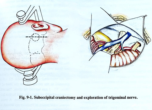
Posterior fossa exploration for the purpose of microvascular decompression of the trigeminal nerve roots is the most widely applied surgical technique. It is performed via a lateral suboccipital craniectomy in the lateral or supine position with the head rotated to the opposite side, and the neck flexed. Venous drainage is improved without danger of air embolism when the head is elevated at a maximum of 20 degrees with respect to the thorax. A straight incision can be done 5 mm medial to the mastoid notch that extends to a maximum of 5 cm superior and 4 cm inferiorly. As an alternative, a "hock-ey-stick incision" may also be employed that is based one-third of the distance from the mastoid to the midline with the short arm onto the mastoid base. A 3 cm craniectomy or craniotomy is made. This bone opening should reveal the borders of the transverse sinus above and the sigmoid sinus laterally. Medial mastoid cells usually are opened and packed with bone wax to prevent postoperative CSF otorrhoea or rhinorrhoea. The dura is opened with a straight incision toward the transverse-sigmoid junction with small flaps reflected superiorly and laterally (Fig. 9-1).
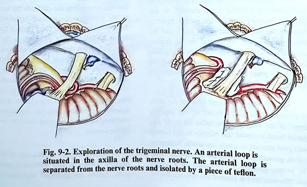
The superolateral border of the cerebellum is retracted inferomedially with a self retaining retractor. The junction of tentorium and petrous bone is followed until the petrous vein is identified. The arachnoid is torn and CSF is aspirated, to further facilitate the retraction. To reach the trigeminal nerve the petrosal vein or some other bridging veins to the sinuses or tentorium veins may need to be coagulated and divided. The retraction continues until the trigeminal nerve is revealed in the cisterna at the place where the roots enter the pons. Traction of the VIIth and VIIIth nerves should be avoided. Brainstem auditory evoked potential monitoring is an useful adjunct for safer retraction and avoidance of VIIIth cranial nerve damage. In this dissection, adherences may be established around the roots of the trigeminal nerve which are then divided. The trigeminal roots and the pontine surface constituting the entry zone are explored for vascular compression by arteries and veins. Vessels are always in proximity, and judgement must be exercised as to whether they are compressive, simple in contact, or not involved at all. When compression or distortion by an arterial loop is established, an attempt to dissect the vessels and to change permanently their relation to the nerve is made. This can be easily done, and to maintain the new position of the arteries, a piece of sponge, muscle, teflon or other isolating material can be placed between them (Fig. 9-2). If the offending vessels are veins, they can be easily coagulated and divided. If neurovascular conflict is not found or the vessels are transfixing the nerve and are unresectable, a partial rhizotomy near the root entry zone is necessary: The lateral two thirds of the portio major of the trigeminal root are divided. The medial cranial part containing the ophthalmic division and proprioceptive part is spared. The craniectomy must be widened and the approach transformed in case of a different pathology with a tumour or vascular lesion. With MRI investigations performed preoperatively, such surprises are extremely rare.
In the postoperative care there are no significant particularities. Usually neuralgic attacks disappear immediately after the operation, but sometimes after microvascular decompression they may gradually decrease in intensity and disappear within a few days.
PERCUTANEOUS RHIZOTOMY BY ELECTROTHERMOCOAGULATION
@@@Several procedures are based on 'trans-foramen ovale' percutaneous methods using a cannula.
The most common are those minimally invasive procedures which permit their performance with simple anaesthesia and no affection of the general condition of the patient; this makes them suitable for elderly and ill patients. The established techniques are: percutaneous rhizotomy by electrocoagulation, glycerol rhizolysis and balloon compression.
Percutaneous rhizotomy continues to be the most widely used percutaneous technique for treatment of trigeminal neuralgia because of its applicability in elderly patients with its low risk compared to craniotomy and microvascular decompression. The use of radiofrequency coagulation permits selective destruction of the poorly myelinated A-delta and unmyelinated C fibers, which are supposed to be nociceptive.
This method is indicated in old patients in whom craniotomy is contraindicated, patients with trigeminal neuralgia associated with multiple sclerosis, and patients with trigeminal neuropathy due to an infiltrating carcinoma.
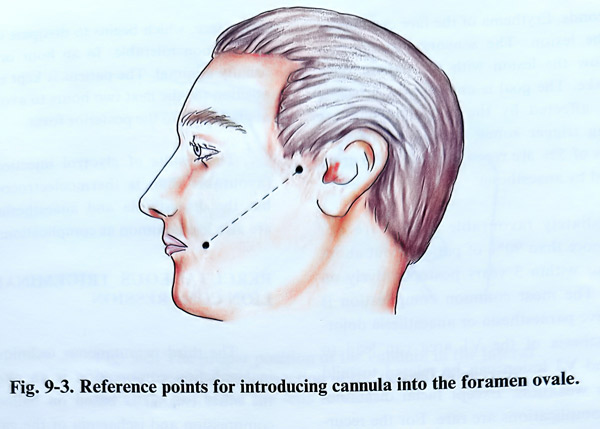
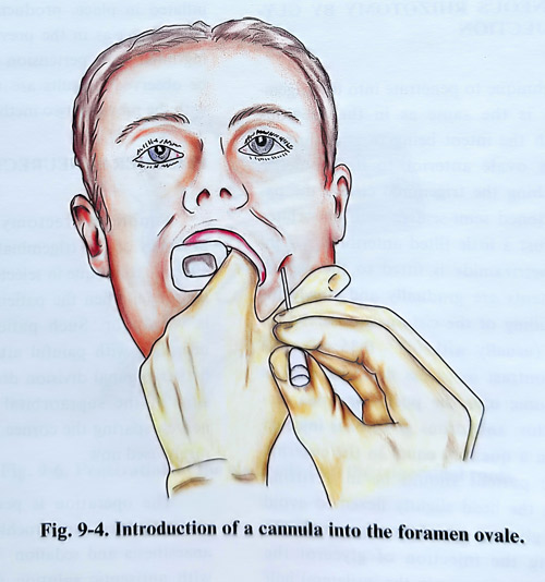
Percutaneous rhizotomy is conducted on the radiographic table, permitting lateral fluoroscopy with the patient supine. After intravenous anaesthesia, a standard 20-gauge cannula at least 100 mm long with a stilette (insulated except at its tip) is introduced into the retro-gasserian portion of the trigeminal nerve. This manipulation is done freehand using three anatomical landmarks on the face (Fig. 9-3). The first point is situated 3 cm anterior to the external auditory meatus, the second point is beneath the medial margin of the pupil, and the third point is 2,5 cm lateral to the lateral commissure of the mouth. The first two points indicate the site of the foramen ovale and the third is the point at which the needle penetrates the skin of the face. An oral airway is placed between the teeth to prevent involuntary biting of the guiding finger of the surgeon. The index finger of the gloved hand is placed just inferior to the lateral pterygoid process to guide the needle trajectory and to prevent mucosal penetration (Fig. 9-4).
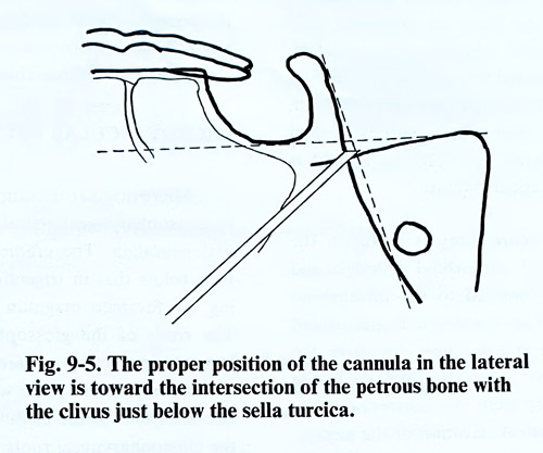
The proper position of the needle on a lateral view (fluoroscopy) is toward the intersection of the petrous bone with the clivus just below the sella turcica (Fig. 9-5). Moving the needle in this region of the skull base reveals the foramen ovale, which should be entered in its medial portion. Penetration of the foramen ovale is usually signalled by a wince and by a brief masseter contraction indicating penetration the mandibular nerve and irritation of the motor branch, which is situated medial to the mandibular nerve in the foramen.
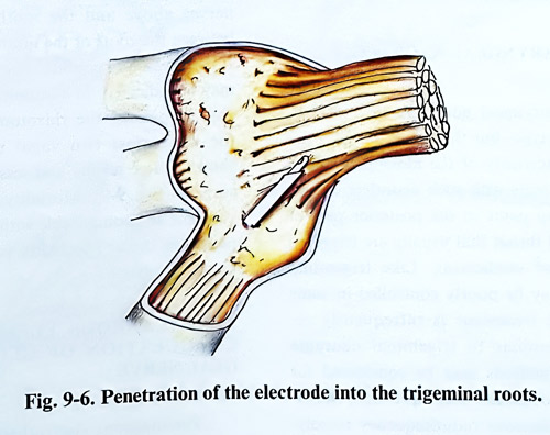
In most patients, proper positioning of the cannula within the trigeminal cistern allows a free flow of CSF through the needle. Exceptions are those with previous trigeminal surgery. The needle is advanced under fluoroscopic guidance to the trigeminal division desired: V3 - 5 mm proximal to the clivus; V2 - at the level of the clivus; V1 - 5 mm beyond the clivus. The stilette is replaced with an electrode, and further localisation is achieved by stimulation. A train of square wave pulses, at 75 cycles per second, with amplitude ranging from 0,1 to 1.0 V may reproduce paroxysms of pain. The electrode is than manipulated until stimulation produces sensory phenomena in the trigeminal division harbour-ing the neuralgic pain or the cutaneous trigger area. Divisions are located by advancing, retracting or rotating the electrode tip according to the presumed somatotopic organisation of the trigeminal ganglion. Third division fibers are found lateral and just proximal to the clivus (Fig. 9-6). First division fibers are located by advancing the electrode beyond the clivus and by rotation medially. If V1 cannot be stimulated, the electrode should be withdrawn, and a new trajectory chosen which is in more lateral to the medial direction. If stimulation with 5 Hz frequency produces masseter contraction, the electrode should be rotated laterally to avoid motor paresis.
After the correct localisation of the electrode is confirmed, and additional anaesthetic given, lesions are generated beginning at 60°C for 60 seconds. Erythema of the face can appear during the lesion. The sensory examination must follow the lesion with the patient once again awake. The goal is analgesia in divisions primarily affected by the neuralgic pain or harbouring trigger zones; hence, lesions with increments of 5% are repeated until these areas are covered by anaesthesia.
Immediate favourable results are obtained in more than 90% of patients, but about 1 in 3 recur within 3 years postoperatively on follow-up. The most common complication is postoperative paraesthesia or anaesthesia dolorosa. Anaesthesia of the Vl area can lead to keratitis and V3 lesion can be related to mild mastication weakness. Except facial dysesthesia, other complications are rare. For the recurrent cases there remains the option of repeating the procedure, but Vl areas remain the most inconvenient for treatment.
PERCUTANEOUS RHIZOTOMY BY GLYCEROL INJECTION
The technique to penetrate into the trigeminal cistern is the same as in the previous method, with the intent being to pass through the foramen ovale anterior to its geometric center. Reaching the trigeminal cistern the patient is positioned semi-seated, with his orbito-meatal line just a little tilted anteriorly; a 1 ml syringe of metrizamide (water-soluble CSF contrast medium) is fitted to the needle, and the contents is gradually and slowly injected until filling of the cistern is observed on the monitor (usually with 0,3 - 0.35 ml). The amount of contrast in excess flows beyond its maximal volume into the posterior fossa cisterns. After this, anhydrous glycerol is injected very slowly in a quantity equal to the cisternal volume. The patient should be in a sitting position with the head slightly flexed to avoid the spread of glycerol out of the trigeminal CSF space. During the injection of glycerol the patient feels a strong pain in the ipsilateral half of the face, which begins to dissipate in minutes and is soon tolerable. In an hour or two it is usually minimal. The patient is kept in a siting position for the next several hours to avoid passage of glycerol into the posterior fossa.
The results of glycerol injection are less favourable than is thermoelectrocoagulation, but the disaesthesia and anaesthesia dolorosa are also less common as complications.
PERCUTANEOUS TRIGEMINAL GANGLION COMPRESSION
The third percutaneous technique, trigeminal ganglion compression is an alternative to the other two. It is based on balloon-induced compression and ischaemia of the ganglion and the rootlets. The technique of inserting the balloon with a thick (14 G) cannula is similar to the approach used in the previous methods. A No 4 Fogartty catheter filled with contrast is inflated in place, producing the same clinical phenomena as in the previous technique. During inflation hypertension and bradicardia may be observed. Results are very similar to those with the previous two methods.
PERIPHERAL NEURECTOMY
Simple neurectomy of the peripheral branches of the trigeminal divisions can be an effective technique in selected cases of trigeminal neuralgia, when the patient's general condition is very poor. Such patients with trigeminal neuralgia and painful attacks confined to the first trigeminal division do well following avulsion of the supraorbital and supratrochlear nerves, sparing the cornea, but this technique is rarely used now.
The operation is performed for the supraorbital and supratrochlear nerves with local anaesthesia and sedation. The skin is prepared with antiseptic solution without shaving the eyebrow. The incision is made along the superior margin of the medial half of the eyebrow.
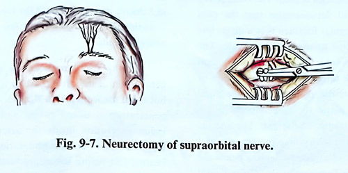
On penetrating the fibers of orbicular oculi muscle, the superior orbital ridge is reached, where the two terminal branches of the ophthalmic nerve pass. These nerves are easily exposed, they are cut and their central part is avulsed (Fig. 9-7). Following avulsion, the wound is closed with a subcuticular suture.
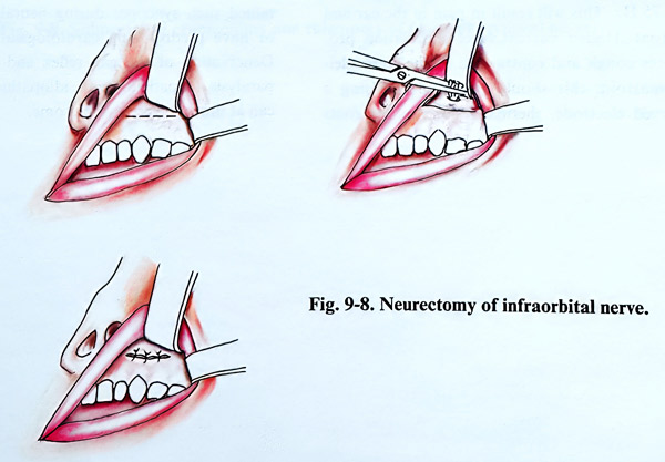
Infraorbital neurectomy is useful in the treatment of typical infraorbital neuralgia and dental neuralgia confined to the infraorbital nerve territory. The operation is performed under anaesthesia and sedation. Elevating the upper lip, an incision is made right above the roots of the anterior teeth in a horizontal direction. The subperiosteal elevation of the mucosa is made upward until the infraorbital foramen through which the nerve comes out is reached The nerve is cut and the proximal end is avulsed (Fig. 9-8).
Mandibular neurectomy offers no advantage over a simple selective V3 percutaneous termoelectrocoagulation.
GLOSSOPHARYNGEAL NEURALGIA
Glossopharyngeal neuralgia is similar to the trigeminal type, but the painful attacks are spread in the territory of the glossopharyngeal nerve. The patients with such neuralgia experience lancinating pains in the posterior part of the tongue and throat that usually are triggered by talking and swallowing. Like trigeminal neuralgia, it may be poorly controlled in some cases; surgical treatment is infrequently re-quired. Very similar to trigeminal neuragia cases, several methods may be considered for treatment: microvascular decompression, rhizotomy and percutaneous radiofrequency rhizolysis. Percutaneous rhizolysis can cause postoperative dysphagia and voice hoarseness, and it is reserved for elderly patients or those with impaired general condition and infiltrating malignancies. Microvascular decompression is the preferred option for the majority of patients, but the lack of obvious neurovascular conflict should lead to selective rhizotomy.
MICROVASCULAR DECOMPRESSION
Microvascular decompression is applicable to glossopharyngeal neuralgia as it is to trigeminal neuralgia. The craniectomy is situated a little below that in trigeminal neuralgia, reaching the foramen magnum without opening it.
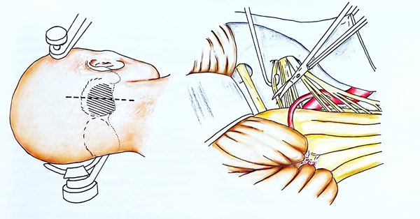
The roots of the glossopharyngeal nerve are explored, retracting upward the inferior pole of the cerebellar hemisphere with its tonsil. On the lateral surface of the medulla oblongata emerge the glossophryngeal roots, arranged one under the other in caudal continuity with the root of the tenth and eleventh nerves. Usually, the roots of the glossopharyngeal nerve are compressed by the inferior posterior cerebellar artery or its branches. Decompression is accomplished by following the most superior root of the ninth nerve, working between the seventh and eighth nerves above and the ninth nerve below or between the roots of the ninth and tenth nerves. If neurovascular conflict is not found, rhizotomy may be indicated. In addition to the glossopharyngeal rootlets, the rhizotomy should involve the uppermost two vagal rootlets; this has shown better results and less recurrences after surgery (Fig. 9-9). Mortality of this surgery is very low and comparable with that in trigeminal neuralgia cases. Morbidity is confined to rare minor dysphagia.
PERCUTANEOUS ELECTROTHERMO-COAGULATION OF GLOSSOPHARYNGEAL NERVE
Percutaneous electrothermocoagulation of the glossopharyngeal nerve is applied at the jugular foramen. A free-hand technique guided by lateral fluoroscopy similar to that described for trigeminal neuralgia is used. The basal X-ray view of the skull demonstrates that the pars nervosa of the jugular foramen is in a direct line with and posterior to the foramen ovale. The electrode entry point is 2,5 cm lateral to the oral commissure. The target is at the intersection of two planes: a sagittal plane through the pupil and a coronal plane through a point 3 cm anterior to the tragus of the ear. The specific targets on the intersection line require a caudal inclination of the electrode approximately 14 degrees below the trajectory to the foramen ovale. On a lateral fluoroscopic image the jugular foramen is situated immediately posterior to the temporomandibular joint and anterior to the occipital condyle. The trajectory in the sagittal plane carries the electrode lateral to the orifice of the carotid canal. Functional localisation is accomplished by stimulation with 100 to 300 mV current using a 1-msec square waves plus at 10 to 75 Hz. This will result in pain in the ear and throat. Higher current level stimulation produces cough and contraction of the sternocleidomastoid; this should be avoided. Using a curved electrode, thermal lesions of the roots are started at 60°C for 60 seconds and repeated at 5 degree increments until the tonsillar pharynx is analgesic and trigger zones fail to reproduce neuralgic pain.
Among the complications which occur, penetration of the carotid artery is a possibility, but is not associated with significant risks unless penetration is unrecognised and attempts at lesion generation inside the artery are made.
Hypotension or bradycardia during a reversible heating test before or during lesion production indicate a spread to vagal fibers and requires repositioning of the electrode tip. Patients suffering from glossopharyngeal syncope (bradycardia, dysrrhythmias, hypotension) can present withserious problems during the procedure, including cardiac arrest.It is recommended that all preventive measures be under-taken, (including placement of a temporary cardiac pacemaker) in patients who have sustained such syncopes during neuralgia attacks or have predisposing cardiological disorders. Denervation of the gag reflex and vocal cord paralysis in patients with idiopathic neuralgia at times can be very troublesome.
10. SURGICAL TREATMENT OF EPILEPSY
Surgical treatment of epilepsy is applied in patients with inadequate control of epileptic seizures by medication i.e. it does not decrease the severity and frequency of the epileptic seizures, and they considerably disable the patient and stop him or her from leading a normal or nearly normal life. No strict rules dictate how many seizures constitute unacceptable control; this must be considered within the patient's vocational, social, and rehabilitation capabilities. For example, three epileptic attacks monthly may be insignificant to a patient who is severely mentally retarded. In contrast, the same number of seizures may be unacceptable for a 14-year old patient with normal intelligence and psychosocial potential. When the seizures are severe with a real danger of injury and the tendency to epileptic status or fast development of mental disturbances, operative treatment can be considered in the event of less frequent seizures and earlier progression.
Special attention must be paid to temporal lobe epilepsy, as it is favourably influenced by surgical treatment and is more resistant to medication than are the other forms of epilepsy. If drugs obviously have no effect, an early operation is recommendable, as it will allow normal education and social adaptation. On the other hand, in children, the attacks change with the maturation of the brain, so that it is very important to know with certainty that the tendency to have seizures is stable, before considering the indications for operation. In children with temporal lobe epilepsy it is better to let them become 15 - 16 years of age, when there is greater certainty that the epileptic focus has clearly been formed. On the other hand, 5 - 10% of patients with temporal lobe epilepsy, obtain spontaneous relief of seizures after 10 - 15 years of age.
The surgical treatment of epilepsy is pathogenic in principle and the etiology has just additional significance for improvement after the operation. However, in some clinical forms of epilepsy the etiology plays a certain role in the selection of patients for operation and is one of the criteria, which must be considered. Among the epilepsies with different etiologies, posttraumatic epilepsy is most favourably influenced by surgical treatment when compared with the others.
EVALUATION OF EPILEPTIC PATIENTS FOR SURGERY
A careful history is necessary, especially regarding evidence of birth trauma, seizures in early life, and other potential causes of epilepsy. Whether other members of the family have epilepsy should be ascertained. The clinical characteristic of epileptic attacks should be known in detail through questioning not only the patients, but also observers because more clinical details of seizures can be obtained from them. Phenomena such as transient dysphasia or postictal paresis or other transient deficits are of considerable value for lateralizing the epileptic focus.
A meticulous neurological examination is required and it may be normal or show deficit from minimal to marked.
Electrophysiological studies are essential for evaluating the patient for surgical treatment.
They can be divided into interictal and ictal recordings performed either extracranially or intracranially. The use of interictal recordings is not recommended for making any major surgical decision in selecting candidates for surgery. However, if interictal epileptic electrographic findings remain consistently restricted to one region of the cortex, like the temporal lobe for a period of years this region most likely harbours the epileptic focus.
Because interictal recordings have high error rate for localizing epileptic foci, long-term EEG monitoring has been used to provide ictal recordings to show the cerebral region of seizure's onset. These recordings can be taken from the scalp or intracranially. The combination of long-term scalp recordings with a video recording of the patient can provide important evidence of the seizure type that characterizes the patient's epilepsy.
Several methods for long-term interictal recordings are available and include deep electrodes, strip electrodes and grid electrodes. Foramen ovale electrodes are also an option.
An intracarotid amytal test to determining speech laterality can be used to select which cortical areas should remain unaffected in operated patients.
Metabolic and blood flow imaging (single photon emission computed tomography and positron emission tomography) have been also considered in the presurgical evaluation, demonstrating high values with ictal phenomena.
Most significantly CT and MRI can provide information for different structural lesions of the brain, because most focal types of epilepsy results from damage or abnormal development in the cortex and some deep structures, as in cortical dysplasia and hyppocampal sclerosis.
ANAESTHESIA
Local anaesthesia is preferable as it allows the establishment of constant contact with the patient during the electrostimulation of the brain cortex. In order to avoid pain at the time of the opening of the dura, local anaesthetic can be injected directly between its two layers by means of a fine needle in the places where the big branches of the middle meningeal artery are located. After the electrostimulation and the electrocorticography have been done, the operation can continue with general anaesthesia. In children and adults patients with mental disorders it is better to perform the operation under general anaesthesia and to decrease the depth of the latter at the time of the electrostimulation.
CORTICAL EXCISION
The patient can be placed on the operating table in different positions, depending on the location of the epileptic focus. However it should always be remembered that the face and the limbs must be free for observation, if a motor response is obtained during the stimulation of the brain cortex.
The extension of the scalp and bone flaps are determined depending of the epileptic focus, for which a topographic map must always be made. This map, made on the patient's head, is exact enough to locate the Sylvian and central fissures at least. Depending on these, the main circumvolutions on the convexity surface of the brain are also found. A wide craniotomy is necessary in order to reveal the epileptic focus and the neighboring regions of the cortex, depending on the clinical, electroencephalographic, CT and MRI data. In the presence of a bone defect situated over the cortex after trauma, after forming the scalp flap, the periosteum is released from the edges of the defect and a separation of the scar tissue from the dura is made. The scars of the dura are excised. If bone enostosis is discovered, the latter is also resected.
The dura is opened in the conventional way, but in cases of traumatic epilepsy, this may prove difficult, owing to its adhesions to the brain tissue. If these adhesions are very dense and are situated in a region, where their separation will induce brain injury and may evoke neurological deficit, it is better to make an electrostimulation and electrocorticography over the dura. When there are obvious morphological changes on the brain cortex, the epileptic focus is usually found in their neighbourhood. Sometimes cysts or scar tissue are revealed in the subarachnoid space in the depth of the brain sulci or brain tissue. At other times deeply situated foreign bodies are located surrounded by fibrosis.
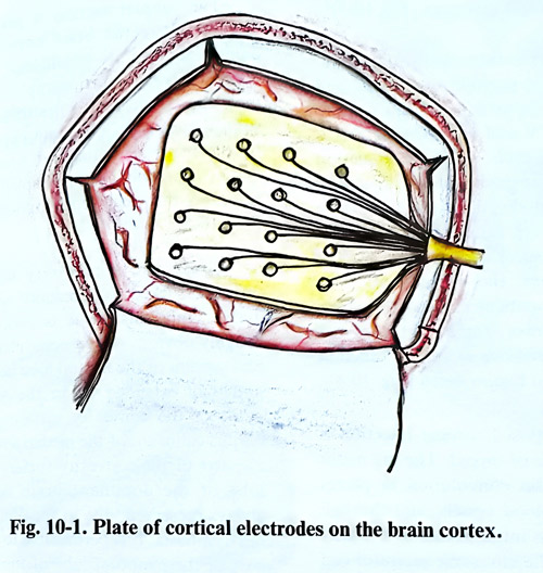
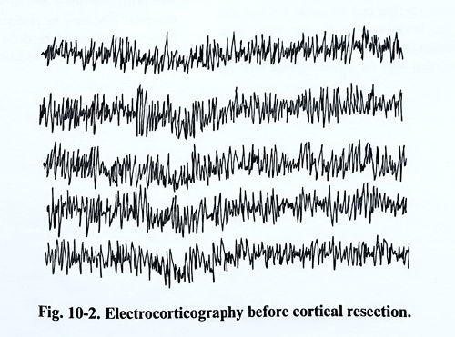
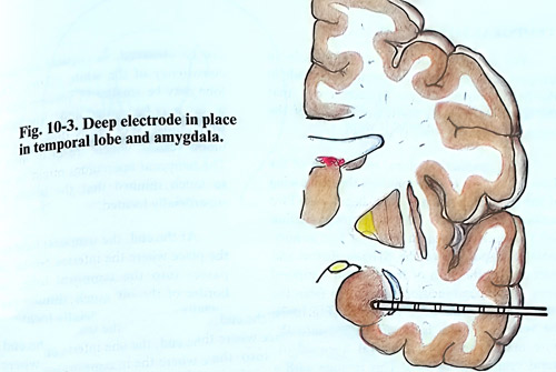
After this the electrocorticography continues as planned. We use contact cortical electrodes mounted on a rubber plate that can record from the exposed cortex of the craniotomy (Fig. 10-1; 10 - 2). They can also be introduced into the subdural space under the border of craniotomy, including on the basal surface of the brain hemisphere. After the electrocorticography study deep electrodes can also be used, especially those directed to the hippocampus and amygdala (Fig. 10-3).
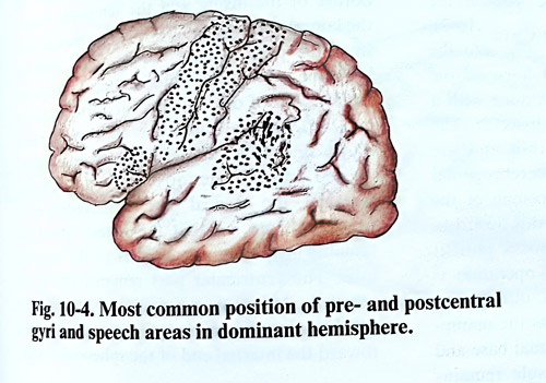
The brain cortex is then electrically stimulated to determine the position of the pre- and postcentral gyri. A 2 m/sec square-wave pulse at 60 Hz, starting at 1 V and increasing by 0.5 V increments following each negative stimulation is used until a motor response is seen, or until a sensory change is felt by the patient. Positive stimulation points are marked. The central sulcus (Rolandic fissure) is identified. In the dominant hemisphere, the speech areas are stimulated while the patient carries out simple verbal tasks. A negative stimulation does not always exclude the presence of speech function in the convolution that is stimulated (Fig. 10-4).
The area of proposed cortical resection is outlined and marked. The pia mater is incised on the brain convolutions in places where there are no blood vessels, and through the incision the suction tip is introduced into the gray matter of the brain. The ultrasonic aspirator can be used for tissue removal. The tissue is aspirated until white matter is reached without penetrating into the depth of the brain sulci. Using optic magnification, the penetration into the brain cortex and its suction can be made through the depth of the brain sulcus. In this way massuve adhesion of the brain tissue to the dura is avoided and the brain surface remains covered by pia-arachnoid.
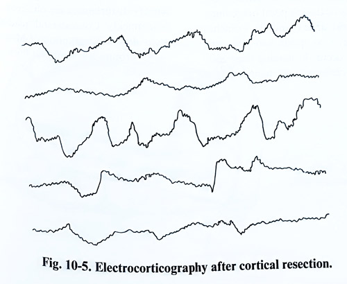
The subpial suction is performed in the regions where the brain cortex is obviously damaged, not relying essentially to electrocorticography and electrostimulation. In a case of posttraumatic epilepsy, subpial suction of the brain cortex is preceded by the removal of the scar tissue and foreign bodies if present. After completing the cortical resection, electrocorticography is repeated (Fig. 10-5).
Great care is necessary when the cortex must be aspirated in proximity with the central gyrus. There is removed only the tissue, which has obvious macroscopic changes. The subpial suction in the frontal lobe is most common, and may extend up to the anterior central gyrus of the convexity surface and up to the corpus callosum of the medial surface. The lower parts of the convexity surface of the frontal lobe of the dominant brain hemisphere are always preserved, due to the danger of provoking dysphasia. It is permissible to remove a large part of the temporal lobe of the non-dominant hemisphere. However, the removal of the posterior half causes hemianopia and this must therefore be preserved. It is permitted to resect the posterior part of the parietal lobe. The occipital lobe may be resected only on one side; however this could be done more freely if the patient has already hemianopia.
TEMPORAL LOBECTOMY
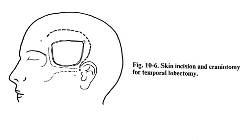
For the resection of the temporal lobe in temporal lobe epilepsy, a craniotomy is made in the frontotemporal region in such a way that the larger part of the convexity surface of the temporal lobe is exposed (Fig. 10-6).
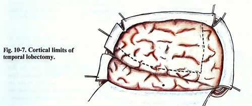
After opening the dura, identification of the Sylvian fissure is easy, as the lesser wing of the sphenoid is indicating its beginning. Two incisions are marked after the electrostimulation and the electrocorticography. The first is horizontal and parallel to the Sylvian fissure and passes along the path of the superior temporal gyrus. The second incision starts 4 cm from the temporal pole and goes vertically (Fig.10-7).
The vertical incision is first deepened into the white matter until the temporal horn of the lateral ventricle is opened. This is done with a fine suction tip and a thin brain dissector. The blood vessels encountered are thin and are coagulated. The appearance of cerebrospinal fluid in the incision shows the opening of the ventricle. The incision is extended downward to the floor of the middle cranial fossa until it reaches the hippocampus. If the operation is performed under local anaesthesia, intravenous anaesthetic is added at this stage as the manipulation around the dura of the cranial base and the tentorium is painful. The insula remains uncovered in its anterior-inferior surface, covered only by the pia of the temporal operculum.
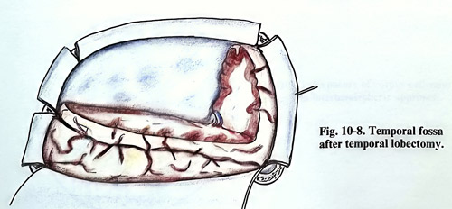
The middle cerebral artery and its branches should not be injured, as spasm and ischaemic complications may occur. In making these incisions, pathological changes in the temporal lobe can be observed, as cortical atrophy or denser consistency of the white matter. The temporal lobe may be smaller in volume and the Sylvian fissure may be located under the sphenoid ridge, which must be carefully observed, as important blood vessels, which are in it, may be injured. The temporal operculum might be atrophic and so much thinned that the insula may appear superficially located. At the end, the temporal lobe stem is cut at the place where the inferior border of the insula passes into the temporal lobe, between the border of the insula and the temporal horn of the lateral ventricle (Fig. 10-8). The section starts from the place, where the temporal horn has been opened and the white matter is interrupted under the roof of the temporal horn, bearing in mind the place where the choroid plexus is attached. In this way the entire temporal horn is opened postero-anteriorly, and before its anterior end, the cortical part of amygdala is exposed and cut, so that the antero-lateral half remains in the amputated part of the temporal lobe. After this manipulation, the incision reaches the pia mater of the temporal pole toward the internal end of the sphenoidal ridge.
As postoperative complications, motor and sensory disturbances are observed, which disappear quickly. Contralateral quadrantopsia may occur due to interruption of Meyer's loop as it passes around the temporal horn of the lateral ventricle.
CALLOSAL SECTION (CALLOSOTOMY)
The role of callosal section in the management of patients with medically intractable epilepsy has been empirically defined. The objective of a corpus callosotomy is not to remove an epileptic focus. The candidates for this procedure do not have a clearly defined and resectable epileptic focus, and callosotomy is performed to prevent the propagation of seizures from one hemisphere to the other. Although the disconnection does not eliminate all seizures, it can prevent the generalized tonic or atonic episodes that are manifested as drop attacks. Controlling drop attacks is essential because patients experiencing them often present with recurrent head and face injuries.
SURGICAL TECHNIQUE
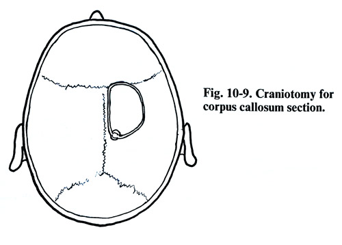
The patient is placed supine on the operating table. Using a U-shaped scalp flap on the right side, a frontal craniotomy is performed near to the midline (Fig. 10-9). The anterior margin of the craniotomy should be just in front of the coronal suture. After the position of the bridging veins is established in this area, one third of the bone flap is planned to be in front of the coronal suture and two thirds behind it.
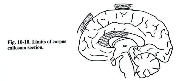
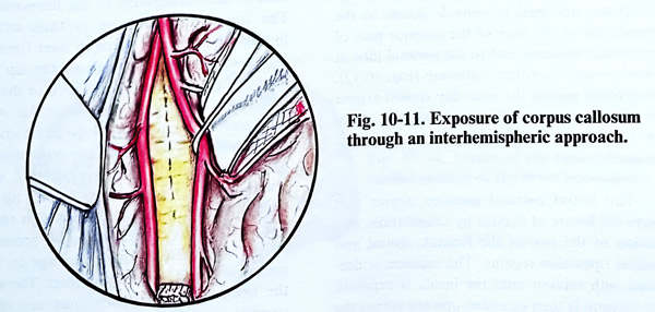
After opening the dura, the right hemisphere and the falx are retracted to expose the pericallosal arteries and the corpus callosum (Fig. 10-10). With bipolar coagulation and gentle suction of the corpus callosum, the ependymal lining of the lateral ventricle is reached, if possible, but not transgressed. The section is continued forward around the genu to divide down to the rostrum. The commissural fibers that link the supplementary motor areas cross in the rostrum and must be interrupted, if drop attacks are to be eliminated. By continuing posteriorly, either the anterior two thirds or the whole of the corpus callosum can be divided (Fig. 10-11).
The callosotomy has a specific group of complications that must be considered. In patients with relatively intact brain function, a set of symptoms may show, including left hand interference with the right hand function that usually slowly resolves. Many patients have reduced verbal and motor activity in the immediate postoperative period after callosal section. These complications are usually transient.
HEMISPHERECTOMY
Hemispherectomy is applied in patients who have sustained such major hemispherical damage that the hemisphere remains nonfunctional and epileptogenic. The seizures arise in the badly damaged cerebral hemisphere and cannot be adequately controlled by tolerable doses of appropriate medication. In most instances, the brain injuries have occurred in infancy or early childhood, resulting from perinatal trauma or from some inflammatory brain disease.
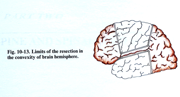
Two types of operation are known. The first is an anatomical hemispherectomy in which the entire brain hemisphere is resected, except the basal ganglia and thalamus. This operation leaves a large, empty space filled with CSF and carries the serious complication of progressive hemosiderosis. This type of hemispherectomy was replaced by the so-called functional hemispherectomy by removing the central cortex and the tip of the temporal lobe but leaving in place the frontal and occipital poles with bridging parietal cortex. These poles are disconnected from the remaining hemisphere by sectioning all the connections to the corpus callosum and basal ganglia.
SURGICAL TECHNIQUE
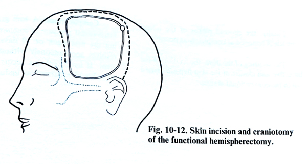
A large U-shaped scalp flap and large craniotomy are used to provide access to the frontal lobe at the level of the anterior part of the corpus callosum, and to the parietal lobe at a level behind the corpus callosum (Fig. 10-12).
The inferior part of the bone flap should expose the inferior aspect of the temporal lobe. After the dura is reflected an electrocorticography is obtained.
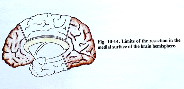
The initial cortical incision begins just above the fissure of Sylvius by coagulation, and incision of the gyri of the frontal, central and parietal opercular regions. The incision is deepened with suction until the insula is exposed. The incision is then extended upward across the frontal and parietal lobes to the midline (Fig. 10-13). The two vertical incisions are then extended on the medial surface of the hemisphere reaching the top of the cingulate gyrus. Leaving the cingulate gyrus in situ, the two vertical incisions are connected just above the cingulate gyrus. The posterior frontal, central, and anterior parietal brain tissue outlined by the cortical incisions is removed 'en bloc' by sectioning the underlying white matter with the suction tip.
The cingulate gyrus is then removed subpially with suction, exposing the anterior cerebral arteries covered by the leptomeninges (Fig. 10-14)
The frontal lobe is then disconnected completely from the upper brain stem and the corpus callosum by suction of white matter in front of the rostrum of the corpus callosum down to the leptomeningeal layer lying on the falx. The same disconnection of the parietal lobe is done by section of the white matter just behind the splenium of the corpus callosum down to the falx and tentorium.
The temporal lobe is completely removed back to the level of the parietal cortical incision.
The cortical incision is initially made just below the fissure of Sylvius and then deepened with suction down to the insula and around the tip of the temporal lobe to the uncus. The vertical incision goes downward to the temporal fossa.
The horizontal incision is then extended through the fusiform gyrus to meet the inferior aspect of the incision around the tip of the temporal pole. The temporal horn of the lateral ventricle is usually opened. After this stage of the operation the temporal lobe is disconnected from its stem and is removed, sectioning any remaining white matter by suction. The amygdaloid nucleus bulging in the medial aspect of the tip of the temporal horn of the ventricle is also removed.
The medial part of amygdala is preserved to protect against inadvertent damage to the hypothalamus and to the optic tract. The pes and the body of the hippocampus are removed completely with any remaining gray matter of the fusiform and hippocampal gyri. Finally, the choroid plexus of the lateral ventricle is removed as completely as possible.
The suturing of the dura, the fixation of the bone flap and closure of the skin flap are carried out in the conventional manner.
Complications. A low-grade increase of intracranial pressure is commonly present during the first week or 10 days, which usually resolves spontaneously without specific treatment. If hydrocephalus occurs a shunting procedure should be carried out without delay.