8. RECONSTRUCTIVE SURGERY OF THE SCALP
GENERAL PRINCIPLES OF SCALP RECONSTRUCTION
Plastic repair of scalp defects is required after injury, surgical removal of malignant tumors or defects caused by radiotherapy, burns, electric burns, etc. In case of a defect, comprising the scalp, the cranial bones, and intracranial structures, the scalp defect is restored first and later on, in a separate surgical session, 3 - 6 months later, plastic repair of the skull is carried out. The close attachment of the skin to galea aponeurotica makes it insufficiently elastic, and defects larger than 2 cm square need plastic repair technique application.
The scalp defect should be assessed regarding the size, local vascularity condition, the nature of the defect, and local tissue availability before taking any decision on which surgical method to apply. A precise measurement of the defect should be done before excision of the scalp lesion. In defects that are already present, light pulling of its edges may show the real size of the scalp deficit. If the blood supply to the defect edges is adequate, free grafting may suffice. If the local blood supply is inadequate because of scar tissue, radiation, or exposed bone, the graft must carry its own circulation. Exposed diploic bone and dura will easily support a free skin graft. The easiest reconstruction usually is accomplished with local tissue. If tissue can be mobilised by flap techniques, this will provide the most efficient closure.
SURGICAL TECHNIQUE
All reconstructive techniques applicable to soft tissues can be used in scalp defect reconstruction.
FLAP ROTATION TECHNIQUE
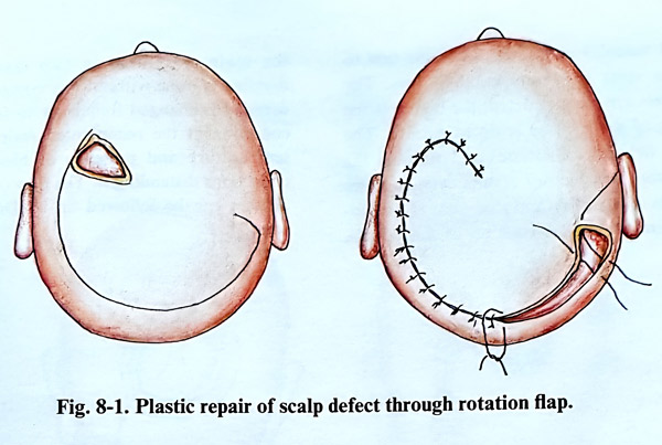
In the flap rotation technique the cover of one scalp defect produces a new defect with an extent which is less than the previous one; the new one must be also covered. For example a defect of 5 by 4 cm can be covered with a rotation flap and the new defect produced is either small enough for easy closure afterward or it does not need to be covered at all. That is achieved with a long curved marginal incision of the rotation flap and the production of a very narrow new defect that can be sutured without tension (Fig. 8-1).
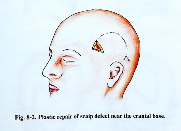
The flap consists of skin, subcutaneous tissue and galea from the neighbourhood of the defect, preserving the pedicle which provides blood supply to the flap. The length of the flap should be no longer than twice its base. The length of the flap, however can be longer in those regions supplied by the major scalp arteries. This applies to the temporal region, with the superficial temporal artery or the occipital artery, accompanied by veins, which provide an abundant circulation. As the course of the arteries is from skullbase to vertex, the flaps should have their base oriented to the origin of these vessels. If there is a scalp defect near to the cranial base, the pedicle of the flap must be lateral to the defect and must have a relatively broader base (Fig. 8-2). The lax fibrous tissue between the galea and periosteum permits easy dissection of the flap. To increase the length of the flap, the galea can be undercut on the internal surface of the flap, with incisions perpendicular to the direction of flap extension. These incisions should not affect the big vessels of the flap; remember that in their initial part they are situated on the surface of the galea when the flap is turned. An additional incision can be made on the base of the flap directed towards the defect. These incisions have a limited depth and should not affect the big arteries of the scalp. A triangular excision of the scalp at the end of the curved incision may be made for better edge adjustment.
FREE SKIN GRAFT
The skin graft is taken by the standard method, excising it with a scalpel or using special dermatomes. The donor place should be cosmetically acceptable as a location. The lower abdominal region, or groin regions are suitable for this purpose.
When the free skin graft is placed on tissue with a normal blood supply and maintained under slight compression over the course of five days, its blood supply begins to be restored from the vessels of the underlying tissue. It is very important during the first days of the postoperative period to maintain the compression without displacement in order to obtain good revascularization. When the skin graft is placed upon an intact periosteum, grafting is usually successful. If the periosteum is removed, the survival of the graft is in doubt. This method does not offer a good cosmetic effect, as the graft is much thinner than the normal scalp, and has no hair. The method is not applicable when there is a defect both of scalp and bone, as second stage bone grafting will not be possible. Therefore free skin grafts in scalp defect repair have a limited application.
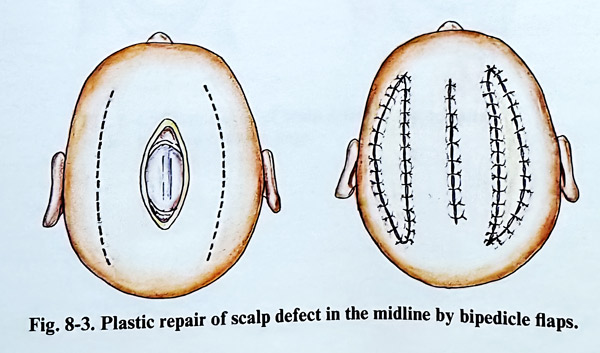
In defects along the midline of the hairy part of the head, which include scalp and bone, repair can be undertaken with bipedicle flaps. This is accomplished by two incisions in a sagittal direction, laterally from the defect, and they must be longer than the anteroposterior length of the defect itself (Fig. 8-3). The scalp and the bone defect situated laterally of the skull vault can be covered by one bipedicle flap. For this purpose an incision parallel to the defect is made in an anterior-posterior direction.
The new scalp defect, obtained after mobilisation of the flap, is covered with a free skin graft.
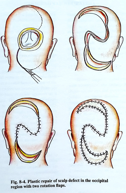
Two rotational flaps can be applied in large defects with a round shape. Two new defects are caused after flap rotation, which are covered with free skin grafts (Fig. 8- 4).
The reconstruction of more complicated defects require an individualised approach and application of more complex plastic surgical techniques.
REIMPLANTATION OF THE SCALP
With the introduction of microvascular anastomosis it became possible to make skin grafts with a pedicle containing artery and vein from the graft territory. For this purpose grafts are produced from skin and subcutaneous tis-sue, taken from the inguinal region and grafted onto scalp defects immediately after the injury and their harvesting.
Using microsurgical technique, it is possible to reimplant an avulsed scalp flap. This reimplantation should be done immediately after admission of the patient and stabilisation of the general condition. The avulsed scalp is preserved in a plastic bag with pieces of ice, until reimplantation is possible.
It is advisable that two surgical teams work simultaneously. One of them should dissect the vessels of the avulsed scalp, as the second debrides the patient's wound, preparing its borders and dissecting the proximal ends of the vessels. The localisation and the dissection of the vessels of the scalp flap and on the border of the wound itself require long and meticulous work. The supraorbital, temporal and occipital arteries with their corresponding veins should be identified. These arteries have diameters from 0.5 to 1,5 mm; after the lacerations they may become spastic. They should be dissected until a short segment of well preserved wall is exposed for the anastomosis. In many cases, direct anastomosis is difficult and a short segment of vein is interposed. This graft is usually taken from the lesser saphenous vein. After dissection of the proximal and distal ends of the vessels is completed, the avulsed scalp is sutured in its place. Microanastomoses are performed between each pair of proximal and distal vascular ends. It is advisable first to suture the veins to diminish blood loss. The more vessels are reconstructed, the higher is the possibility of the avulsed scalp to survive. The abundant network of arteries and veins in the scalp permits satisfactory results even in cases where only one artery and one vein is anastomosed. After the reconstruction of the vessels, the scalp is sutured in two layers and dressing is done without any compression. The dressing is changed frequently to follow up the condition of the reimplanted scalp: its colour, temperature and presence of blood or other collections distending it. The flow of the feeding arteries can be followed up by Doppler ultra-sound.