9. TRIGEMINAL AND GLOSSOPHARYNGEAL NEURALGIA
TRIGEMINAL NEURALGIA
Trigeminal neuralgia is a painful condition of the face. The main characteristics of trigeminal neuralgia include pain that is paroxysmal and lancinating, with the pain confined to trigeminal sensory territory on one side of the face, and a pain that is provoked by cutaneous trigger points and oral activities. Every pain involving the trigeminal territory but lacking the other characteristics is termed atypical.
It has been demonstrated that compression on the trigeminal roots at the entry zone from adjacent vessels is a causal factor in most of the cases of typical trigeminal neuralgia. Four per cent of patients have multiple sclerosis as the cause of neuralgia. The history and the examinations should be directed towards the exclusion of patients with multiple sclerosis. On rare occasions, a CT scan or MRI may detect a tumour as the cause of trigeminal pain, but these patients rarely present with classical “tic douloureux”. Most have either atypical pain, trigeminal sensory or motor deficit, or another neurological deficit outside the trigeminal territory.
Trigeminal neuralgia does not respond to conventional analgesic medication. The three most effective drugs in order of preference are carbamazepine, baclofen, and diphenylhydantoin. Appropriate medical therapy results in a 90% initial resolution rate, which diminishes to a 25% rate of sustained long-term efficacy. The loss of positive effect, undesirable side effects, or complications, which include bone marrow suppression and hepatotoxicity are indications for surgical treatment.
Several principles have been implemented and clinically proved as effective in the surgical treatment of this condition. They are vascular decompression and selective lesions at the root entry zone (REZ), compression, glycerol or radiofrequency rhizolysis at the Gasserian ganglion, and central selective lesions at the nucleus tractus spinalis. Some of the procedures have achieved much wider acceptance because of effectiveness and good tolerance by the patients.
SURGICAL TREATMENT
Several surgical techniques have gained wide acceptance; the factors determining the choice are the age and general condition of the patient, the nerve division affected, and the surgeon's experience and preferences. Microsurgical decompression is the procedure recommended by the majority of surgeons; however, as an 'open' surgical procedure it is associated with minimal, but existing mortality. Partial sensory rhizotomy remains as an option when no micro-vascular or other type of insult to the nerve root and its entry zone can be blamed. Those patients who are expected to have for some reason even slightly increased surgical risks should be recommended for the currently available percutaneous trans-foramen ovale methods. As a compensation for their higher rates of pain recurrence, they are repeatable and their risks are insignificant.
We can recommend at present a posterior fossa approach and microvascular decompression for all young patients and those without serious concomitant illness, and a partial sensory rhizotomy for the extremely few patients, who do not show compression or whose compression cannot be relieved with preserving the nerve. All other patients are submitted to glycerol rhizolysis, or radiofrquency rhizolysis, or balloon compression.
POSTERIOR FOSSA EXPLORATION
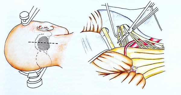
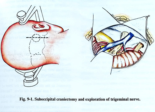
Posterior fossa exploration for the purpose of microvascular decompression of the trigeminal nerve roots is the most widely applied surgical technique. It is performed via a lateral suboccipital craniectomy in the lateral or supine position with the head rotated to the opposite side, and the neck flexed. Venous drainage is improved without danger of air embolism when the head is elevated at a maximum of 20 degrees with respect to the thorax. A straight incision can be done 5 mm medial to the mastoid notch that extends to a maximum of 5 cm superior and 4 cm inferiorly. As an alternative, a "hock-ey-stick incision" may also be employed that is based one-third of the distance from the mastoid to the midline with the short arm onto the mastoid base. A 3 cm craniectomy or craniotomy is made. This bone opening should reveal the borders of the transverse sinus above and the sigmoid sinus laterally. Medial mastoid cells usually are opened and packed with bone wax to prevent postoperative CSF otorrhoea or rhinorrhoea. The dura is opened with a straight incision toward the transverse-sigmoid junction with small flaps reflected superiorly and laterally (Fig. 9-1).
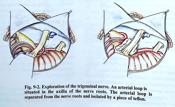
The superolateral border of the cerebellum is retracted inferomedially with a self retaining retractor. The junction of tentorium and petrous bone is followed until the petrous vein is identified. The arachnoid is torn and CSF is aspirated, to further facilitate the retraction. To reach the trigeminal nerve the petrosal vein or some other bridging veins to the sinuses or tentorium veins may need to be coagulated and divided. The retraction continues until the trigeminal nerve is revealed in the cisterna at the place where the roots enter the pons. Traction of the VIIth and VIIIth nerves should be avoided. Brainstem auditory evoked potential monitoring is an useful adjunct for safer retraction and avoidance of VIIIth cranial nerve damage. In this dissection, adherences may be established around the roots of the trigeminal nerve which are then divided. The trigeminal roots and the pontine surface constituting the entry zone are explored for vascular compression by arteries and veins. Vessels are always in proximity, and judgement must be exercised as to whether they are compressive, simple in contact, or not involved at all. When compression or distortion by an arterial loop is established, an attempt to dissect the vessels and to change permanently their relation to the nerve is made. This can be easily done, and to maintain the new position of the arteries, a piece of sponge, muscle, teflon or other isolating material can be placed between them (Fig. 9-2). If the offending vessels are veins, they can be easily coagulated and divided. If neurovascular conflict is not found or the vessels are transfixing the nerve and are unresectable, a partial rhizotomy near the root entry zone is necessary: The lateral two thirds of the portio major of the trigeminal root are divided. The medial cranial part containing the ophthalmic division and proprioceptive part is spared. The craniectomy must be widened and the approach transformed in case of a different pathology with a tumour or vascular lesion. With MRI investigations performed preoperatively, such surprises are extremely rare.
In the postoperative care there are no significant particularities. Usually neuralgic attacks disappear immediately after the operation, but sometimes after microvascular decompression they may gradually decrease in intensity and disappear within a few days.
PERCUTANEOUS RHIZOTOMY BY ELECTROTHERMOCOAGULATION
@@@Several procedures are based on 'trans-foramen ovale' percutaneous methods using a cannula.
The most common are those minimally invasive procedures which permit their performance with simple anaesthesia and no affection of the general condition of the patient; this makes them suitable for elderly and ill patients. The established techniques are: percutaneous rhizotomy by electrocoagulation, glycerol rhizolysis and balloon compression.
Percutaneous rhizotomy continues to be the most widely used percutaneous technique for treatment of trigeminal neuralgia because of its applicability in elderly patients with its low risk compared to craniotomy and microvascular decompression. The use of radiofrequency coagulation permits selective destruction of the poorly myelinated A-delta and unmyelinated C fibers, which are supposed to be nociceptive.
This method is indicated in old patients in whom craniotomy is contraindicated, patients with trigeminal neuralgia associated with multiple sclerosis, and patients with trigeminal neuropathy due to an infiltrating carcinoma.
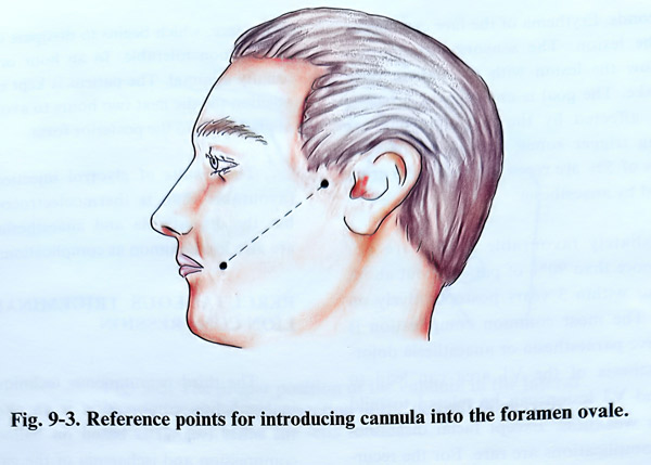
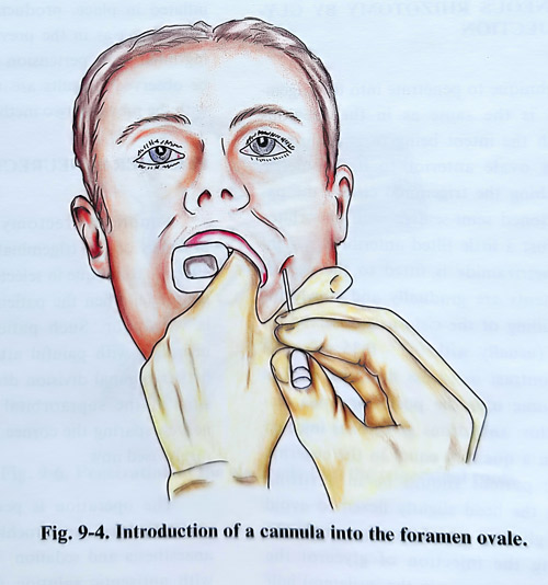
Percutaneous rhizotomy is conducted on the radiographic table, permitting lateral fluoroscopy with the patient supine. After intravenous anaesthesia, a standard 20-gauge cannula at least 100 mm long with a stilette (insulated except at its tip) is introduced into the retro-gasserian portion of the trigeminal nerve. This manipulation is done freehand using three anatomical landmarks on the face (Fig. 9-3). The first point is situated 3 cm anterior to the external auditory meatus, the second point is beneath the medial margin of the pupil, and the third point is 2,5 cm lateral to the lateral commissure of the mouth. The first two points indicate the site of the foramen ovale and the third is the point at which the needle penetrates the skin of the face. An oral airway is placed between the teeth to prevent involuntary biting of the guiding finger of the surgeon. The index finger of the gloved hand is placed just inferior to the lateral pterygoid process to guide the needle trajectory and to prevent mucosal penetration (Fig. 9-4).
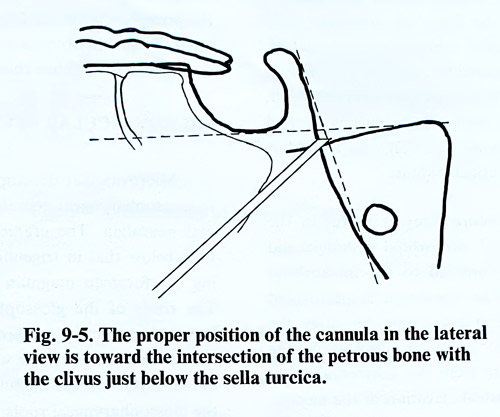
The proper position of the needle on a lateral view (fluoroscopy) is toward the intersection of the petrous bone with the clivus just below the sella turcica (Fig. 9-5). Moving the needle in this region of the skull base reveals the foramen ovale, which should be entered in its medial portion. Penetration of the foramen ovale is usually signalled by a wince and by a brief masseter contraction indicating penetration the mandibular nerve and irritation of the motor branch, which is situated medial to the mandibular nerve in the foramen.
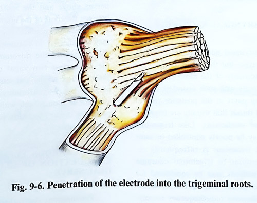
In most patients, proper positioning of the cannula within the trigeminal cistern allows a free flow of CSF through the needle. Exceptions are those with previous trigeminal surgery. The needle is advanced under fluoroscopic guidance to the trigeminal division desired: V3 - 5 mm proximal to the clivus; V2 - at the level of the clivus; V1 - 5 mm beyond the clivus. The stilette is replaced with an electrode, and further localisation is achieved by stimulation. A train of square wave pulses, at 75 cycles per second, with amplitude ranging from 0,1 to 1.0 V may reproduce paroxysms of pain. The electrode is than manipulated until stimulation produces sensory phenomena in the trigeminal division harbour-ing the neuralgic pain or the cutaneous trigger area. Divisions are located by advancing, retracting or rotating the electrode tip according to the presumed somatotopic organisation of the trigeminal ganglion. Third division fibers are found lateral and just proximal to the clivus (Fig. 9-6). First division fibers are located by advancing the electrode beyond the clivus and by rotation medially. If V1 cannot be stimulated, the electrode should be withdrawn, and a new trajectory chosen which is in more lateral to the medial direction. If stimulation with 5 Hz frequency produces masseter contraction, the electrode should be rotated laterally to avoid motor paresis.
After the correct localisation of the electrode is confirmed, and additional anaesthetic given, lesions are generated beginning at 60°C for 60 seconds. Erythema of the face can appear during the lesion. The sensory examination must follow the lesion with the patient once again awake. The goal is analgesia in divisions primarily affected by the neuralgic pain or harbouring trigger zones; hence, lesions with increments of 5% are repeated until these areas are covered by anaesthesia.
Immediate favourable results are obtained in more than 90% of patients, but about 1 in 3 recur within 3 years postoperatively on follow-up. The most common complication is postoperative paraesthesia or anaesthesia dolorosa. Anaesthesia of the Vl area can lead to keratitis and V3 lesion can be related to mild mastication weakness. Except facial dysesthesia, other complications are rare. For the recurrent cases there remains the option of repeating the procedure, but Vl areas remain the most inconvenient for treatment.
PERCUTANEOUS RHIZOTOMY BY GLYCEROL INJECTION
The technique to penetrate into the trigeminal cistern is the same as in the previous method, with the intent being to pass through the foramen ovale anterior to its geometric center. Reaching the trigeminal cistern the patient is positioned semi-seated, with his orbito-meatal line just a little tilted anteriorly; a 1 ml syringe of metrizamide (water-soluble CSF contrast medium) is fitted to the needle, and the contents is gradually and slowly injected until filling of the cistern is observed on the monitor (usually with 0,3 - 0.35 ml). The amount of contrast in excess flows beyond its maximal volume into the posterior fossa cisterns. After this, anhydrous glycerol is injected very slowly in a quantity equal to the cisternal volume. The patient should be in a sitting position with the head slightly flexed to avoid the spread of glycerol out of the trigeminal CSF space. During the injection of glycerol the patient feels a strong pain in the ipsilateral half of the face, which begins to dissipate in minutes and is soon tolerable. In an hour or two it is usually minimal. The patient is kept in a siting position for the next several hours to avoid passage of glycerol into the posterior fossa.
The results of glycerol injection are less favourable than is thermoelectrocoagulation, but the disaesthesia and anaesthesia dolorosa are also less common as complications.
PERCUTANEOUS TRIGEMINAL GANGLION COMPRESSION
The third percutaneous technique, trigeminal ganglion compression is an alternative to the other two. It is based on balloon-induced compression and ischaemia of the ganglion and the rootlets. The technique of inserting the balloon with a thick (14 G) cannula is similar to the approach used in the previous methods. A No 4 Fogartty catheter filled with contrast is inflated in place, producing the same clinical phenomena as in the previous technique. During inflation hypertension and bradicardia may be observed. Results are very similar to those with the previous two methods.
PERIPHERAL NEURECTOMY
Simple neurectomy of the peripheral branches of the trigeminal divisions can be an effective technique in selected cases of trigeminal neuralgia, when the patient's general condition is very poor. Such patients with trigeminal neuralgia and painful attacks confined to the first trigeminal division do well following avulsion of the supraorbital and supratrochlear nerves, sparing the cornea, but this technique is rarely used now.
The operation is performed for the supraorbital and supratrochlear nerves with local anaesthesia and sedation. The skin is prepared with antiseptic solution without shaving the eyebrow. The incision is made along the superior margin of the medial half of the eyebrow.
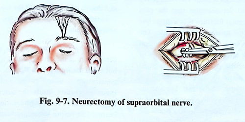
On penetrating the fibers of orbicular oculi muscle, the superior orbital ridge is reached, where the two terminal branches of the ophthalmic nerve pass. These nerves are easily exposed, they are cut and their central part is avulsed (Fig. 9-7). Following avulsion, the wound is closed with a subcuticular suture.
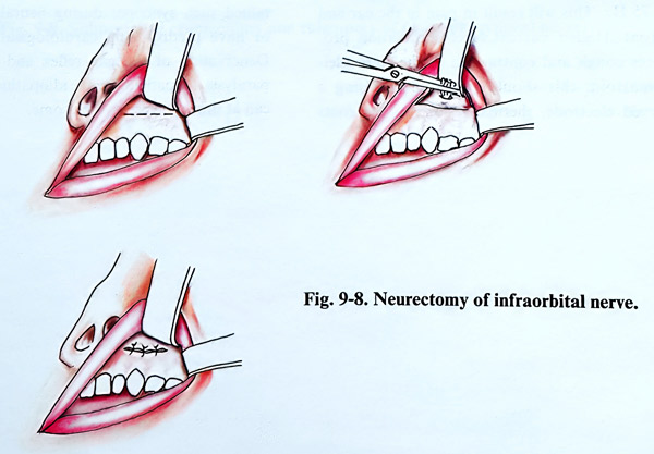
Infraorbital neurectomy is useful in the treatment of typical infraorbital neuralgia and dental neuralgia confined to the infraorbital nerve territory. The operation is performed under anaesthesia and sedation. Elevating the upper lip, an incision is made right above the roots of the anterior teeth in a horizontal direction. The subperiosteal elevation of the mucosa is made upward until the infraorbital foramen through which the nerve comes out is reached The nerve is cut and the proximal end is avulsed (Fig. 9-8).
Mandibular neurectomy offers no advantage over a simple selective V3 percutaneous termoelectrocoagulation.
GLOSSOPHARYNGEAL NEURALGIA
Glossopharyngeal neuralgia is similar to the trigeminal type, but the painful attacks are spread in the territory of the glossopharyngeal nerve. The patients with such neuralgia experience lancinating pains in the posterior part of the tongue and throat that usually are triggered by talking and swallowing. Like trigeminal neuralgia, it may be poorly controlled in some cases; surgical treatment is infrequently re-quired. Very similar to trigeminal neuragia cases, several methods may be considered for treatment: microvascular decompression, rhizotomy and percutaneous radiofrequency rhizolysis. Percutaneous rhizolysis can cause postoperative dysphagia and voice hoarseness, and it is reserved for elderly patients or those with impaired general condition and infiltrating malignancies. Microvascular decompression is the preferred option for the majority of patients, but the lack of obvious neurovascular conflict should lead to selective rhizotomy.
MICROVASCULAR DECOMPRESSION
Microvascular decompression is applicable to glossopharyngeal neuralgia as it is to trigeminal neuralgia. The craniectomy is situated a little below that in trigeminal neuralgia, reaching the foramen magnum without opening it.
The roots of the glossopharyngeal nerve are explored, retracting upward the inferior pole of the cerebellar hemisphere with its tonsil. On the lateral surface of the medulla oblongata emerge the glossophryngeal roots, arranged one under the other in caudal continuity with the root of the tenth and eleventh nerves. Usually, the roots of the glossopharyngeal nerve are compressed by the inferior posterior cerebellar artery or its branches. Decompression is accomplished by following the most superior root of the ninth nerve, working between the seventh and eighth nerves above and the ninth nerve below or between the roots of the ninth and tenth nerves. If neurovascular conflict is not found, rhizotomy may be indicated. In addition to the glossopharyngeal rootlets, the rhizotomy should involve the uppermost two vagal rootlets; this has shown better results and less recurrences after surgery (Fig. 9-9). Mortality of this surgery is very low and comparable with that in trigeminal neuralgia cases. Morbidity is confined to rare minor dysphagia.
PERCUTANEOUS ELECTROTHERMO-COAGULATION OF GLOSSOPHARYNGEAL NERVE
Percutaneous electrothermocoagulation of the glossopharyngeal nerve is applied at the jugular foramen. A free-hand technique guided by lateral fluoroscopy similar to that described for trigeminal neuralgia is used. The basal X-ray view of the skull demonstrates that the pars nervosa of the jugular foramen is in a direct line with and posterior to the foramen ovale. The electrode entry point is 2,5 cm lateral to the oral commissure. The target is at the intersection of two planes: a sagittal plane through the pupil and a coronal plane through a point 3 cm anterior to the tragus of the ear. The specific targets on the intersection line require a caudal inclination of the electrode approximately 14 degrees below the trajectory to the foramen ovale. On a lateral fluoroscopic image the jugular foramen is situated immediately posterior to the temporomandibular joint and anterior to the occipital condyle. The trajectory in the sagittal plane carries the electrode lateral to the orifice of the carotid canal. Functional localisation is accomplished by stimulation with 100 to 300 mV current using a 1-msec square waves plus at 10 to 75 Hz. This will result in pain in the ear and throat. Higher current level stimulation produces cough and contraction of the sternocleidomastoid; this should be avoided. Using a curved electrode, thermal lesions of the roots are started at 60°C for 60 seconds and repeated at 5 degree increments until the tonsillar pharynx is analgesic and trigger zones fail to reproduce neuralgic pain.
Among the complications which occur, penetration of the carotid artery is a possibility, but is not associated with significant risks unless penetration is unrecognised and attempts at lesion generation inside the artery are made.
Hypotension or bradycardia during a reversible heating test before or during lesion production indicate a spread to vagal fibers and requires repositioning of the electrode tip. Patients suffering from glossopharyngeal syncope (bradycardia, dysrrhythmias, hypotension) can present withserious problems during the procedure, including cardiac arrest.It is recommended that all preventive measures be under-taken, (including placement of a temporary cardiac pacemaker) in patients who have sustained such syncopes during neuralgia attacks or have predisposing cardiological disorders. Denervation of the gag reflex and vocal cord paralysis in patients with idiopathic neuralgia at times can be very troublesome.