HYDATID CYSTS
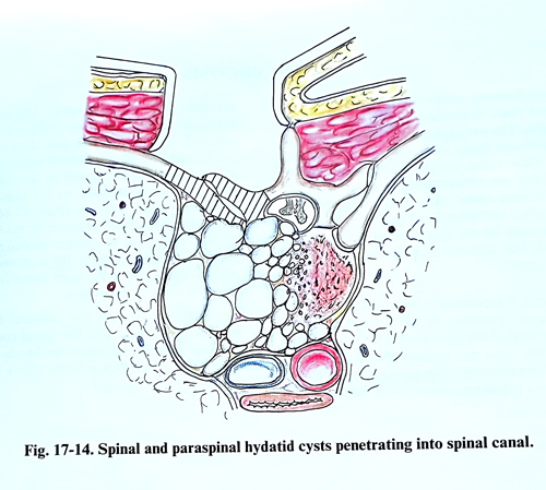
Vertebral hydatidosis is more common in adults. The parasites are microvesicular, invasive and progressive. The initial involvement of the vertebra is in the thoracic, lumbar or sacral area. This cystic lesion infiltrates the bone tissue, penetrates into the epidural space and often forms a large cavity in the paravertebral space, filled with parasitic cysts. When the parasites are located in the thoracic vertebrae, the cavity is situated in the posterior mediastinum, and in a case of lumbar localisation - in the retroperitoneal space. Very rarely there can be a case of an isolated parasitic cyst with subdural localisa-tion. Spinal cord involvement is due to compression. A plain X-ray, CT, MRI are routine investigations confirming the location and spread of the parasites.
Till now no chemotherapeutic agent has been able to replace surgery. As an adjuvant, mebendazole combined with surgical treatment has given good results in selected cases. Complete surgical removal is, however, curative.
Extraspinal cysts should be dealt with by radical excision, and the cavities swabbed out with 5% formalin to kill any live parasitic tissue. As formalin cannot be used on neural tissue, 20% hypertonic saline has been used with variable results.
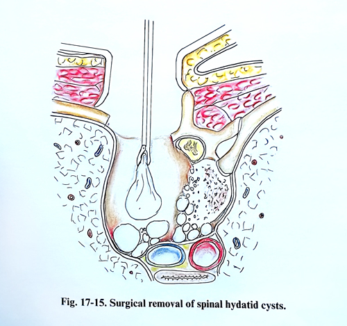
The surgical approach is planned according the localisation and the spread of the parasites. Because hydatidosis of the spine invades the bone and epidural space, and forms a large collection to one side of the paravertebral space, a costotransversectomy with lateral opening of the spinal canal is an approach of choice (Figs. 17-14; 17-15). The bone infiltrated by the parasites looks whitish and porous and in its structure small parasites are seen of different sizes - from those hardly visible to some at a millimetre in diameter. In the epidural space the parasitic cysts are bigger and are easily removed. It is recommended that the parasites be removed without tearing their capsule and it is important not to open the dura. After a good cleaning of the epidural space, the part of the vertebral body infiltrated by parasites is removed until normal bone tissue is reached. The extravertebral parasitic collection is often in communication with the epidural space, and during the cleaning is penetrated this collection too. All parasites inside this collection should be cleaned completely. Sometimes this is an easy task when the fibrotic wall of the collection is smooth. In other cases the parasitic collection is with a septum and many protruding pockets, which makes complete cleaning very difficult.
After surgical decompression of the spinal cord in cases of spinal hydatidosis, improvement of the neurological condition soon takes place. As the radical removal of the parasites from the bone tissue is rarely achieved, their growth develops rather fast, and ordinarily in the course of a year manifestations of spinal cord compression appear again.