Legacy: "Atlast of Neurosurgery" / L.Karaguiosov, A. Ramadan, K.Karaguiosov / Kiwait/ 1998
PREV> 16. Arteriovenous Malformations of the Spinal Cord
NEXT< 18. Functional Surgery of the Spinal Cord
17. INFECTIONS OF THE SPINE AND SPINAL CORD
Infections of the spine and spinal cord that need surgical treatment are rare compared to the frequency of cranial and intracranial infections. According to the aetiology they are of different categories: granulomatous, pyogenic, parasitic. Tuberculosis is very rare in developed countries. In many other countries tuberculous spondylitis with affection of the spinal cord is still a serious problem and on many occasions needs surgical treatment. Pyogenic haematogenous osteomyelitis, spinal epidural abscess and infections of the intervertebral discs on many occasions need surgical treatment. Subdural and intramedullary abscesses are very rare, and surgical treatment is essential to avoid permanent damage of the spinal cord and nerve roots. Parasitic infections in endemic areas are common. In the present era of migrating populations, it is possible to find a parasitic disease such as hydatid cyst or cysticercosis in a patient with a suspected spinal tumour.
TUBERCULOUS SPONDYLIS
Active tuberculous spondylitis causes a neurological deficit by pressure and toxic effects from pus, granulation tissue, bony sequestration, intervertebral disc material and bony dislocation. Direct penetration of the dura is rare; where it does occur, it causes tuberculous meningitis or meningomyelitis.
Healed disease also causes a neurological deficit by the remaining internal bony ridge or fibrous tissue constriction of the spinal cord.
Clinical presentations include signs and symptoms of infection: fever, malaise and weight loss. Evidence of tuberculous involvement in other organs may be present. Other manifestations include localised spinal pain in the early stage, followed by spinal deformity later. Muscle spasm is common with limited spinal movement. Back pain is present and aggravated by movement. With presence of a kyphotic deformity, adjacent segments in the spine develop compensatory lordosis.
Neurological manifestations develop slowly and progressively, but occasionally they can start suddenly. Neurological deficit is expressed to a varying degree. as ambulation may be difficult, and in advanced cases complete paraplegia or quadriplegia is present.
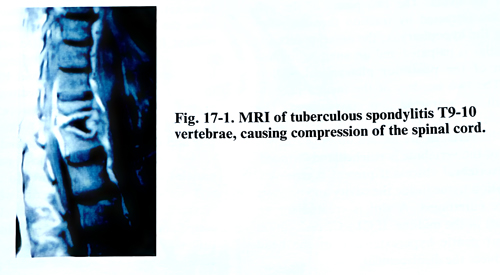
Diagnostic X-rays can demonstrate destruction, paravertebral abscesses, and deformities of the spine. Myelographic studies confirm spinal cord compression. A CT scan will define the extent of the paraspinal abscess and calcifications, together with the bone destruction. MRI provides additional data on the pathological changes within the spine and the spinal canal (Fig. 17-1). The definitive diagnosis is based on a combination of clinicoradiological features, and in particular positive histology of specimens obtained at operation.
SURGICAL MANAGEMENT
A complete course of antituberculous chemotherapy still remains the basis of treatment for all forms of active spinal tuberculosis. In cases with a neurological deficit, especially if it is progressing, surgical treatment is required. Operation essentially consists of excision of the disease focus and grafting. Abscess and granulation tissue evacuation will very often rapidly alleviate pain and improve the clinical condition of the patient. Alleviation of pressure on neural structures will enhance early neurological recovery, this being an absolute indication for surgery. The thorough debridement and placement of a bone graft under axial compression will enhance early fusion. The increasing deformity is prevented with early fusion, and late spinal cord compression from a bony bar is also prevented.
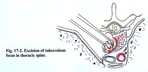
The operation consists of two main elements: 1. Eradication of the tuberculous focus by removing pus, sequestration of disc, bony fragments, and granulation tissue until the normal bone tissue is reached. 2. A slightly oversized graft is put under compression in an attempt to open the kyphosis during its insertion (Figs. 17-2; 17-3). As bone grafts, either a rib removed during thoracotomy, or the iliac crest are used. After placing the first bony piece, the rest of the bony defect is packed firmly with additional pieces. The next stage of the operation is posterior spinal fusion if anterior grafts are to replace two or more vertebral bodies. In cases of neurological deficit in patients with healed disease, the offending anterior bone ridge and fibrosis compressing the spinal cord should be removed.
As the destruction focus usually is situated in the vertebral bodies, the surgical approach should be anterior or anterolateral. The C1 and C2 vertebra are approached, usually transorally (Fig. 17-4). Infection in the oropharyngeal cavity should be treated before surgery. Tracheostomy is usually necessary for the anaesthesia.
The neck is extended and a special retractor maintains the mouth open with the tongue depressed. The soft palate is incised in the midline and the incision passes in one side of the base of the uvula. The two pieces of the soft palate are retracted by traction threads. After packing the hypopharynx, the anterior tubercle of the atlas is palpated and an anterior midline incision of the posterior pharyngeal wall is made. The two borders of the incised pharyngeal wall are retracted and stay-sutured. The prevertebral fascia and the fibrous tissue anterior to the vertebra are incised and the anterior surface of the vertebrae is reached and stripped.
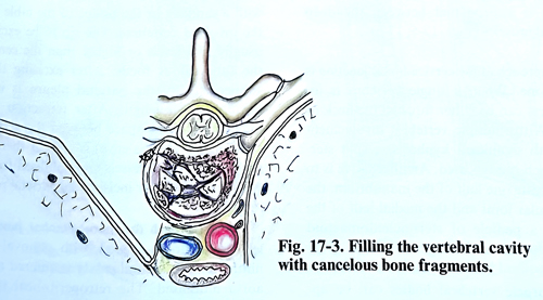
The prevertebral abscess if present is removed. Granulation tissue inside the cavity and necrotic bone are curettaged. A slot is created for an inlay graft in the midline. If C1 - C2 dislocation is present gentle hyperextension of the head often reduces the displacement.
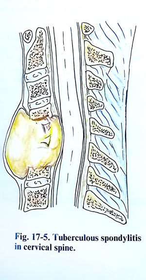
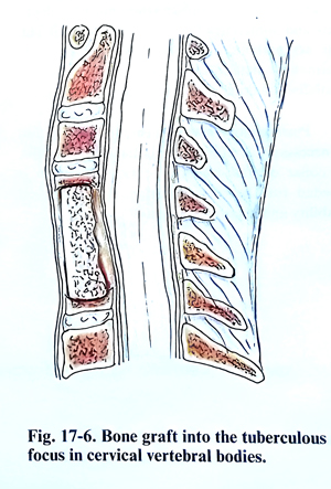
The C3 - C7 vertebrae are approached by penetrating between the neurovascular bundle in the neck laterally and the pharynx, larynx and trachea medially. A transverse skin incision in a small tuberculous focus along the skin creases is preferable from the cosmetic point of view. A vertical incision along the sternomastoid muscle may be necessary, if an extensive exposure over multiple levels is needed. The superficial cervical fascia is divided and the omohyoid muscle is identified and retracted or divided. The carotid sheath is retracted laterally and the larynx, pharynx, trachea and oesophagus - medially. The prevertebral fascia is incised vertically in the midline, taking care not to damage the sympathetic trunks. The tuberculous focus is approached between the deep prevertebral muscles (Figs. 17-5; 17-6).
The approach of the cervico-thoracic junction is a difficult one. When a single vertebra is in-volved, access can be either through the neck or the chest. With multiple vertebrae involvement together with significant kyphosis, a split sternum approach can be applied. An alternative is to excise or elevate one half of the manubrium, the sternoclavicular joint and the medial half of the clavicle on a pedicle of sternocleidomastoid muscle.
The thoracic vertebral bodies can be approached through a costotransversectomy or thoracotomy for lesions from T2 to TI2.
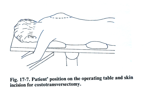
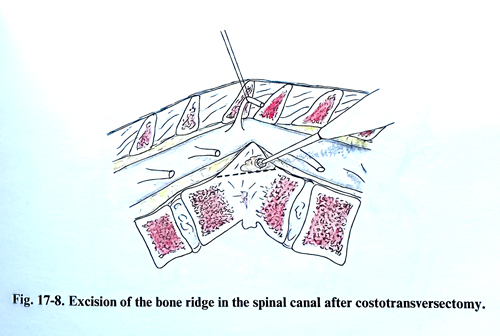
In costotransversectomy, the cutaneous incision takes the form of an arch, beginning from the midline 10 cm above the affected vertebra, and at the same level 10 cm laterally from the mid-line, ending on the midline again 10 cm below the vertebral pathology. The convexity of the arch is on the side of the more expressed neurological deficit. A flap is formed from the skin and the subcutaneous tissue and fascia, and turned medially. The superficial paravertebral muscles are cut perpendicularly to the midline, and are separated from the rib, which will be resected. The paravertebral muscles are dissected and separated upward and downward. In this way the transverse processes of 3 - 4 vertebrae together with the medial parts of the respective ribs are exposed. The rib corresponding to the level of the tuberculous focus is stripped from the periosteum and is resected from 6 to 8 cm. After that the transverse process and the head of the rib are resected too, and the lateral aspect of the vertebra is reached. These manipulations have to be done carefully, in order not to open the pleural cavity (Figs. 17-7; 17-8).
A thoracotomy approach, usually on the right side for a lesion from C7 to T4 and the left side for a lesion from T4 to T12, is preferred. On the left side, the aorta is a useful landmark and is less liable to be damaged as compared to the vena cava on the right side. The patient is in a lateral position on the operative table, usually with a sandbag or the bridge of the table under the involved vertebrae. The rib to be excised is usually two levels or higher than the centre of the tuberculous focus. After excising the rib subperiosteally the parietal pleura is incised throughout its exposed length. After retraction of the lung forward, the space between the great vessels (aorta and vena cava) is seen. If there is no abscess, the great vessels are mobilised from the vertebral body after incising the parietal pleura.
The approach the thoracolumbar junction: a left thoracotomy is used, with removal of the ninth rib. The pleural cavity is entered and the aorta mobilised. The retroperitoneal space is entered, usually at the costal cartilage of the ninth rib. The parietal peritoneum is mobilised from the undersurface of the diaphragm and retracted antero-inferiorly. The diaphragm is then cut circumferentially, leaving a 1 cm edge so that approximation is possible during wound closure. Approaching the vertebral bodies, the psoas muscle must be mobilised from the vertebral bodies.
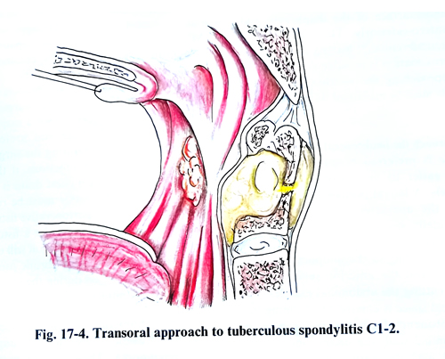
To approach the lumbosacral spine (L2 - S1) is preferred the left side because the major arterial vessels are easier to deal with than are the venous vessels. A lateral position of the patient is used on the operative table. An oblique incision is made posteriorly, starting at the midpoint between the iliac crest and the costal margin and extending to the midpoint between the umbilicus and the anterior superior iliac spine. After cutting the abdominal muscles, the retroperitoneal space is entered by stripping the parietal peritoneum from the posterior and lateral abdominal wall; the ureter, and kidney are reflected medially with the rest of the abdominal content. In approaching the lumbosacral junction, the common and external iliac vessels are mobilised. The iliolumbar artery must be identified and sometimes ligated. If damaged or cut, the sympathetic chain does not provoke any permanent deterioration. The psoas muscle is mobilised and its origin detached.
Postoperative management. Immobilisation is necessary for at least the initial three months.
A collar or halo-jacket type immobilisation is needed for the cervical spine, depending on stability and length of the graft.
Complications. During the operation, vascular, visceral or neural damages may occur.
Minor vascular tears can be repaired or the vessel can be ligated if it is appropriate. A major vascular accident may occur if the anatomy is grossly distorted and marked fibrosis makes the dissection difficult. A vascular surgeon's assistance may be necessary if such a major complication occurs. The lung and ureter are the structures vulnerable during the surgery. After freeing the lung from adhesions, if there is proper air filling, insertion of a chest drain is sufficient. Any damage of the ureter must be repaired to prevent leakage or a postoperative fistula. Dural tears require closure; otherwise a fistula and/or cerebrospinal fluid accumulation will occur. The lumbar plexus overlying a fibrotic psoas muscle may also be damaged during the removal of an abscess or mobilisation of the muscle.
Post-thoracotomy complications include pleural effusion, haemothorax, lung collapse and pneumonia. These can be prevented by good postoperative care and chest physiotherapy. Retroperitoneal dissection in the dorsolumbar and lumbar region often results in transient paralytic ileus.
Minor postoperative neurological complications include transient Horner's syndrome due to cervical sympathetic damage. The appearance of postoperative deterioration of the spinal cord function (paraparesis or tetraparesis) is due to either spinal cord damage or spinal cord compression by slippage of a bone graft.
PYOGENIC VERTEBRAL OSTEOMYELITIS
Pyogenic vertebral osteomyelitis has usually a haematogenous origin. Among the pyogenic bacteria, staphylococcus aureus and coliform bacilli are most common. Compromise of the spinal cord occurs when an associated extradural abscess develops or when a bony deformity and vertebral collapse compromise the spinal canal.
Back pain is the most common initial symptom. The objective findings are minimal and limited to local tenderness and to restriction of spinal movements. Fever, leucocytosis, and weight loss occur in a minority of patients. Local pain may progress to radicular pain over periods of weeks to several months.
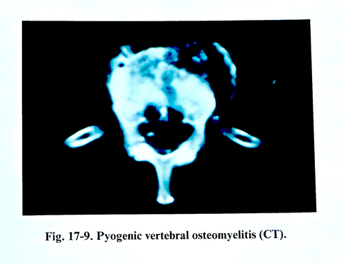
When compromise of the spinal cord occurs, X-ray studies, CT, MRI and/or myelography are necessary. The thoracic area is the most frequent site of osteomyelitis. If the patient is neurologically intact and the bone destruction is minimal, medical treatment is appropriate, especially if the responsible organism can be identified by needle biopsy or by blood culture. The appropriate antibiotic is given intravenously for up to 6 weeks, and if the bone destruction is arrested and pain subsides, the patient can be mobilised in a body jacket. With any progressive clinical manifestations of cord compression, surgical treatment is indicated.
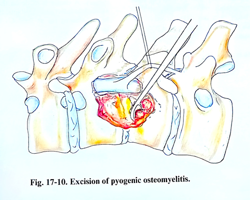
Costotransversectomy is a procedure of choice (Figs. 17-9; 17-10). The removal of the involved part of the vertebral bodies is carried out after removing transverse processes and pedicles (in a thoracic lesion including the medial portion of the corresponding rib). Anterior operations are best for those rare cases in which the patients have progressive paralysis from osteomyelitis of the cervical spine.
When spinal instability is present, long-term immobilisation by casts, halo devices, or braces must be provided.
SPINAL EXTRADURAL ABSCESS
Abscesses of the spinal extradural space can result from septic emboli to the extradural fatty tissue. Alternatively it may derive from a spread of infection from a paravertebral focus site via an intervertebral foramen, or by direct extension from spinal osteomyelitis. Rarely they can be caused by penetrating injuries or as a complication of surgery or lumbar puncture.
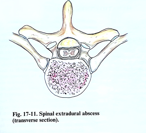
Spinal epidural abscesses are usually located dorsally to the dural sac and are situated mainly in the thoracic part of the spine (Fig. 17-11).
Staphylococcus aureus is the most common causative organism for spinal epidural abscesses, although streptococcus and/or pseudomonas may be also responsible. Fungus granulomas such as actinomycosis and blastomycosis are occasionally encountered.
Initial symptoms are local, with root pain as well as fever and other general manifestations of infection. Motor and sensory deficits occurring distal to spinal cord compression and sphincter troubles may also develop. In some patients deterioration occurs quickly, within a matter of hours or a few days. In others the disease has a prolonged course over a period of days and weeks.
Patients suspected of having a spinal extra-dural abscess need a careful clinical and neurological evaluation. Spinal X-rays are indicated. but are often normal, unless paravertebral abscesses or osteomyelitis are demonstrated. Myelography should be done promptly; it can demonstrate the upper and lower limits of the abscess. CT and MRI give additional diagnostic information about the location and the spread of the abscess.
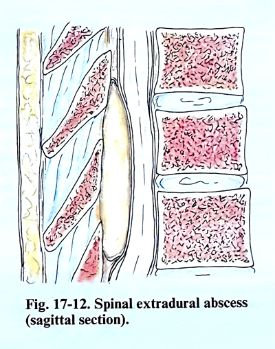
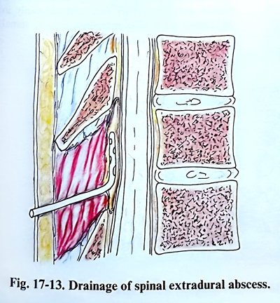
Prompt decompressive laminectomy and removal of the abscess should be carried out. In acute abscesses, frankly purulent material will be evacuated (Figs. 17-12; 17-13). In the chronic forms only granulation tissue can be found, which might be mistaken for a malignant neoplasm (metastasis). Complete removal is usually possible. Bacteriological culture and smears should be obtained. In cases of acute purulence, local antibiotics, and external drainage of the wound should be applied for several days.
If an extradural abscess is suspected pre-operatively, antibiotics should be given before surgery. A choice is made on the basis of the antecedent infections. Antibiotic therapy should be maintained for at least 4 - 6 weeks after surgery.
HYDATID CYSTS
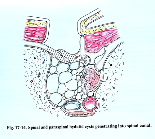
Vertebral hydatidosis is more common in adults. The parasites are microvesicular, invasive and progressive. The initial involvement of the vertebra is in the thoracic, lumbar or sacral area. This cystic lesion infiltrates the bone tissue, penetrates into the epidural space and often forms a large cavity in the paravertebral space, filled with parasitic cysts. When the parasites are located in the thoracic vertebrae, the cavity is situated in the posterior mediastinum, and in a case of lumbar localisation - in the retroperitoneal space. Very rarely there can be a case of an isolated parasitic cyst with subdural localisa-tion. Spinal cord involvement is due to compression. A plain X-ray, CT, MRI are routine investigations confirming the location and spread of the parasites.
Till now no chemotherapeutic agent has been able to replace surgery. As an adjuvant, mebendazole combined with surgical treatment has given good results in selected cases. Complete surgical removal is, however, curative.
Extraspinal cysts should be dealt with by radical excision, and the cavities swabbed out with 5% formalin to kill any live parasitic tissue. As formalin cannot be used on neural tissue, 20% hypertonic saline has been used with variable results.
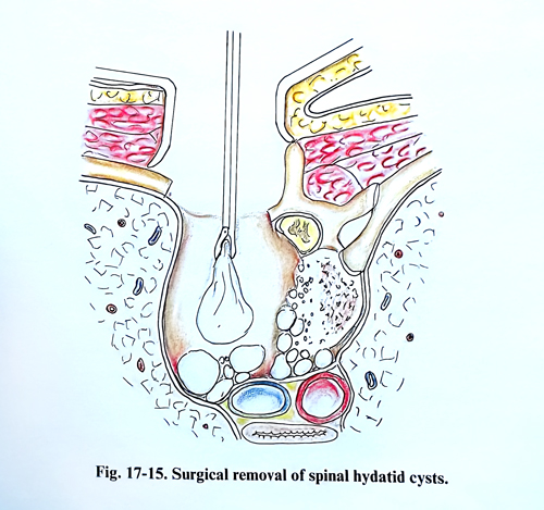
The surgical approach is planned according the localisation and the spread of the parasites. Because hydatidosis of the spine invades the bone and epidural space, and forms a large collection to one side of the paravertebral space, a costotransversectomy with lateral opening of the spinal canal is an approach of choice (Figs. 17-14; 17-15). The bone infiltrated by the parasites looks whitish and porous and in its structure small parasites are seen of different sizes - from those hardly visible to some at a millimetre in diameter. In the epidural space the parasitic cysts are bigger and are easily removed. It is recommended that the parasites be removed without tearing their capsule and it is important not to open the dura. After a good cleaning of the epidural space, the part of the vertebral body infiltrated by parasites is removed until normal bone tissue is reached. The extravertebral parasitic collection is often in communication with the epidural space, and during the cleaning is penetrated this collection too. All parasites inside this collection should be cleaned completely. Sometimes this is an easy task when the fibrotic wall of the collection is smooth. In other cases the parasitic collection is with a septum and many protruding pockets, which makes complete cleaning very difficult.
After surgical decompression of the spinal cord in cases of spinal hydatidosis, improvement of the neurological condition soon takes place. As the radical removal of the parasites from the bone tissue is rarely achieved, their growth develops rather fast, and ordinarily in the course of a year manifestations of spinal cord compression appear again.
CYSTICERCOSIS
Involvement of the spinal cord is far less common than is involvement of the brain and meninges. The parasites can be found within the subarachnoid space, and even within the spinal cord. They cause compression of the spinal cord, and an inflammatory reaction in the subarachnoid space with arachnoid adhesions, which affect the spinal cord and the nerve roots. Cysticercosis of the spinal canal is usually not isolated, but accompanies cysticercosis in the cranial cavity.
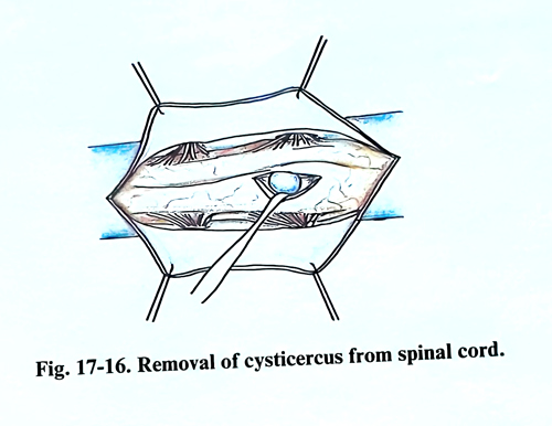
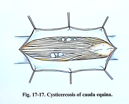
Surgical treatment is indicated in cases of a progressive syndrome of spinal cord affection. The removal of the cisticerci from the subarachnoid space does not present special difficulties. After laminectomy, the dura is opened and arachnoid adhesions are revealed, among which parasitic cysts are also seen. The arachnoid adhesions are carefully torn and the cysts easily removed, as they are free and their capsule is not attached to the spinal cord meninges. When it is a case of cysts located in the spinal cord tissue, the technique is the same as with an intramedullary tumour (Pigs. 17-16; 17-17), A small longitudinal spinal cord incision is made on the protrusion provoked by the parasite. After reaching it, the separation of the parasite from the spinal cord tissue is easy, as there are no adhesions.
After removal of the cisticerci from the subarachnoid space and the spinal cord tissue, meningoradicular irritation can be observed for some days, which then subsides relatively fast.