
Legacy: "Atlast of Neurosurgery" / L.Karaguiosov, A. Ramadan, K.Karaguiosov / Kiwait/ 1998
Read PREVIOUS: 1. General Surgical Techniques in Cranial and Intracranial Lesions
Read NEXT: 3. Head Injury - chapter 1
2. Head Injury
In head injury, brain damage results from the initial impact (primary brain damage) and from the development of secondary complications (secondary brain damage). Cerebral contusion and laceration as well as diffuse injury of the brain parenchyma are the major components of the primary brain damage. Secondary brain damage is caused by intracranial haematomas, brain swelling and shifting, ischaemia or infection. The main goal of treatment is prevention or rapid treatment of secondary brain damage, and providing the patient with optimal conditions for recovery from the primary damage. The role of surgery consists of the urgent evacuation of traumatic mass lesions, relieving or preventing intracranial hypertension and brain shifts, repair of wounds and depressed skull fractures for prevention of infection, as well as at a later stage closure of CSF fistulae and cranioplasty. To be efficient, surgery in traumatic mass lesions must be really urgent and before the patient deteriorates and irreversible secondary brain damage appears. Intracranial pressure monitoring can be very helpful in the evaluation of indications for surgery and for the patient's prognosis.
SCALP LACERATIONS
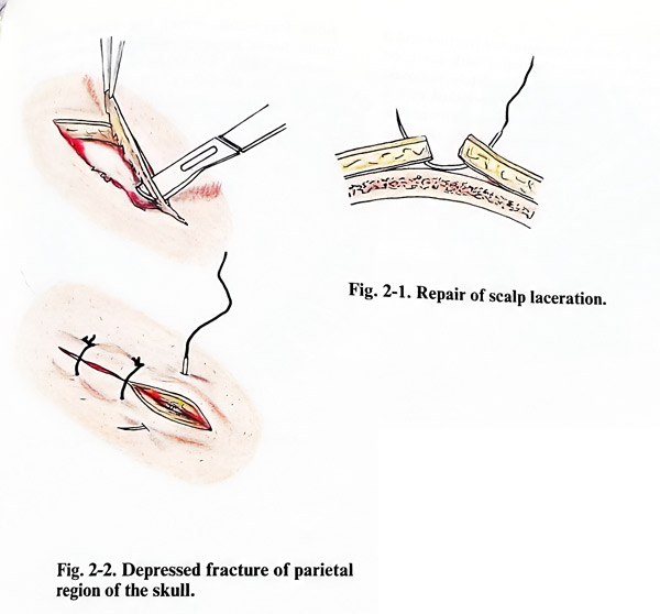
Sufficient hair must be shaved around the wound to allow proper visualisation of all lacerated skin. The scalp edges should be infiltrated with local anaesthetic if the patient's perception of pain is preserved. Then the wound is explored, searching for foreign bodies and for any damage of underlying layers, especially visible fractures, CSF leak or brain debris and also irrigating it with sterile saline. If the damage is considered to be limited to the epicranial tissues, the damaged tissue is debrided to allow closure with minimal tension, and any necrotic parts are removed (Fig. 2-1). Bleeding is often better controlled by coagulation or by bending the galea outwards, rather than by attempting direct clamping of vessels. Excessive tension of the wound edges should be avoided.
Single-layer scalp sutures with approximation of the galea complete the treatment. However, should there be a need to adapt and reduce tension of the edges, the galea should be inverted and interrupted stitches placed. In case of scalp loss, it is possible to control the situation by the undermining or advancement of flaps, sometimes turning them to reduce the skin defect. Any large loss of scalp requires specialised techniques of plastic surgery.
DEPRESSED SKULL FRACTURES
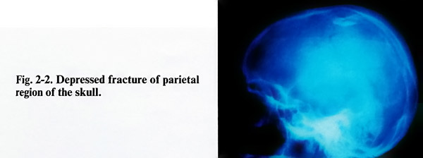
In simple skull fractures, i. e. without laceration of the scalp, surgical repair is indicated, when there is depression of bone fragments into the cranial cavity. The degree of depression of the bone fragments must be precisely determined by skull X-rays and especially by reference to a view that is tangential to the area of depression. CT images with a window visualising bone are extremely helpful to show clearly the depression. The operation should be performed as early as possible, when the patient's general condition permits it with an acceptably low risk (Fig. 2-2).
The goals of the operation are: 1. Elevation of the bone fragments, which have penetrated intracranially; 2. Evacuation of the haematomas and necrotic intracranial tissues; 3. Restoration of the anatomical integrity of the dura, bone and epicranial layers. Three methods can berecommended:
First method.
The incision of the scalp is either in the form of a horseshoe or 'S' - shaped. The skin, and the subcutaneous tissues with the galea are separated and lifted as a flap, leaving the bone covered only with the pericranium.
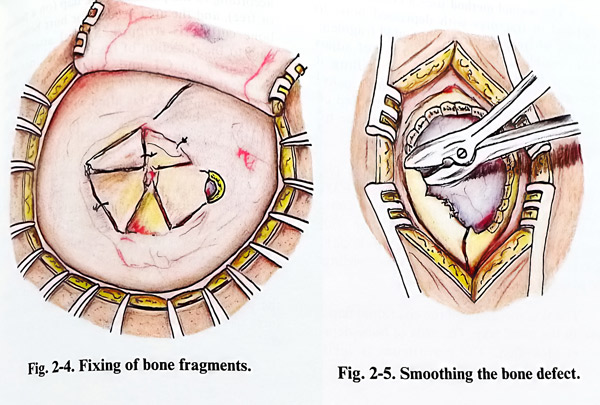
Bone fragments are seen frequently with torn pericranium over them. There is always a certain amount of haemorrhage below the periosteum, or if it is torn, even below the galea. All fragments which are small and free from periosteum are removed together with the necrotic brain tissue. Larger fragments are elevated with their pericranium, making possible an inspection of the dura and the brain. Very often some of the bone fragments cannot be easily removed, because they are indented in the cranial cavity, and firmly fixed, as the edge of the inner table remains under the surrounding normal bones. A burr hole just at the border of the fracture, but within normal bone is made. Through the burr hole, and after a small amount of nibbling, the fixed indented fragments are elevated. The elevator can be based on the edge of the intact bone and used as a lever. If the dura is intact, the bone defect is covered using the large bone fragments, fixing them to the margins of the defect by wiring (Fig. 2-4). At the end the scalp layers are closed.
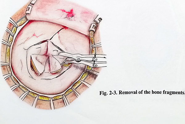
When there is a comminuted fracture and it is impossible to close the defect with the bone fragments, the latter are completely removed and the edges of the defect are smoothed with a bone nibbler. In such a case, cranioplasty can be performed at the same surgical session (Fig. 2-5)
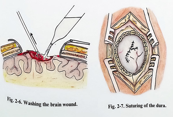
When the skull fractures are associated with tearing of the dura and brain injury, the bone fragments, which are found inside the brain tissue must be removed along with the necrotic parts. So as to inspect the cerebral surface well, the defect of the dura is widened by radial incisions. If additional necrotic tissues and clots are found, they are removed by suction and a stream of saline (Fig. 2-6; 2-7).
The surgical wound is closed in separate layers: dura, pericranium, galea and skin.
The second method uses a craniotomy. It is applied in fractures with depressed bone fragments, where access to the indented fragments is not possible without much sacrifice of adjacent normal bone in the process of drilling and nibbling it. Therefore, a craniotomy enclosing the fracture in the centre of the bone flap induces to the patient less injury to the dura and the brain and there is less definitive bone removal.
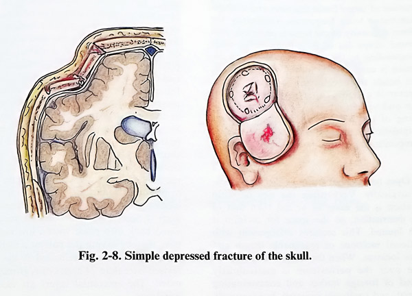
Surgery is planned by defining the places for burr holes and bone cutting lines, always in intact bone, but not too far away from the fracture (Caution: the lifting of the flap should not provoke additional fracture of bone). A suitable epicranial flap is tailored, according to the place of craniotomy (Fig. 2-8).
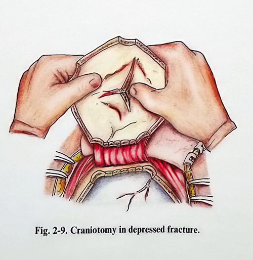
The skin incision and the epicranial flap are done in the usual way. The area of bone depression is identified. The periosteum is incised according to the planned bone flap (on a pedicle or free, and the area needed for burr holes and bone cutting is stripped. Special attention is paid to the detection of fracture lines, not seen previously on CT and X-rays, as they can change the planned bone flap. After drilling and bone cutting, the flap is lifted with much care in separating the dura from the inner table. The fragments usually remain attached to the periosteum and are lifted easily. With the flap already in hand, fragments are usually repositioned with no difficulties (Fig. 2-9). The fragments should be stable and fixed in the bone defect. However, if a defect remains, it is closed in the other routine ways. Then the craniotomy can be closed in the standard way.This method is applied less frequently than the previous one, and can be suitable for fractures in children, where bone elasticity is greater.
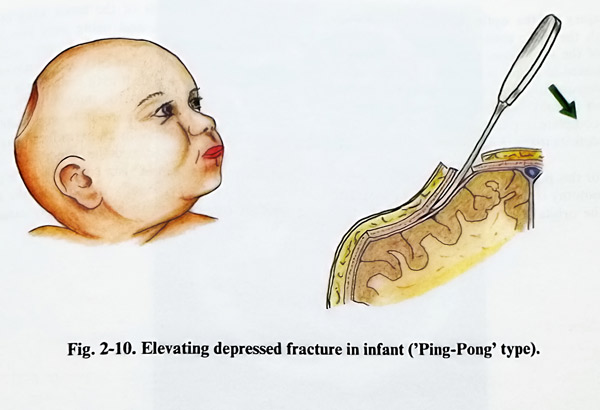
The third method is applied in depressed fractures in infants. The great elasticity of the cranial vault in infants does not allow the formation of larger linear fractures and multiple comminuted bone fragments. Ordinarily a uniform concavity of the bone is observed, with a round shape (as in a Ping-Pong ball) that can correspond to the area of impact. This depression is corrected by means of a burr hole, made beside the edge of the concavity. A fine dissector is carefully inserted in the epidural space and with a lever-like movement, repositioning of the bone is obtained. When this manipulation is applied early after the injury, the repositioning of the bone is easier (Fig. 2-10).
Open (compound) fractures should be treated surgically. Very often the size of scalp laceration is the same as or less than that of bone destruction, so the space for revision is much more limited. This requires enlargement with additional incisions of reasonable length and proper location. When the scalp is retracted, the space over the periosteum is meticulously
cleaned of foreign bodies and contaminating material, and copiously irrigated. Intracranially indented fragments are not removed before incising and stripping periosteum expose the edges of intact bone. Only when the surgeon has sufficient space to control sudden major bleeding may the fragments be removed.
Here the technique employed in closed (simple) depressed fractures can be followed. Nasal sinuses that have been exposed are stripped of mucose. If possible, a galeal flap can be pulled over the end of the exposed sinus and tacked to the dura below to seal the opening. Intact, normal-appearing dura should be left unopened; however if it appears tense or bluish in colour, it is incised and the underlying brain examined. Intracranial haematoma or necrotic brain tissue is removed. A dural laceration should be trimmed and extended with additional incisions to permit removal of foreign material, haematoma, contused brain tissue and to perform haemostasis. Dura is closed in a water-tight fashion with pericranium or fascia graft if necessary. Bone fragments can be wired back into place with a low risk of infection, thereby saving the patient a further operation. This is contraindicated if the wound is revised later than 24 hours or is grossly contaminated. The epicranial layers are closed in the usual way.
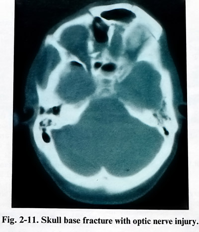
Injury of the optic nerve in its passage through the optic canal is common with fractures of the skull base. Surgical decompression is indicated if vision is present after the injury but subsequently deteriorates. Surgical decompression is unlikely to be of benefit in treating stable complete or partial visual defect or partial visual deficits that have improved.
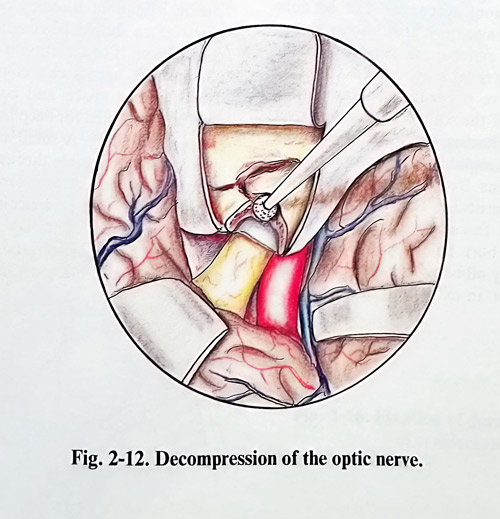
For this purpose a frontal or frontotemporal craniotomy is made and the dura is separated from the orbital roof. With the drill the orbital roof, anterior to the lesser wing of the sphenoid is removed, and using the finest bone instruments, the upper wall of optic canal is resected. Then all fragments are removed. A high-speed diamond drill with continuous irrigation is very convenient for unroofing the optic canal. In the case, where it is impossible to relieve the optic nerve in an extradural way, the dura may be opened and the upper wall of the optic canal resected under visual control of the nerve (Figs. 2-11; 2-12). Finally the optic nerve sheet and the ring of Zinn are longitudinally divided.
TRAUMATIC MASS LESIONS
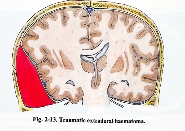
Extradural, subdural or intracerebral collections, especially those with rapid increase of volume, can easily lead to raising and decompensation of the ICP with all the notorious consequences of brain compression, shifts and herniation. Therefore, these collections need early (usually urgent) evacuation. Usually it is the case of blood collection, but sometimes CSF accumulated in the subdural space can have the effect of a mass lesion. Localised brain contusion with perifocal oedema provoking a mass effect can also need surgical evacuation (Fig. 2-13).
ACUTE EXTRADURAL AND SUBDURAL НАЕМАТОМА
The goals of surgical treatment are to decrease intracranial pressure by removing the haematoma as early as possible, especially in patients with an impaired level of consciousness, and to prevent rebleeding.
The approach to the haematoma depends on its size, location and the available facilities when dealing with it as an emergency.
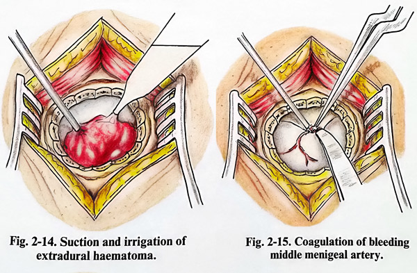
In case of a small acute extradural and subdural haematoma, especially one located in the temporal region, evacuation can be done with a limited craniectomy. It is also an acceptable approach in extreme emergencies when the surgical facilities available are limited. In such a case the operative procedure begins with a vertical incision of the scalp 5 - 6 cm long over the area of the haematoma. As most often haematomas are located in the temporal region, the incision begins from the zygomatic arch projecting quite vertically. The temporal muscle is split along its fascicles, the periosteum is separated, and a burr hole is opened. The hole is enlarged by nibbling to a craniectomy of 3-4 cm. In case of an extradural haematoma a clot of dark blood extrudes through the craniectomy. The clots should be removed with saline jet irrigation and gentle suction (Fig. 2-14). More aggressive suction usually provokes additional bleeding. A saline wash is also used to detect persistent bleeding. Such bleeding is controlled by bipolar coagulation if the dural bleeding point is accessible, or by topical haemostatic agents (oxidised cellulose or other available substances) and stitches to fix the dura to the periosteum at the edge of the craniectomy. In cases of major arterial bleeding from basal dura or fractures injuring the middle meningeal artery, the craniectomy can be extended more towards the skull base and the dura retracted. This can permit better control of the middle meningeal artery, particularly when the injury is close to its intracranial entrance (Fig. 2-15). This situation may require packing of the foramen spinosum with bone wax or bone wax mixed with cotton fibres to control haemorrhage. If the CT does not show any subdural collection or brain contusion, the dura should be left intact.
In case of an acute subdural haematoma, the craniectomy will expose a bluish and tense dura. Its opening precipitates the extrusion of dark clots. These clots are removed by gentle suction or traction with cup forceps and saline stream irrigation. Bleeding points on the cerebral surface should be coagulated with bipolar coagulation. If coagulation is ineffective, topical haemostatic agents can control the bleeding.
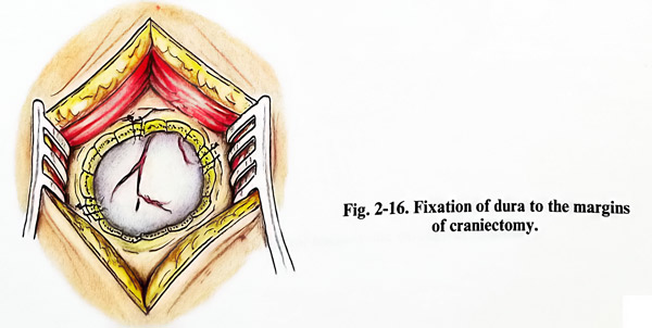
The subdural space and cortical surface are carefully inspected for deep haematomas, contusions, or bleeding. A very small amount of clot that is not easily accessible through the craniectomy is best not removed, as it can provoke unnecessary bleeding that is difficult to control. After bleeding has been controlled, the dura is closed in a watertight fashion and tacked up to the margins of the craniectomy (Fig. 2-16).
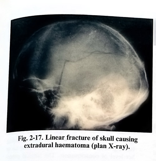
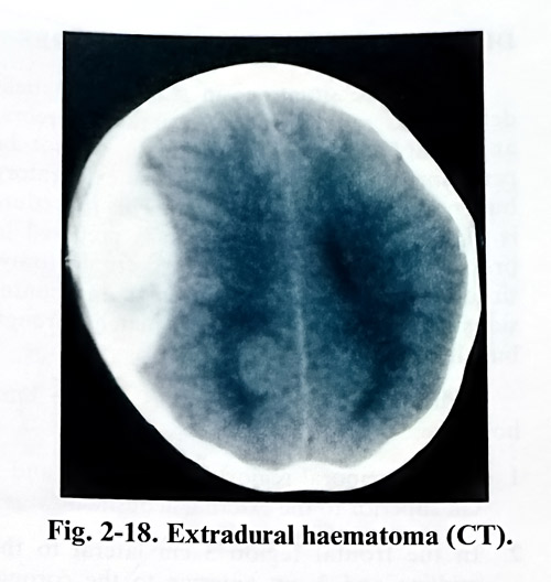
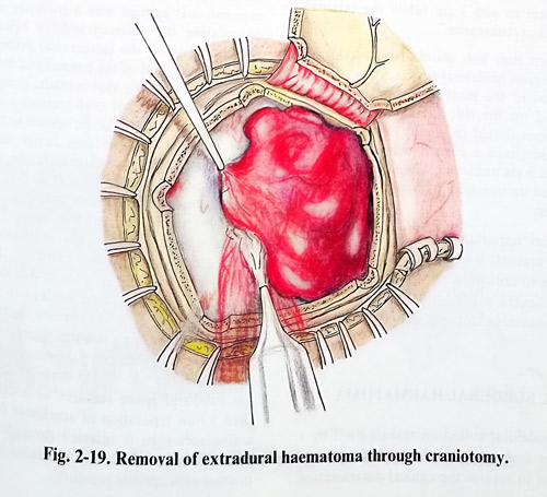
Craniotomy gives much greater access for removal of acute extradural and subdural haematomas. The size and location of the craniotomy should ideally cover the whole area of the haematoma and the epicranial flap should be planned accordingly. This facilitates an approach to the most peripheral part of the haematoma. Extradural haematomas are managed with less blood loss and safer haemostasis due to better exposure of the dura. If the dura must be opened, then it can be done with better visibility and control of manipulation. In subdural haematomas the removal of clots and haemostasis is also easier and safer. The underlying brain can be carefully inspected in all the area covered by the haematoma, and the detection of the bleeder is easier, whether it is from a brain laceration, rupture of a vein draining into the sagittal sinus, petrous sinus or sylvian veins.
Occasionally a ruptured cortical artery can cause the haematoma. Hence, because of its advantages therefore, whenever it is possible, a craniotomy must be preferred over a limited craniectomy (Figs. 2- 17; 2-18; 2-19).
DIAGNOSTIC SURGICAL PROCEDURES
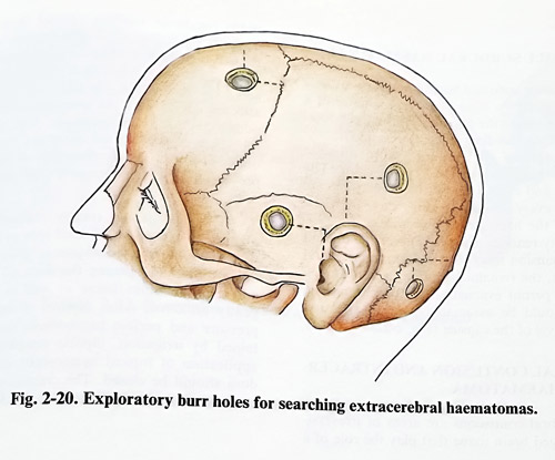
There are situations in which the patient deteriorates rapidly and CT, or even cerebral angiography are not available or cannot be performed in time. In such a case exploratory burr holes are the better option. This procedure is of value only if the surgeon is prepared to proceed with a craniotomy or craniectomy, since acute traumatic haematomas and contusions cannot be dealt with adequately through burr holes only (Fig. 2-20).
The most convenient places for burrholes for hematoma detection are:
1 In the temporal region 3 cm anterior and 3 cm superior to the external acoustic meatus.
2 In the frontal region 3 cm lateral to themidline and 3 cm anterior to the coronal suture. This suture coincides with the line which connects the midpoints of the zygomatic arches and is perpendicular to them.
3 In the parietal region 3 cm superior to and 3 cm posterior to the tip of the auricle.
4 When a haematoma is suspected in the posterior cranial fossa, a burr hole is made 3 cm lateral to and 3 cm below the external occipital protuberance.
The first burr hole should be made in the temporal region ipsilateral to the dilated pupil, contralateral to the most abnormal motor response, and on the side of a fracture. The scalp incisions should be placed to permit their incorporation into a formal craniotomy if a haematoma is encountered. When the procedure is completed on one side, it should be repeated on the other.
Another important procedure, considered routine at present, is invasive ICP monitoring.
Its increase to critical levels in the head-injured patient indicates a need to take corresponding measures to decrease it again.
CHRONIC SUBDURAL HAEMATOMA
This subdural collection reveals itself by a mass effect and its prompt evacuation is the only option to reverse the clinical deterioration of the patient. Once the patient is showing symptoms and signs of increased intracranial pressure or deterioration of the focal deficit, surgery should not be delayed. The evacuation of a chronic subdural haematoma is planned by deciding the place of one or two burr holes, as it is usually fluid. They should be over the thickest part of the haematoma, but the overlying incision, a straight one, should be in hairy skin. The burr holes and skin incisions should permit incorporation into a craniotomy for a wider approach. If there are bilateral haematomas, the larger should be evacuated first, but both sides should be done in a single operative session.
General or local anaesthesia is used according to the general condition of the patient and his or her level of consciousness. The surgical procedure is relatively simple but close attention must be paid to these patients, especially the elderly, because of other significant medical problems.
With the patient in the usual supine position, the preplanned burr hole(s) should be made on the affected side. After the dura is exposed, it is opened with a cross-like incision, coagulating the place of incision beforehand.
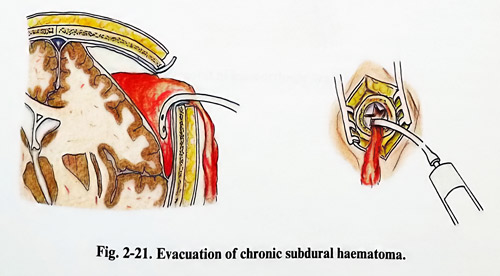
Depending upon the intracranial pressure, the dark brown liquid of the haematoma may burst through the opening that is made. Sometimes a thick brown, black, or even greenish membrane below the dura must also be opened to reach the fluid part. The haematoma cavity should be irrigated with warm saline until the subdural space yields only clear fluid (Fig. 2-21). In big haematomas sometimes complete evacuation is achieved through a second or more burr holes. The depth of the subdural space must be inspected and its reduction should be attempted by lowering of the upper half of the table so that the patient remains positioned for a while with the head slightly-down. This manoeuvre increases the intracranial venous pressure, therefore also the ICP. If the cerebral cortex comes up to the dura, the burr holes are closed without drainage. If the subdural space remains as a cavity more than 5 mm separation of arachnoid from dura, a fine soft tube is inserted through the more posterior burr hole and is connected to a bag at normal atmospheric pressure.
INFANTILE SUBDURAL HAEMATOMA
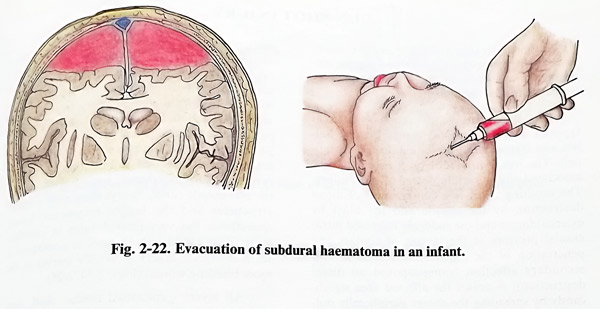
Infantile subdural haematomas are usually bilateral and very extensive. The existence of a fontanelle allows the puncturing of the subdural space with a needle, which is inserted in the lateral angle of the fontanelle and directed laterally tangential to the cranial vault. The puncture and evacuation of the haematoma are repeated every second or third day, until the cavity of the haematoma disappears. If a residual cavity remains (the cerebral mantle doesn't show expansion towards the dura) after several punctures, the remaining option is craniotomy. This will permit evacuation of the haematoma which should be associated with fenestration and removal of the capsule (Fig. 2-22).
CEREBRAL CONTUSION AND INTRACEREBRAL HAEMATOMA
Cerebral contusions are areas of irreversibly damaged brain tissue that induce and present with brain swelling around them. Quite often they are associated with intrinsic haemorrhages (i.e. haemorrhagic contusions, "burst lobe" syndrome). When such cerebral contusion acts as a mass lesion, the patient's condition is affected not only by the contusion, but also by additional surrounding brain compression. Temporal lobes are more affected than are the frontal with this condition. Therefore, early resection of this nidus prevents further compression, shift and herniation.
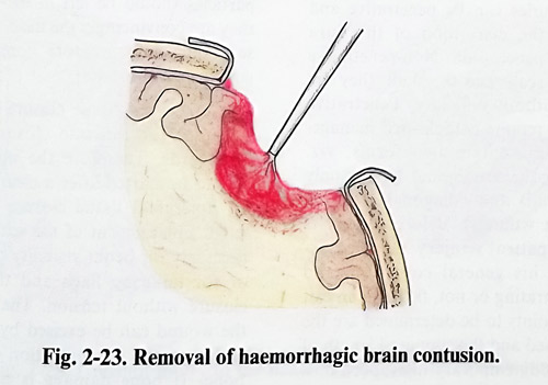
The craniotomy (rarely craniectomy) is planned to expose the affected area or lobe, in a way similar to the techniques in gliomas. After craniotomy and opening the dura all lacerated and necrotic brain tissue as well as clots are gently removed. After control of intracranial pressure and perfect haemostasis (usually obtained by irrigation, bipolar coagulation, and application of topical haemostatic agents), the dura should be closed. The craniotomy should be closed in the routine way (Fig. 2-23).
GUN-SHOT INJURY
Surgical treatment of gun-shot injury aims at the preservation of life and the prevention of complications at the least consequent neurological deficit for the patient.
Gun-shot injuries are caused by different types of missiles, the most common being bullets and fragments of exploding devices (shrapnels). The injury produced by a missile can be associated with a certain degree of blast injury too.The resulting effect is a combination of direct destruction by the missile and the blast by external forces, and the sudden increase of intracranial pressure at the instant of contact and penetration of the missile intracranially. This secondary affection (superimposed on direct destruction), increases the affected area significantly by spreading the energy peripherally outward from the missile canal. The higher the velocity of the missile, the greater the secondary effects we observe. They are considered to affect by two factors: shock waves and temporary cavitation.
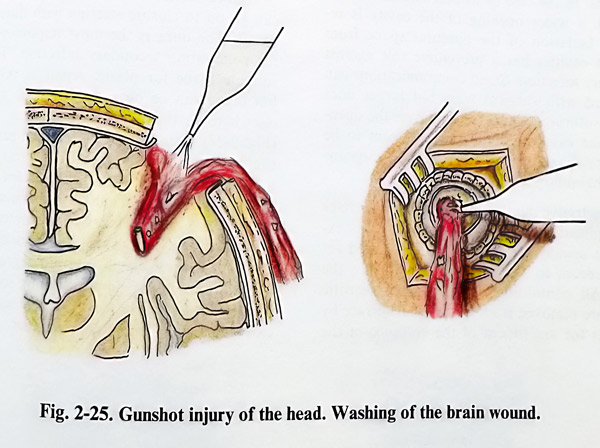
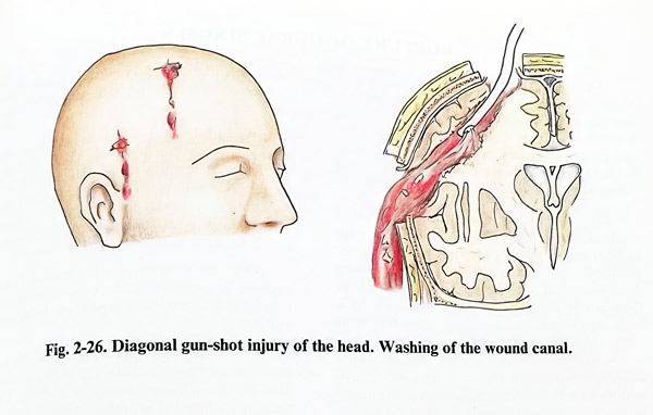
Gun-shot injuries can be penetrative and non-penetrative, the disruption of the dura being the sign of penetration. Non-penetrative injuries affect the scalp and the skull; they are often repaired without difficulty. Penetrative injuries, however, require complicated management, and they present in two forms: "blind" (retaining the missile and having only an entrance wound) and "diagonal" (having entrance and exit wounds). Before surgery is undertaken, the patient and the injury must be assessed regarding general condition, and whether it is penetrating or not, i.e. is there an exit wound. Other points to be determined are the type of weapon used and the supposed length of the trajectory. A contemporary high-speed missile (from conventional weapons) provokes severe generalized brain damage. Therefore, in consideration of the severe generalised damage and the need of early prevention of complications, surgical treatment should be considered quite always as an emergency. In patients requiring resuscitation or with unstable vital signs, surgery is postponed until stabilisation. However, if skull X-rays and particularly CT equipment are available in hospital, their use should precede the surgery. CT scans provide the best information about the presence of haemorrhages, bone fragments and the real missile canal. X-rays show only metallic structures and the bigger fragments. Local anaesthesia has very limited value in gun-shot injury, being used only in cases of nonextensive scalp injuries. Surgery is usually started at the wound wuth more intense bleeding (Figs. 2-24: 2-25).
All layers - epicranial tissues, skull, dura and brain - require separate attention for debridement and if necessary, repair. Another general principle is the special care taken for the prophylaxis of infection by removing all contaminating foreign bodies, as well as necrotic tissues from all layers. No bone fragments and particles should be left in the missile canal, as they are evidently the most frequent cause of secondary inflammatory complications (especially brain abscesses).
Epicranial tissue closure has essential importance for the safe healing of the wound afterwards. Therefore the initial debridement should be started after a clear preliminary plan for epicranial tissue closure has been formulated. Enlargement of the scalp wound may be necessary for better visibility of the layers below or for tailoring flaps, and thereby permitting closure without tension. The necrotic edges of the wound can be excised by a few millimetres and retracted for inspection of periosteum and bone. If bone damage is not confirmed, the wound is closed in the usual way.
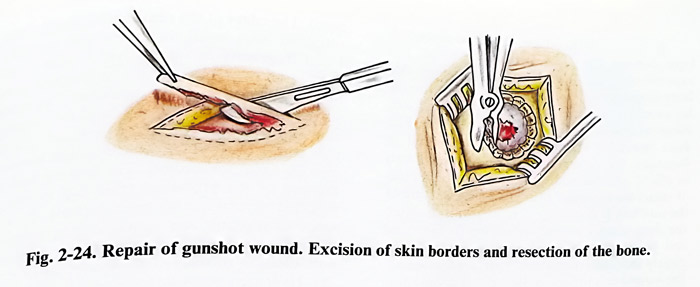
If X-ray/CT investigations have disclosed bone damage, the edges of the bone defect are exposed with additional periosteal incisions. All bone fragments and edges of the defect should be considered contaminated. Fragments are removed and edges nibbled and smoothened. The dura must be exposed by bone edge nibbling, and if damaged, the exposure should be large enough to afford additional access of a few millimetres of intact dura around the defect. The starting of the nibbling is done with care so that not to injure the dura further by not separating its lacerated end from the bone. If the dura is intact and clearly indicates the absence of a haematoma, the wound can be closed. Because of contamination and increased ICP, cranioplasty can be postponed. If the paranasal sinuses, middle ear or orbits are also damaged, all precautions should be taken to avoid leaving any communication with the epidural space. Sinuses are cleaned of mucosa, fragments, foreign bodies and clots. In the case of frontal sinuses and the mastoid, a wider opening of the cavity is re-quired. Isolation of the epidural space from adjacent cavities has a preventive role against secondary infection. Small communications can be closed with surgical wax, but larger ones require suturing a periosteal flap over the opening. Some cases need the surgical team to be joined by maxillofacial, ENT, plastic surgeons or ophthalmologists.
Penetrative injuries carry much higher risks of complications. Once the dural defect is exposed, it usually has to be slightly enlarged for better access to the missile canal inside the brain. All contaminated debris and necrotic tissues are removed from the cortical surface by saline jet for assessment of the entrance of the canal. The edges of the canal entrance are gently retracted with small dissectors and spatulas.
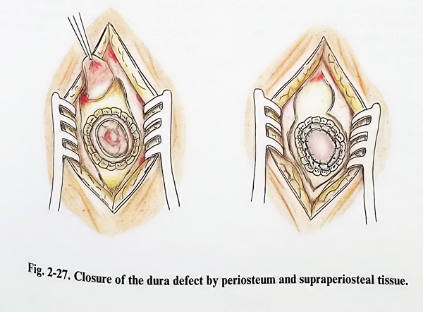
Fixed fragments or foreign bodies seen in the depth after the retraction can be removed using microinstruments and magnification. The saline stream is introduced into the canal for cleaning clots, debris and fragments. A fine, soft, elastic tube with a blunt tip or catheter can be inserted into the canal for flushing. The flushing fluid at the end of the procedure (this step is always repeated several times) must indicate the absence of necrotic and contaminating particles being flushed out of the canal. This will also be an indication of successful haemostasis (Fig. 2-26). Hydrogen peroxide (diluted 1:2 v/v) can be used for flushing and haemostasis. If the distance between entrance and exit wounds is not large, and if it does not traverse ventricles, brain stem, basal ganglia, central and midline structures, the flushing fluid can drain through the exit wound only.Once haemostasis is assured, the surgeon can go on to closure starting with dural plastic repair. The dura is the most important barrier for penetrating secondary infection. The most suitable tissue for plastic repair is periosteum, but fascia can be obtained from the wound or elsewhere in the body for the same purposes (Fig. 2-27). Usually a patch of intact periosteum, slightly exceeding the size of the dural defect, is taken from a part of the scalp distant from the injury wound. It is fixed to the already excised edges of the dural defect by interrupted sutures. Watertightness in closure is always obligatory.
Usually the presence of a certain degree of brain oedema prevents epidural bleeding, so no duro-periosteal sutures are required. The scalp is then closed in the standard manner.
RUPTURE OF DURAL SINUSES
The special attention that is given to a dural sinus rupture is due to the importance of fast and efficient control of the massive bleeding it usually provokes. It leads to rapid blood loss and proper neurosurgical management is essential for preserving the patient's life and reducing subsequent complications.
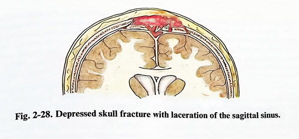
Dural sinus rupture occurs either as a result of a head injury (by the penetrative agent or bone fragment) or as an intraoperative complication, when manipulating lesions involving dural sinuses. If bleeding is massive at the site of the accident, it is uncontrollable and mortality is high. However, the injuring bone fragment often temporarily occludes the rupture and it can be its removal that suddenly precipitates the massive bleeding. In fractures and penetrative injuries over the external projection of the major sinuses (sagittal and lateral), surgical repair can be always complicated by venous sinus rupture, requiring skilful repair (Fig. 2-28).
There are some general rules in dural sinus repairs. They aim to stop any bleeding that may occur after already existing tamponade and to decrease intracranial venous pressure by different means, mainly by lifting the patient's head higher than the right atrium and avoiding venous compression on the neck. If the case is one of a head injury, wound revision should be undertaken after all surgical technical means and instrumentation are available for proper handling of this condition. The patient's condition after the injury must be stabilised, and endotracheal intubation with general anaesthesia is required.
Wounds in head injuries are revised in a way not to touch or remove the occluding fragment or injuring agent until the last movement, when sufficient space (and the dura surrounding the rupture) has been exposed. Almost always this will require additional skin incisions and bone removal on both sides of the sinus. Once the sinus rupture is identified, then a decision is required on the existing options to control it. These options depend on the location and size of the tear.
The location determines the possibility for partial or complete occlusion of the sinus blood flow for the efficient control of bleeding. The sinus is occluded if no other option exists and only in those areas and segments of the sinuses, where it will not lead to severe congestion, incompatible with normal function. It is a rule not to occlude sagittal, lateral and sigmoid sinuses (at times except for the anterior third of the sagittal). Most often ruptures of sinuses in head injuries affect those located superficially on the convexity of the skull - the sagittal and the lateral. In these cases it is important to know which of the walls of the sinus are affected. Tears on the exyernal walls are controlled more easily, but access to the other walls requires some brain retraction.
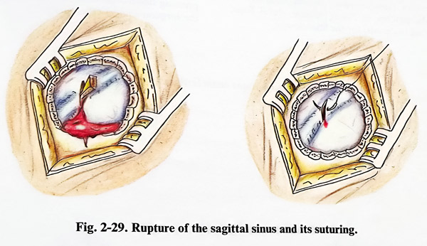

On the size of the tear depend the chances to close the defect of the sinus wall only by suture. Regrettably this can be achieved only in small and linear tears (Fig. 2-29). Suturing of wider tears narrows the sinus. When there is a hole on one of the walls, a tissue patch or flap is needed for closure.
Before removing the temporary occlusion, the surgeon should have a clear idea of what has to be done. It should be decided which method will be used and then prepare in advance everything necessary (tamponades, topical haemostatic agents, patches of fascia, muscle, dural flaps, suturing materials) for the final closure.
The patient's positioning includes the general rules of management with special attention paid to head and neck elevation avoiding any twisting of the neck. The site of the rupture should preferably the highest area of the head (for better access and lower venous pressure).
The site of the rupture is exposed surgically in the already mentioned way after which the inserted into the sinus fragment is removed and the massive venous blood stream is stopped immediately placing a piece of gelatine sponge, oxidised cellulose or muscle over the tear, and pressing down with an instrument or even with the surgeon's finger (Figs. 2-30).
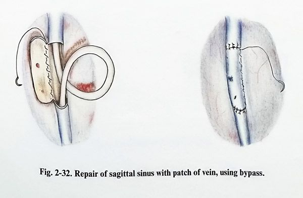
The suture of the rupture is better done with 4/0 to 6/0 silk or synthetic (but not mono-filament) mounted on an atraumatic round needle, starting it slightly inside the normal wall of the sinus. Suturing progresses with a delicate push of the tamponade behind and uncovering the edges of the tear, helping with the suction. The suture is completed and tied also after reaching normal sinus wall at the other end of the tear. If a small leak persists, it is covered by oxidised cellulose.
Repair of sinus wall defects starts with a tamponade, using a piece of muscle or oxidised cellulose. If the affected wall is the external one, the tamponade can be fixed with a patch of fascia sutured to adjacent dura. This can be achieved also by turning a dural flap over the sinus tamponade and fixing it with sutures on the opposite side of the sinus (Fig. 2-31). Sometimes only the external dural layer is used as a flap. A simple defect on the lateral wall is also tamponed and later covered by a flap fixed to the falx below the sinus.
However, a more complex defect of the sinus walls requires more sophisticated closure with sutures and tamponades, covered by patches and flaps, sometimes needing temporary interruption of the flow. If sinus reconstruction has to be done two options exist: using the wall of the disrupted sinus or using a saphenous venous graft harvested from the lower limb (do not forget venous valves - they should not be present in the graft). Sinus reconstruction in segments not permitting occlusion requires temporary bypassing Fig. 2-32).
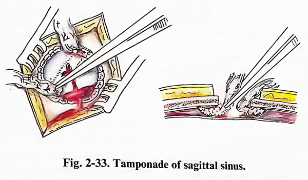
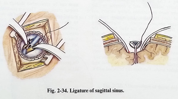
Sinus ligation is the only and last option in complete destruction of the sinus and impossible repair. It is tolerated only in the anterior part of the sagittal sinus. The procedure is simple (the suture passes through dura, falx or tentorium, surrounding the sinus), and the consequences are usually uncertain and irreversible (Figs. 2-33; 2-34).
CSF FISTULA
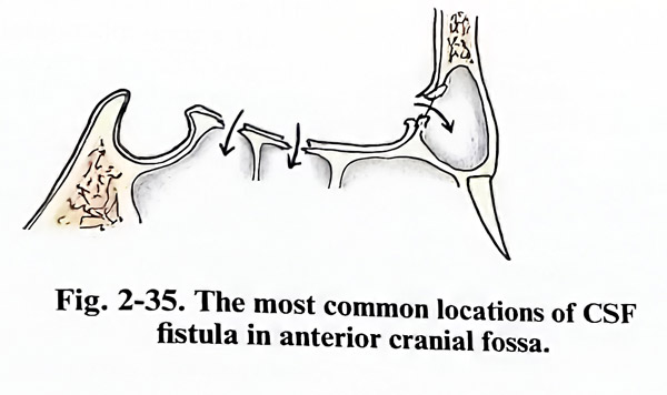
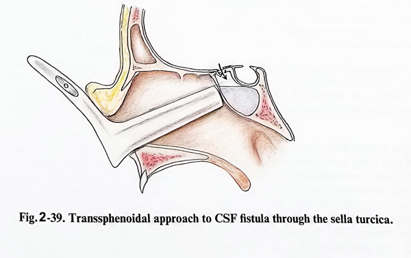
Every CSF fistula originates at a defect of the skull base with torn arachnoid at this defect. It can be either traumatic or spontaneous. The leak of CSF follows a path from the torn arachnoid and skull base to the aerated cavities in the cranial bones - frontal, ethmoid, sphenoid or petro-mastoid. The clinical manifestation of rhino- or otorrhoea can indicate the location of the fistula. However extremely precise imaging work-up of the case should clearly show the fistula as a skull base defect to serve as a planning basis for surgery. The usual locations of traumatic fistulae are through the posterior walls of the frontal sinuses, the cribriform palate to the ethmoid, the sella to the sphenoid and the superior, rarely posterior wall of the petrous bone to tympanic and mastoid cavities. Spontaneous or post-traumatic CSF leaks differ slightly, having more specific locations around the sphenoid sinus and the cribriform palate. The approaches used at present depend on all these locations. They are transcranial to the skull base or transbasal, and the most common of the last is the transsphenoidal (Fig. 2-35).
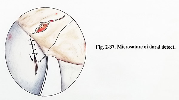
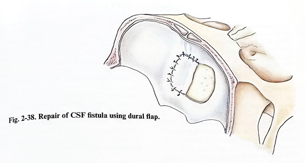
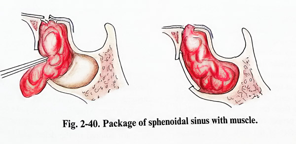
There are several common principles in closing a CSF fistula. The closure should be absolutely watertight and a decrease in CSF pressure must be obtained during surgery and several days thereafter. Its location must be absolutely precise, especially in relatively long linear fractures through the skull base. At the place of the fistula the arachnoid is clearly and firmly adherent to the dura and penetrates into the defect. The defects (according to their location and depth) can be exposed either only extradurally or both: extra- and intradurally. The fistula needs to be repaired at dural and bone defect levels. The dura can be closed with a microsuture and/or patched and also glued, preferably both. When an intradural approach is undertaken, the dural defect frequently needs enlargement for better closure of the underlaying bone defect, and later on it requires plastic closure. The bone defect is gently curreted from granulation tissue, then filled with suitable bone particles, fixed as wedges and overpacked with muscle pieces. Additional glue application can secure against unwanted displacement of the bone and muscle pieces. In recent years acrylic glues have been replaced by fibrin based products. They are better tolerated by adjacent tissues.
Lowering CSF pressure during surgery makes the closure easier, as the operative field remains dry. This is simply obtained by elevating the head's position. It is common to apply measures for lowering CSF pressure and antibiotic prophylaxis.
Craniotomies approach the area in the standard and most convenient way; there must be appropriate access to the skull base area of interest and this is ensured by placing the basal burr hole very precisely.
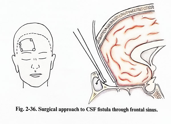
Fistulas communicating with the frontal sinus can be approached through an unilateral frontal craniotomy. They are practically always located on the posterior wall of the sinus. After an epicranial flap complying with cosmetic requirements (incision in the hairy skin) a small craniotomy is done just superior to the affected frontal sinus, but not opening its cavity (Fig. 2-36). The distance from the craniotomy edge to the fistula is usually relatively short (1-2 cm) and the site is reached extradurally. The defect in the dura and the posterior sinus wall are identified and closed in the previously mentioned way.
Wider and bifrontal approaches are preferred in fistulas located in the midline area of the anterior cranial fossa (cribriform and sellar areas, as manipulation on both sides of the midline is required. Therefore scalp incisions are bicoronal (anterior hairline) and bone flaps bifrontal. Unless the fistula is very near to the edge of the craniotomy, intradural repair is recommended because of the many tears provoked in the cribriform area by the extradural approach (detachment of dura from skull base). Falx and superior sagittal sinus rarely require transsection (Fig. 2-38).
The area of the fistula is identified. If it is in the area of the cribriform plate, there are more difficulties, and additional care must be taken to preserve olfactory bulbs from unnecessary manipulation. Otherwise the fistula is localized by the observation of unusual arachnoid adherences to a particular place on the dura, usually related to skull base defect. The olfactory bulbs can be sacrificed if that is needed for the efficient closure of the fistula.
The sellar floor is more difficult to approach, and if a transsphenoidal approach has not been chosen, it can be packed with muscle under the chiasma and glued. In all other locations bone and dural defects are closed in the standard way, and dural cover is easier by reflecting a flap of intact dura from the unaffected surroundings.
CSF fistulas through the sella are easily approached by the transsphenoidal route (Figs. 2-39; 2-40).
Fistulas between the middle cranial fossa and tympanic cavity are exposed by temporobasal craniotomies. The fistula is reached after temporal lobe retraction through an intradural approach and closed in the same way as in other locations.
Approach is different if the communication is between the posterior cranial fossa and the tympanic or mastoid cavities. The area can be approached either transtentorially or by a lateral suboccipital craniectomy. Closure is done also in the common way.
CRANIOPLASTY
Cranioplasty is the treatment of choice for replacement and repair of the cranial bones when they have been irreversibly damaged and removed. It should provide as much as possible the natural qualities of skull bone. These essential qualities present certain requirements for the plastic material in use which are mainly related to the biomechanical properties of the material, its tissue compatibility and its plasticity to satisfy the cosmetic needs. Many materials for cranioplasty have been created and used, but none of them has fully survived the test of time, except the autologous bone graft. Other cranioplastic materials are used when the autologous bone graft is not suitable due to considerations related to a particular case. Most often bone is substituted by metals and organic plastics. Cranioplasty can be primary (at the time of treating the skull lesion) or secondary when an already existing defect is repaired after complete healing of the scalp wound.
Cranioplasty is done under general anaesthesia. The general principles of surgical technique include the manner of exposure of the cranial defect and the preparation of the plastic material.
The surgical field is prepared and draped, but in such a way as to allow proper evaluation of the resulting cosmetic effect by the surgeon. This requires that wider areas be left uncovered by towels and protected only by sterile adhesive films (widely available commercially). Exposure of the defect is done with great attention to the epicranial tissues, preserving their healing properties as much as possible. All preexisting incision scars are used again for exposure; haemostasis should not impair the vascular supply. The periosteum is best preserved and sutured later as a separate layer 1-2 cm outside the margins of the cranial defect. We recommend the use of binocular loupes at least as magnification when separating tissues. Major care is taken at separation of scarring tissues from the dura, as its integrity is crucial for the proper cranial repair further on.
Every small defect of the dura, if accidentally provoked, should be closed meticulously. At fixation of the plastic material and closure of the wound, the layers should not be left separated, as also not allowing the collection of fluids close to the cranioplasty. The plastic material is fenestrated to allow drainage of fluids out of the epidural space. Postoperative wound care is also dedicated to the avoidance of this complication.
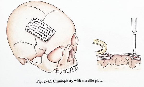
Wound suction drains are left in the epidural space only if the surgeon is strongly convinced of their need, as there is a serious danger that they might offer a pathway for postoperative infection. The same concern relates to the suturing of the layers, usually at two or three levels; the stitches should not be positioned near to the plastic material (Figs. 2-41; 2-42).
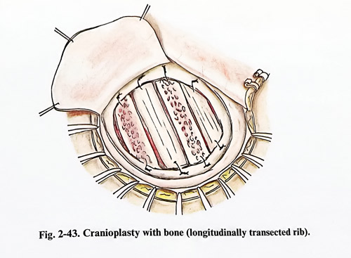
Bone as plastic material can be obtained from iliac crests, ribs, fibular and tibial bone shafts, or neighbouring to the defective one cranial bones of the patient. Bone has the best tissue compatibility properties; its subsequent ossification recovers the biomechanical properties of the skull. However, many difficulties are faced in attempting to reshape it for results satisfactory from an aesthetic point of view.
Difficulties arise from the need of a bone graft to be shaped as a segment of a sphere (for defects of the convexity), and they are more when defects are near the skull base or facial bones have to be repaired. Sometimes the required shape is achieved by using more than one bone fragment; when the defect is filled and the shape of the corresponding part of the skull regained, fragments are fixed by sutures or wiring to the refreshed edges of the defect. This refreshment (very gentle nibbling to obtain bleeding cancellous bone) as well as the subsequent cover with preserved periosteum (of optimal size and vitality) improve osteogenesis. Then closure of the galea and skin takes place (Figs. 2-43; 2-44)
The other common technique of cranial defect repair is alloplasty. Two materials are currently employed: metals and bone cement on an acrylic or other type base. Metals - preferably like titanium or other alloys with low paramagnetic properties - are tailored to the defect and fixed with small conical screws to the surrounding normal bone. Thin, standard prefabricated plates and meshes can also be used, as they can be assembled to properly cover the defect or assemble the bone fragments used for the closure.
Assembling small elements, especially when they are malleable, gives better cosmetic results. No additional wound closure technique and postoperative wound care are needed.
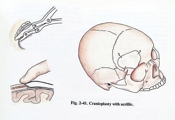
Plastics and bone cement are easiest to use and to shape; their disadvantage lies in problems associated with tissue compatibility. The material is usually composed of two components - base and hardener; they are mixed in the advised proportions. After mixing, the substance slowly hardens; before this occurs, it must be modelled to the desired shape. When the optimal fit for the defect has been achieved, it is fixed with sutures or wires to the defect edges, and the wound can then be closed.
Read PREVIOUS: 1. General Surgical Techniques in Cranial and Intracranial Lesions
Read NEXT: 3. Head Injury - chapter 1