TRAUMATIC MASS LESIONS
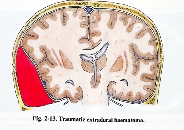
Extradural, subdural or intracerebral collections, especially those with rapid increase of volume, can easily lead to raising and decompensation of the ICP with all the notorious consequences of brain compression, shifts and herniation. Therefore, these collections need early (usually urgent) evacuation. Usually it is the case of blood collection, but sometimes CSF accumulated in the subdural space can have the effect of a mass lesion. Localised brain contusion with perifocal oedema provoking a mass effect can also need surgical evacuation (Fig. 2-13).
ACUTE EXTRADURAL AND SUBDURAL НАЕМАТОМА
The goals of surgical treatment are to decrease intracranial pressure by removing the haematoma as early as possible, especially in patients with an impaired level of consciousness, and to prevent rebleeding.
The approach to the haematoma depends on its size, location and the available facilities when dealing with it as an emergency.
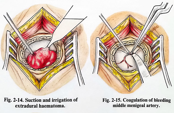
In case of a small acute extradural and subdural haematoma, especially one located in the temporal region, evacuation can be done with a limited craniectomy. It is also an acceptable approach in extreme emergencies when the surgical facilities available are limited. In such a case the operative procedure begins with a vertical incision of the scalp 5 - 6 cm long over the area of the haematoma. As most often haematomas are located in the temporal region, the incision begins from the zygomatic arch projecting quite vertically. The temporal muscle is split along its fascicles, the periosteum is separated, and a burr hole is opened. The hole is enlarged by nibbling to a craniectomy of 3-4 cm. In case of an extradural haematoma a clot of dark blood extrudes through the craniectomy. The clots should be removed with saline jet irrigation and gentle suction (Fig. 2-14). More aggressive suction usually provokes additional bleeding. A saline wash is also used to detect persistent bleeding. Such bleeding is controlled by bipolar coagulation if the dural bleeding point is accessible, or by topical haemostatic agents (oxidised cellulose or other available substances) and stitches to fix the dura to the periosteum at the edge of the craniectomy. In cases of major arterial bleeding from basal dura or fractures injuring the middle meningeal artery, the craniectomy can be extended more towards the skull base and the dura retracted. This can permit better control of the middle meningeal artery, particularly when the injury is close to its intracranial entrance (Fig. 2-15). This situation may require packing of the foramen spinosum with bone wax or bone wax mixed with cotton fibres to control haemorrhage. If the CT does not show any subdural collection or brain contusion, the dura should be left intact.
In case of an acute subdural haematoma, the craniectomy will expose a bluish and tense dura. Its opening precipitates the extrusion of dark clots. These clots are removed by gentle suction or traction with cup forceps and saline stream irrigation. Bleeding points on the cerebral surface should be coagulated with bipolar coagulation. If coagulation is ineffective, topical haemostatic agents can control the bleeding.
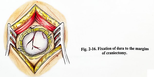
The subdural space and cortical surface are carefully inspected for deep haematomas, contusions, or bleeding. A very small amount of clot that is not easily accessible through the craniectomy is best not removed, as it can provoke unnecessary bleeding that is difficult to control. After bleeding has been controlled, the dura is closed in a watertight fashion and tacked up to the margins of the craniectomy (Fig. 2-16).
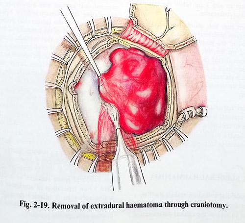
Craniotomy gives much greater access for removal of acute extradural and subdural haematomas. The size and location of the craniotomy should ideally cover the whole area of the haematoma and the epicranial flap should be planned accordingly. This facilitates an approach to the most peripheral part of the haematoma. Extradural haematomas are managed with less blood loss and safer haemostasis due to better exposure of the dura. If the dura must be opened, then it can be done with better visibility and control of manipulation. In subdural haematomas the removal of clots and haemostasis is also easier and safer. The underlying brain can be carefully inspected in all the area covered by the haematoma, and the detection of the bleeder is easier, whether it is from a brain laceration, rupture of a vein draining into the sagittal sinus, petrous sinus or sylvian veins.
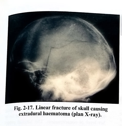
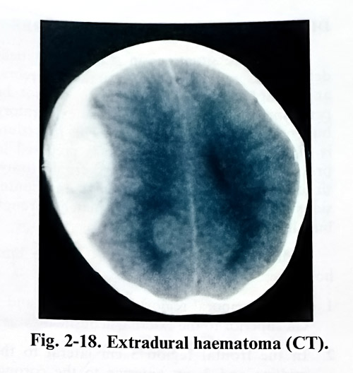
Occasionally a ruptured cortical artery can cause the haematoma. Hence, because of its advantages therefore, whenever it is possible, a craniotomy must be preferred over a limited craniectomy (Figs. 2- 17; 2-18; 2-19).
DIAGNOSTIC SURGICAL PROCEDURES
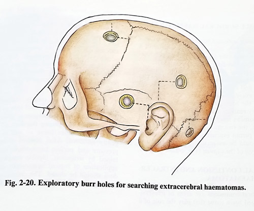
There are situations in which the patient deteriorates rapidly and CT, or even cerebral angiography are not available or cannot be performed in time. In such a case exploratory burr holes are the better option. This procedure is of value only if the surgeon is prepared to proceed with a craniotomy or craniectomy, since acute traumatic haematomas and contusions cannot be dealt with adequately through burr holes only (Fig. 2-20).
The most convenient places for burrholes for hematoma detection are:
1 In the temporal region 3 cm anterior and 3 cm superior to the external acoustic meatus.
2 In the frontal region 3 cm lateral to themidline and 3 cm anterior to the coronal suture. This suture coincides with the line which connects the midpoints of the zygomatic arches and is perpendicular to them.
3 In the parietal region 3 cm superior to and 3 cm posterior to the tip of the auricle.
4 When a haematoma is suspected in the posterior cranial fossa, a burr hole is made 3 cm lateral to and 3 cm below the external occipital protuberance.
The first burr hole should be made in the temporal region ipsilateral to the dilated pupil, contralateral to the most abnormal motor response, and on the side of a fracture. The scalp incisions should be placed to permit their incorporation into a formal craniotomy if a haematoma is encountered. When the procedure is completed on one side, it should be repeated on the other.
Another important procedure, considered routine at present, is invasive ICP monitoring.
Its increase to critical levels in the head-injured patient indicates a need to take corresponding measures to decrease it again.
CHRONIC SUBDURAL HAEMATOMA
This subdural collection reveals itself by a mass effect and its prompt evacuation is the only option to reverse the clinical deterioration of the patient. Once the patient is showing symptoms and signs of increased intracranial pressure or deterioration of the focal deficit, surgery should not be delayed. The evacuation of a chronic subdural haematoma is planned by deciding the place of one or two burr holes, as it is usually fluid. They should be over the thickest part of the haematoma, but the overlying incision, a straight one, should be in hairy skin. The burr holes and skin incisions should permit incorporation into a craniotomy for a wider approach. If there are bilateral haematomas, the larger should be evacuated first, but both sides should be done in a single operative session.
General or local anaesthesia is used according to the general condition of the patient and his or her level of consciousness. The surgical procedure is relatively simple but close attention must be paid to these patients, especially the elderly, because of other significant medical problems.
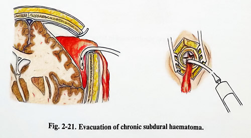
With the patient in the usual supine position, the preplanned burr hole(s) should be made on the affected side. After the dura is exposed, it is opened with a cross-like incision, coagulating the place of incision beforehand.
Depending upon the intracranial pressure, the dark brown liquid of the haematoma may burst through the opening that is made. Sometimes a thick brown, black, or even greenish membrane below the dura must also be opened to reach the fluid part. The haematoma cavity should be irrigated with warm saline until the subdural space yields only clear fluid (Fig. 2-21). In big haematomas sometimes complete evacuation is achieved through a second or more burr holes. The depth of the subdural space must be inspected and its reduction should be attempted by lowering of the upper half of the table so that the patient remains positioned for a while with the head slightly-down. This manoeuvre increases the intracranial venous pressure, therefore also the ICP. If the cerebral cortex comes up to the dura, the burr holes are closed without drainage. If the subdural space remains as a cavity more than 5 mm separation of arachnoid from dura, a fine soft tube is inserted through the more posterior burr hole and is connected to a bag at normal atmospheric pressure.
INFANTILE SUBDURAL HAEMATOMA
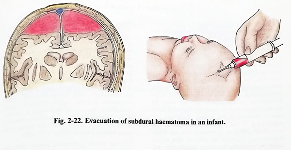
Infantile subdural haematomas are usually bilateral and very extensive. The existence of a fontanelle allows the puncturing of the subdural space with a needle, which is inserted in the lateral angle of the fontanelle and directed laterally tangential to the cranial vault. The puncture and evacuation of the haematoma are repeated every second or third day, until the cavity of the haematoma disappears. If a residual cavity remains (the cerebral mantle doesn't show expansion towards the dura) after several punctures, the remaining option is craniotomy. This will permit evacuation of the haematoma which should be associated with fenestration and removal of the capsule (Fig. 2-22).
CEREBRAL CONTUSION AND INTRACEREBRAL HAEMATOMA
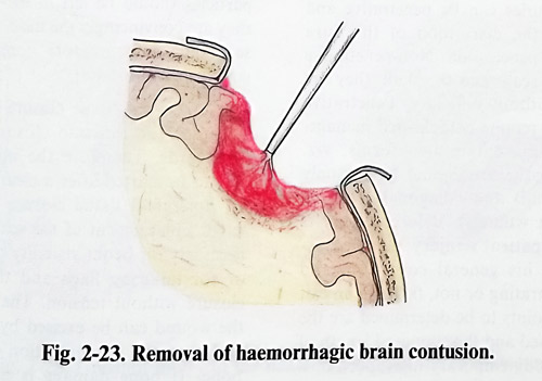
Cerebral contusions are areas of irreversibly damaged brain tissue that induce and present with brain swelling around them. Quite often they are associated with intrinsic haemorrhages (i.e. haemorrhagic contusions, "burst lobe" syndrome). When such cerebral contusion acts as a mass lesion, the patient's condition is affected not only by the contusion, but also by additional surrounding brain compression. Temporal lobes are more affected than are the frontal with this condition. Therefore, early resection of this nidus prevents further compression, shift and herniation.
The craniotomy (rarely craniectomy) is planned to expose the affected area or lobe, in a way similar to the techniques in gliomas. After craniotomy and opening the dura all lacerated and necrotic brain tissue as well as clots are gently removed. After control of intracranial pressure and perfect haemostasis (usually obtained by irrigation, bipolar coagulation, and application of topical haemostatic agents), the dura should be closed. The craniotomy should be closed in the routine way (Fig. 2-23).