
CRANIOPLASTY
Cranioplasty is the treatment of choice for replacement and repair of the cranial bones when they have been irreversibly damaged and removed. It should provide as much as possible the natural qualities of skull bone. These essential qualities present certain requirements for the plastic material in use which are mainly related to the biomechanical properties of the material, its tissue compatibility and its plasticity to satisfy the cosmetic needs. Many materials for cranioplasty have been created and used, but none of them has fully survived the test of time, except the autologous bone graft. Other cranioplastic materials are used when the autologous bone graft is not suitable due to considerations related to a particular case. Most often bone is substituted by metals and organic plastics. Cranioplasty can be primary (at the time of treating the skull lesion) or secondary when an already existing defect is repaired after complete healing of the scalp wound.
Cranioplasty is done under general anaesthesia. The general principles of surgical technique include the manner of exposure of the cranial defect and the preparation of the plastic material.
The surgical field is prepared and draped, but in such a way as to allow proper evaluation of the resulting cosmetic effect by the surgeon. This requires that wider areas be left uncovered by towels and protected only by sterile adhesive films (widely available commercially). Exposure of the defect is done with great attention to the epicranial tissues, preserving their healing properties as much as possible. All preexisting incision scars are used again for exposure; haemostasis should not impair the vascular supply. The periosteum is best preserved and sutured later as a separate layer 1-2 cm outside the margins of the cranial defect. We recommend the use of binocular loupes at least as magnification when separating tissues. Major care is taken at separation of scarring tissues from the dura, as its integrity is crucial for the proper cranial repair further on.
Every small defect of the dura, if accidentally provoked, should be closed meticulously. At fixation of the plastic material and closure of the wound, the layers should not be left separated, as also not allowing the collection of fluids close to the cranioplasty. The plastic material is fenestrated to allow drainage of fluids out of the epidural space. Postoperative wound care is also dedicated to the avoidance of this complication.
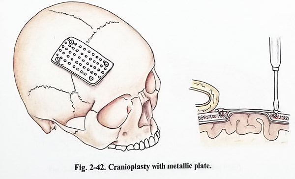
Wound suction drains are left in the epidural space only if the surgeon is strongly convinced of their need, as there is a serious danger that they might offer a pathway for postoperative infection. The same concern relates to the suturing of the layers, usually at two or three levels; the stitches should not be positioned near to the plastic material (Figs. 2-41; 2-42).
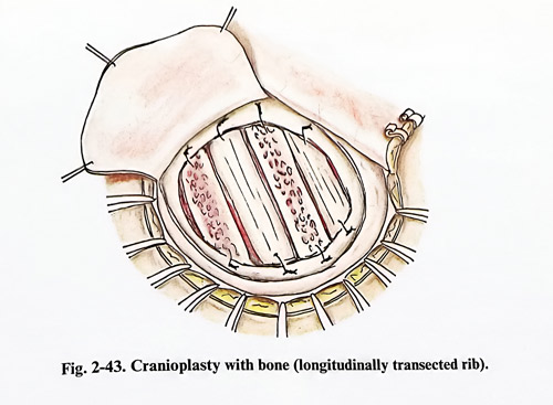
Bone as plastic material can be obtained from iliac crests, ribs, fibular and tibial bone shafts, or neighbouring to the defective one cranial bones of the patient. Bone has the best tissue compatibility properties; its subsequent ossification recovers the biomechanical properties of the skull. However, many difficulties are faced in attempting to reshape it for results satisfactory from an aesthetic point of view.
Difficulties arise from the need of a bone graft to be shaped as a segment of a sphere (for defects of the convexity), and they are more when defects are near the skull base or facial bones have to be repaired. Sometimes the required shape is achieved by using more than one bone fragment; when the defect is filled and the shape of the corresponding part of the skull regained, fragments are fixed by sutures or wiring to the refreshed edges of the defect. This refreshment (very gentle nibbling to obtain bleeding cancellous bone) as well as the subsequent cover with preserved periosteum (of optimal size and vitality) improve osteogenesis. Then closure of the galea and skin takes place (Figs. 2-43; 2-44)
The other common technique of cranial defect repair is alloplasty. Two materials are currently employed: metals and bone cement on an acrylic or other type base. Metals - preferably like titanium or other alloys with low paramagnetic properties - are tailored to the defect and fixed with small conical screws to the surrounding normal bone. Thin, standard prefabricated plates and meshes can also be used, as they can be assembled to properly cover the defect or assemble the bone fragments used for the closure.
Assembling small elements, especially when they are malleable, gives better cosmetic results. No additional wound closure technique and postoperative wound care are needed.
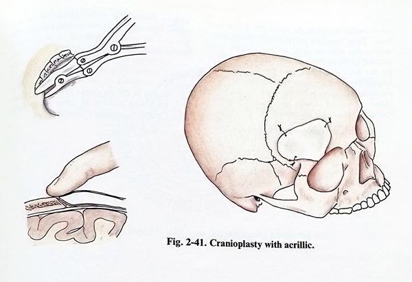
Plastics and bone cement are easiest to use and to shape; their disadvantage lies in problems associated with tissue compatibility. The material is usually composed of two components - base and hardener; they are mixed in the advised proportions. After mixing, the substance slowly hardens; before this occurs, it must be modelled to the desired shape. When the optimal fit for the defect has been achieved, it is fixed with sutures or wires to the defect edges, and the wound can then be closed.
Read PREVIOUS: 1. General Surgical Techniques in Cranial and Intracranial Lesions
Read NEXT: 3. Head Injury - chapter 1