RUPTURE OF DURAL SINUSES
The special attention that is given to a dural sinus rupture is due to the importance of fast and efficient control of the massive bleeding it usually provokes. It leads to rapid blood loss and proper neurosurgical management is essential for preserving the patient's life and reducing subsequent complications.
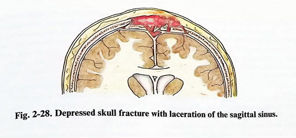
Dural sinus rupture occurs either as a result of a head injury (by the penetrative agent or bone fragment) or as an intraoperative complication, when manipulating lesions involving dural sinuses. If bleeding is massive at the site of the accident, it is uncontrollable and mortality is high. However, the injuring bone fragment often temporarily occludes the rupture and it can be its removal that suddenly precipitates the massive bleeding. In fractures and penetrative injuries over the external projection of the major sinuses (sagittal and lateral), surgical repair can be always complicated by venous sinus rupture, requiring skilful repair (Fig. 2-28).
There are some general rules in dural sinus repairs. They aim to stop any bleeding that may occur after already existing tamponade and to decrease intracranial venous pressure by different means, mainly by lifting the patient's head higher than the right atrium and avoiding venous compression on the neck. If the case is one of a head injury, wound revision should be undertaken after all surgical technical means and instrumentation are available for proper handling of this condition. The patient's condition after the injury must be stabilised, and endotracheal intubation with general anaesthesia is required.
Wounds in head injuries are revised in a way not to touch or remove the occluding fragment or injuring agent until the last movement, when sufficient space (and the dura surrounding the rupture) has been exposed. Almost always this will require additional skin incisions and bone removal on both sides of the sinus. Once the sinus rupture is identified, then a decision is required on the existing options to control it. These options depend on the location and size of the tear.
The location determines the possibility for partial or complete occlusion of the sinus blood flow for the efficient control of bleeding. The sinus is occluded if no other option exists and only in those areas and segments of the sinuses, where it will not lead to severe congestion, incompatible with normal function. It is a rule not to occlude sagittal, lateral and sigmoid sinuses (at times except for the anterior third of the sagittal). Most often ruptures of sinuses in head injuries affect those located superficially on the convexity of the skull - the sagittal and the lateral. In these cases it is important to know which of the walls of the sinus are affected. Tears on the exyernal walls are controlled more easily, but access to the other walls requires some brain retraction.
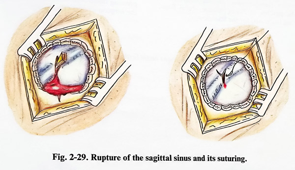

On the size of the tear depend the chances to close the defect of the sinus wall only by suture. Regrettably this can be achieved only in small and linear tears (Fig. 2-29). Suturing of wider tears narrows the sinus. When there is a hole on one of the walls, a tissue patch or flap is needed for closure.
Before removing the temporary occlusion, the surgeon should have a clear idea of what has to be done. It should be decided which method will be used and then prepare in advance everything necessary (tamponades, topical haemostatic agents, patches of fascia, muscle, dural flaps, suturing materials) for the final closure.
The patient's positioning includes the general rules of management with special attention paid to head and neck elevation avoiding any twisting of the neck. The site of the rupture should preferably the highest area of the head (for better access and lower venous pressure).
The site of the rupture is exposed surgically in the already mentioned way after which the inserted into the sinus fragment is removed and the massive venous blood stream is stopped immediately placing a piece of gelatine sponge, oxidised cellulose or muscle over the tear, and pressing down with an instrument or even with the surgeon's finger (Figs. 2-30).
The suture of the rupture is better done with 4/0 to 6/0 silk or synthetic (but not mono-filament) mounted on an atraumatic round needle, starting it slightly inside the normal wall of the sinus. Suturing progresses with a delicate push of the tamponade behind and uncovering the edges of the tear, helping with the suction. The suture is completed and tied also after reaching normal sinus wall at the other end of the tear. If a small leak persists, it is covered by oxidised cellulose.
Repair of sinus wall defects starts with a tamponade, using a piece of muscle or oxidised cellulose. If the affected wall is the external one, the tamponade can be fixed with a patch of fascia sutured to adjacent dura. This can be achieved also by turning a dural flap over the sinus tamponade and fixing it with sutures on the opposite side of the sinus (Fig. 2-31). Sometimes only the external dural layer is used as a flap. A simple defect on the lateral wall is also tamponed and later covered by a flap fixed to the falx below the sinus.
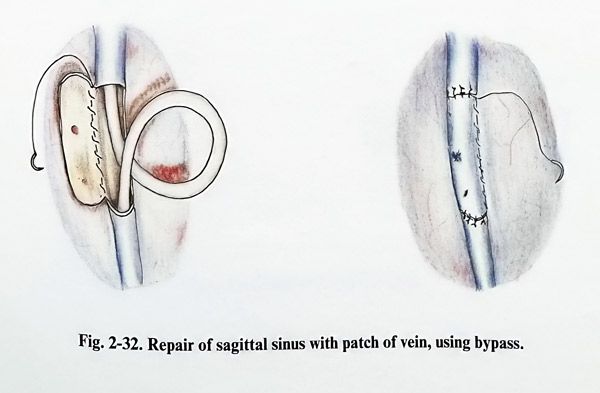
However, a more complex defect of the sinus walls requires more sophisticated closure with sutures and tamponades, covered by patches and flaps, sometimes needing temporary interruption of the flow. If sinus reconstruction has to be done two options exist: using the wall of the disrupted sinus or using a saphenous venous graft harvested from the lower limb (do not forget venous valves - they should not be present in the graft). Sinus reconstruction in segments not permitting occlusion requires temporary bypassing Fig. 2-32).
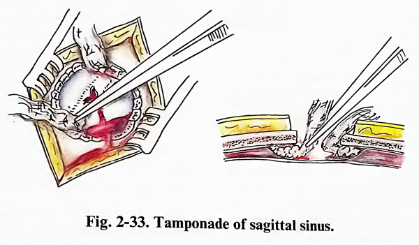
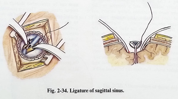
Sinus ligation is the only and last option in complete destruction of the sinus and impossible repair. It is tolerated only in the anterior part of the sagittal sinus. The procedure is simple (the suture passes through dura, falx or tentorium, surrounding the sinus), and the consequences are usually uncertain and irreversible (Figs. 2-33; 2-34).
CSF FISTULA
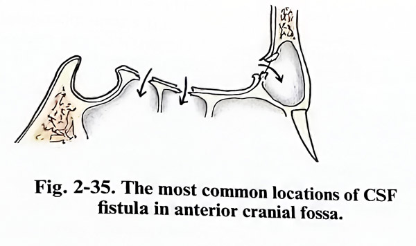
Every CSF fistula originates at a defect of the skull base with torn arachnoid at this defect. It can be either traumatic or spontaneous. The leak of CSF follows a path from the torn arachnoid and skull base to the aerated cavities in the cranial bones - frontal, ethmoid, sphenoid or petro-mastoid. The clinical manifestation of rhino- or otorrhoea can indicate the location of the fistula. However extremely precise imaging work-up of the case should clearly show the fistula as a skull base defect to serve as a planning basis for surgery. The usual locations of traumatic fistulae are through the posterior walls of the frontal sinuses, the cribriform palate to the ethmoid, the sella to the sphenoid and the superior, rarely posterior wall of the petrous bone to tympanic and mastoid cavities. Spontaneous or post-traumatic CSF leaks differ slightly, having more specific locations around the sphenoid sinus and the cribriform palate. The approaches used at present depend on all these locations. They are transcranial to the skull base or transbasal, and the most common of the last is the transsphenoidal (Fig. 2-35).
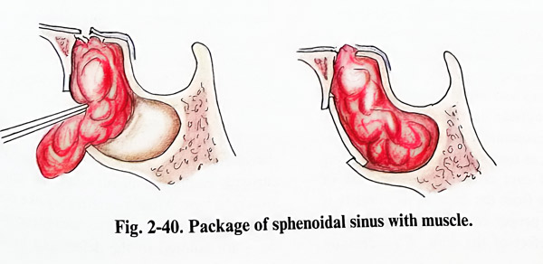
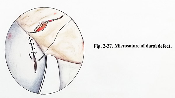
There are several common principles in closing a CSF fistula. The closure should be absolutely watertight and a decrease in CSF pressure must be obtained during surgery and several days thereafter. Its location must be absolutely precise, especially in relatively long linear fractures through the skull base. At the place of the fistula the arachnoid is clearly and firmly adherent to the dura and penetrates into the defect. The defects (according to their location and depth) can be exposed either only extradurally or both: extra- and intradurally. The fistula needs to be repaired at dural and bone defect levels. The dura can be closed with a microsuture and/or patched and also glued, preferably both. When an intradural approach is undertaken, the dural defect frequently needs enlargement for better closure of the underlaying bone defect, and later on it requires plastic closure. The bone defect is gently curreted from granulation tissue, then filled with suitable bone particles, fixed as wedges and overpacked with muscle pieces. Additional glue application can secure against unwanted displacement of the bone and muscle pieces. In recent years acrylic glues have been replaced by fibrin based products. They are better tolerated by adjacent tissues.
Lowering CSF pressure during surgery makes the closure easier, as the operative field remains dry. This is simply obtained by elevating the head's position. It is common to apply measures for lowering CSF pressure and antibiotic prophylaxis.
Craniotomies approach the area in the standard and most convenient way; there must be appropriate access to the skull base area of interest and this is ensured by placing the basal burr hole very precisely.
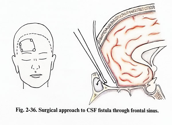
Fistulas communicating with the frontal sinus can be approached through an unilateral frontal craniotomy. They are practically always located on the posterior wall of the sinus. After an epicranial flap complying with cosmetic requirements (incision in the hairy skin) a small craniotomy is done just superior to the affected frontal sinus, but not opening its cavity (Fig. 2-36). The distance from the craniotomy edge to the fistula is usually relatively short (1-2 cm) and the site is reached extradurally. The defect in the dura and the posterior sinus wall are identified and closed in the previously mentioned way.
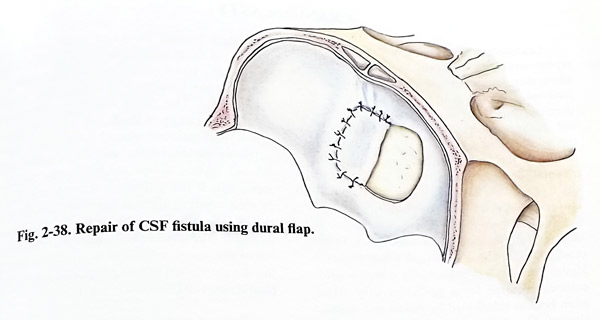
Wider and bifrontal approaches are preferred in fistulas located in the midline area of the anterior cranial fossa (cribriform and sellar areas, as manipulation on both sides of the midline is required. Therefore scalp incisions are bicoronal (anterior hairline) and bone flaps bifrontal. Unless the fistula is very near to the edge of the craniotomy, intradural repair is recommended because of the many tears provoked in the cribriform area by the extradural approach (detachment of dura from skull base). Falx and superior sagittal sinus rarely require transsection (Fig. 2-38).
The area of the fistula is identified. If it is in the area of the cribriform plate, there are more difficulties, and additional care must be taken to preserve olfactory bulbs from unnecessary manipulation. Otherwise the fistula is localized by the observation of unusual arachnoid adherences to a particular place on the dura, usually related to skull base defect. The olfactory bulbs can be sacrificed if that is needed for the efficient closure of the fistula.
The sellar floor is more difficult to approach, and if a transsphenoidal approach has not been chosen, it can be packed with muscle under the chiasma and glued. In all other locations bone and dural defects are closed in the standard way, and dural cover is easier by reflecting a flap of intact dura from the unaffected surroundings.
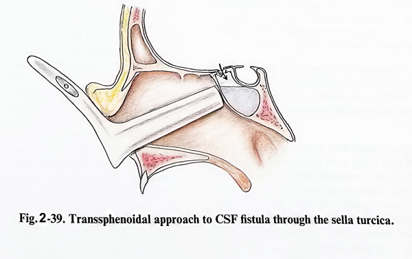
CSF fistulas through the sella are easily approached by the transsphenoidal route (Figs. 2-39; 2-40).
Fistulas between the middle cranial fossa and tympanic cavity are exposed by temporobasal craniotomies. The fistula is reached after temporal lobe retraction through an intradural approach and closed in the same way as in other locations.
Approach is different if the communication is between the posterior cranial fossa and the tympanic or mastoid cavities. The area can be approached either transtentorially or by a lateral suboccipital craniectomy. Closure is done also in the common way.