DEPRESSED SKULL FRACTURES
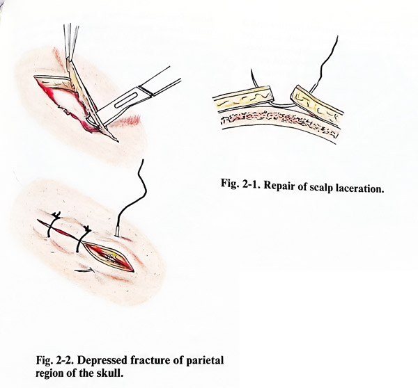
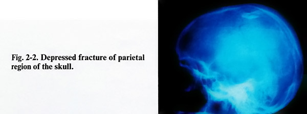
In simple skull fractures, i. e. without laceration of the scalp, surgical repair is indicated, when there is depression of bone fragments into the cranial cavity. The degree of depression of the bone fragments must be precisely determined by skull X-rays and especially by reference to a view that is tangential to the area of depression. CT images with a window visualising bone are extremely helpful to show clearly the depression. The operation should be performed as early as possible, when the patient's general condition permits it with an acceptably low risk (Fig. 2-2).
The goals of the operation are: 1. Elevation of the bone fragments, which have penetrated intracranially; 2. Evacuation of the haematomas and necrotic intracranial tissues; 3. Restoration of the anatomical integrity of the dura, bone and epicranial layers. Three methods can berecommended:
First method.
The incision of the scalp is either in the form of a horseshoe or 'S' - shaped. The skin, and the subcutaneous tissues with the galea are separated and lifted as a flap, leaving the bone covered only with the pericranium.
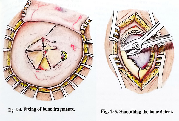
Bone fragments are seen frequently with torn pericranium over them. There is always a certain amount of haemorrhage below the periosteum, or if it is torn, even below the galea. All fragments which are small and free from periosteum are removed together with the necrotic brain tissue. Larger fragments are elevated with their pericranium, making possible an inspection of the dura and the brain. Very often some of the bone fragments cannot be easily removed, because they are indented in the cranial cavity, and firmly fixed, as the edge of the inner table remains under the surrounding normal bones. A burr hole just at the border of the fracture, but within normal bone is made. Through the burr hole, and after a small amount of nibbling, the fixed indented fragments are elevated. The elevator can be based on the edge of the intact bone and used as a lever. If the dura is intact, the bone defect is covered using the large bone fragments, fixing them to the margins of the defect by wiring (Fig. 2-4). At the end the scalp layers are closed.
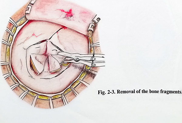
When there is a comminuted fracture and it is impossible to close the defect with the bone fragments, the latter are completely removed and the edges of the defect are smoothed with a bone nibbler. In such a case, cranioplasty can be performed at the same surgical session (Fig. 2-5)
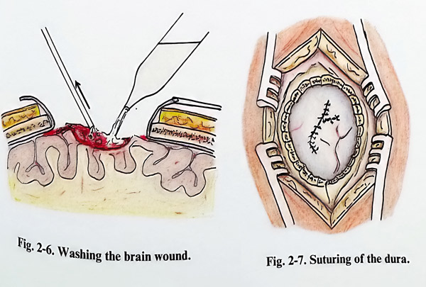
When the skull fractures are associated with tearing of the dura and brain injury, the bone fragments, which are found inside the brain tissue must be removed along with the necrotic parts. So as to inspect the cerebral surface well, the defect of the dura is widened by radial incisions. If additional necrotic tissues and clots are found, they are removed by suction and a stream of saline (Fig. 2-6; 2-7).
The surgical wound is closed in separate layers: dura, pericranium, galea and skin.
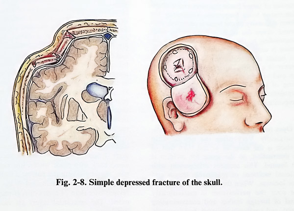
The second method uses a craniotomy. It is applied in fractures with depressed bone fragments, where access to the indented fragments is not possible without much sacrifice of adjacent normal bone in the process of drilling and nibbling it. Therefore, a craniotomy enclosing the fracture in the centre of the bone flap induces to the patient less injury to the dura and the brain and there is less definitive bone removal.
Surgery is planned by defining the places for burr holes and bone cutting lines, always in intact bone, but not too far away from the fracture (Caution: the lifting of the flap should not provoke additional fracture of bone). A suitable epicranial flap is tailored, according to the place of craniotomy (Fig. 2-8).
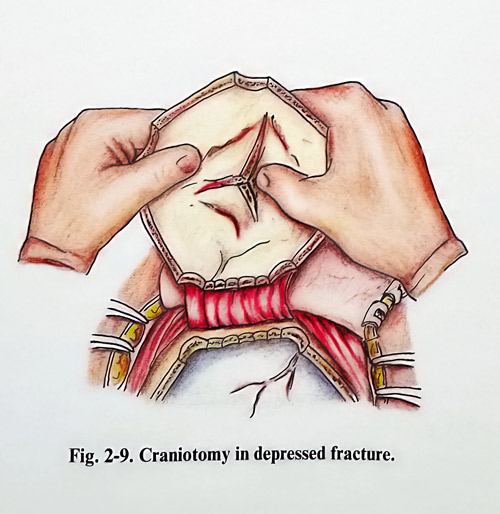
The skin incision and the epicranial flap are done in the usual way. The area of bone depression is identified. The periosteum is incised according to the planned bone flap (on a pedicle or free, and the area needed for burr holes and bone cutting is stripped. Special attention is paid to the detection of fracture lines, not seen previously on CT and X-rays, as they can change the planned bone flap. After drilling and bone cutting, the flap is lifted with much care in separating the dura from the inner table. The fragments usually remain attached to the periosteum and are lifted easily. With the flap already in hand, fragments are usually repositioned with no difficulties (Fig. 2-9). The fragments should be stable and fixed in the bone defect. However, if a defect remains, it is closed in the other routine ways. Then the craniotomy can be closed in the standard way.This method is applied less frequently than the previous one, and can be suitable for fractures in children, where bone elasticity is greater.
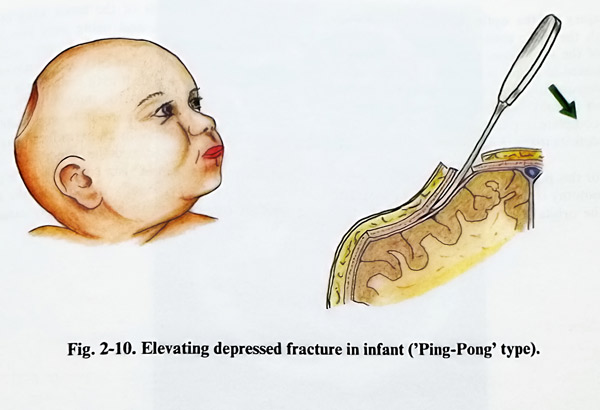
The third method is applied in depressed fractures in infants. The great elasticity of the cranial vault in infants does not allow the formation of larger linear fractures and multiple comminuted bone fragments. Ordinarily a uniform concavity of the bone is observed, with a round shape (as in a Ping-Pong ball) that can correspond to the area of impact. This depression is corrected by means of a burr hole, made beside the edge of the concavity. A fine dissector is carefully inserted in the epidural space and with a lever-like movement, repositioning of the bone is obtained. When this manipulation is applied early after the injury, the repositioning of the bone is easier (Fig. 2-10).
Open (compound) fractures should be treated surgically. Very often the size of scalp laceration is the same as or less than that of bone destruction, so the space for revision is much more limited. This requires enlargement with additional incisions of reasonable length and proper location. When the scalp is retracted, the space over the periosteum is meticulously
cleaned of foreign bodies and contaminating material, and copiously irrigated. Intracranially indented fragments are not removed before incising and stripping periosteum expose the edges of intact bone. Only when the surgeon has sufficient space to control sudden major bleeding may the fragments be removed.
Here the technique employed in closed (simple) depressed fractures can be followed. Nasal sinuses that have been exposed are stripped of mucose. If possible, a galeal flap can be pulled over the end of the exposed sinus and tacked to the dura below to seal the opening. Intact, normal-appearing dura should be left unopened; however if it appears tense or bluish in colour, it is incised and the underlying brain examined. Intracranial haematoma or necrotic brain tissue is removed. A dural laceration should be trimmed and extended with additional incisions to permit removal of foreign material, haematoma, contused brain tissue and to perform haemostasis. Dura is closed in a water-tight fashion with pericranium or fascia graft if necessary. Bone fragments can be wired back into place with a low risk of infection, thereby saving the patient a further operation. This is contraindicated if the wound is revised later than 24 hours or is grossly contaminated. The epicranial layers are closed in the usual way.
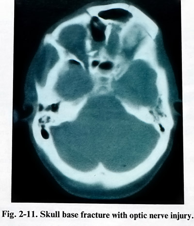
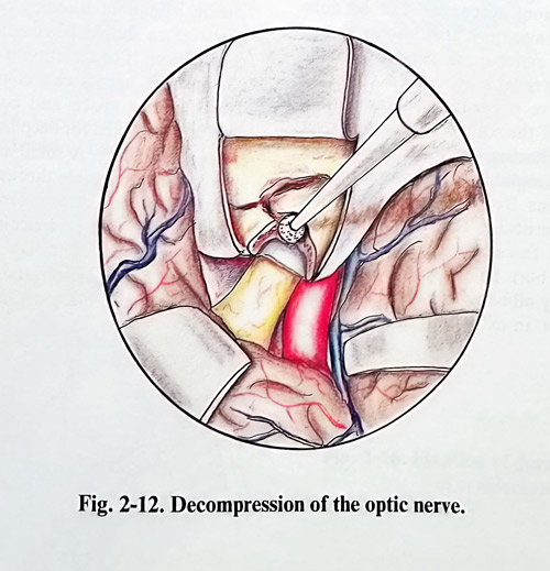
Injury of the optic nerve in its passage through the optic canal is common with fractures of the skull base. Surgical decompression is indicated if vision is present after the injury but subsequently deteriorates. Surgical decompression is unlikely to be of benefit in treating stable complete or partial visual defect or partial visual deficits that have improved.
For this purpose a frontal or frontotemporal craniotomy is made and the dura is separated from the orbital roof. With the drill the orbital roof, anterior to the lesser wing of the sphenoid is removed, and using the finest bone instruments, the upper wall of optic canal is resected. Then all fragments are removed. A high-speed diamond drill with continuous irrigation is very convenient for unroofing the optic canal. In the case, where it is impossible to relieve the optic nerve in an extradural way, the dura may be opened and the upper wall of the optic canal resected under visual control of the nerve (Figs. 2-11; 2-12). Finally the optic nerve sheet and the ring of Zinn are longitudinally divided.