GUN-SHOT INJURY
Surgical treatment of gun-shot injury aims at the preservation of life and the prevention of complications at the least consequent neurological deficit for the patient.
Gun-shot injuries are caused by different types of missiles, the most common being bullets and fragments of exploding devices (shrapnels). The injury produced by a missile can be associated with a certain degree of blast injury too.The resulting effect is a combination of direct destruction by the missile and the blast by external forces, and the sudden increase of intracranial pressure at the instant of contact and penetration of the missile intracranially. This secondary affection (superimposed on direct destruction), increases the affected area significantly by spreading the energy peripherally outward from the missile canal. The higher the velocity of the missile, the greater the secondary effects we observe. They are considered to affect by two factors: shock waves and temporary cavitation.
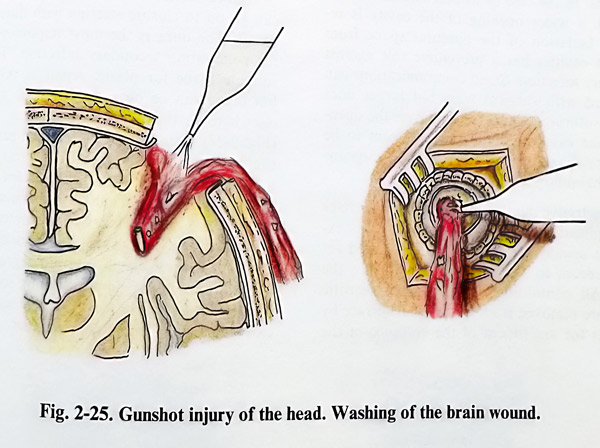
Gun-shot injuries can be penetrative and non-penetrative, the disruption of the dura being the sign of penetration. Non-penetrative injuries affect the scalp and the skull; they are often repaired without difficulty. Penetrative injuries, however, require complicated management, and they present in two forms: "blind" (retaining the missile and having only an entrance wound) and "diagonal" (having entrance and exit wounds). Before surgery is undertaken, the patient and the injury must be assessed regarding general condition, and whether it is penetrating or not, i.e. is there an exit wound. Other points to be determined are the type of weapon used and the supposed length of the trajectory. A contemporary high-speed missile (from conventional weapons) provokes severe generalized brain damage. Therefore, in consideration of the severe generalised damage and the need of early prevention of complications, surgical treatment should be considered quite always as an emergency. In patients requiring resuscitation or with unstable vital signs, surgery is postponed until stabilisation. However, if skull X-rays and particularly CT equipment are available in hospital, their use should precede the surgery. CT scans provide the best information about the presence of haemorrhages, bone fragments and the real missile canal. X-rays show only metallic structures and the bigger fragments. Local anaesthesia has very limited value in gun-shot injury, being used only in cases of nonextensive scalp injuries. Surgery is usually started at the wound wuth more intense bleeding (Figs. 2-24: 2-25).
All layers - epicranial tissues, skull, dura and brain - require separate attention for debridement and if necessary, repair. Another general principle is the special care taken for the prophylaxis of infection by removing all contaminating foreign bodies, as well as necrotic tissues from all layers. No bone fragments and particles should be left in the missile canal, as they are evidently the most frequent cause of secondary inflammatory complications (especially brain abscesses).
Epicranial tissue closure has essential importance for the safe healing of the wound afterwards. Therefore the initial debridement should be started after a clear preliminary plan for epicranial tissue closure has been formulated. Enlargement of the scalp wound may be necessary for better visibility of the layers below or for tailoring flaps, and thereby permitting closure without tension. The necrotic edges of the wound can be excised by a few millimetres and retracted for inspection of periosteum and bone. If bone damage is not confirmed, the wound is closed in the usual way.
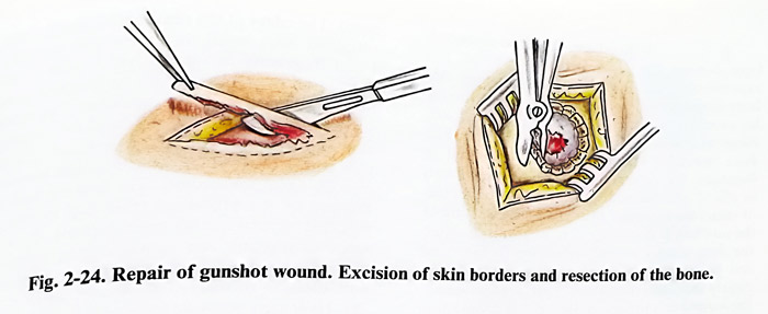
If X-ray/CT investigations have disclosed bone damage, the edges of the bone defect are exposed with additional periosteal incisions. All bone fragments and edges of the defect should be considered contaminated. Fragments are removed and edges nibbled and smoothened. The dura must be exposed by bone edge nibbling, and if damaged, the exposure should be large enough to afford additional access of a few millimetres of intact dura around the defect. The starting of the nibbling is done with care so that not to injure the dura further by not separating its lacerated end from the bone. If the dura is intact and clearly indicates the absence of a haematoma, the wound can be closed. Because of contamination and increased ICP, cranioplasty can be postponed. If the paranasal sinuses, middle ear or orbits are also damaged, all precautions should be taken to avoid leaving any communication with the epidural space. Sinuses are cleaned of mucosa, fragments, foreign bodies and clots. In the case of frontal sinuses and the mastoid, a wider opening of the cavity is re-quired. Isolation of the epidural space from adjacent cavities has a preventive role against secondary infection. Small communications can be closed with surgical wax, but larger ones require suturing a periosteal flap over the opening. Some cases need the surgical team to be joined by maxillofacial, ENT, plastic surgeons or ophthalmologists.
Penetrative injuries carry much higher risks of complications. Once the dural defect is exposed, it usually has to be slightly enlarged for better access to the missile canal inside the brain. All contaminated debris and necrotic tissues are removed from the cortical surface by saline jet for assessment of the entrance of the canal. The edges of the canal entrance are gently retracted with small dissectors and spatulas.
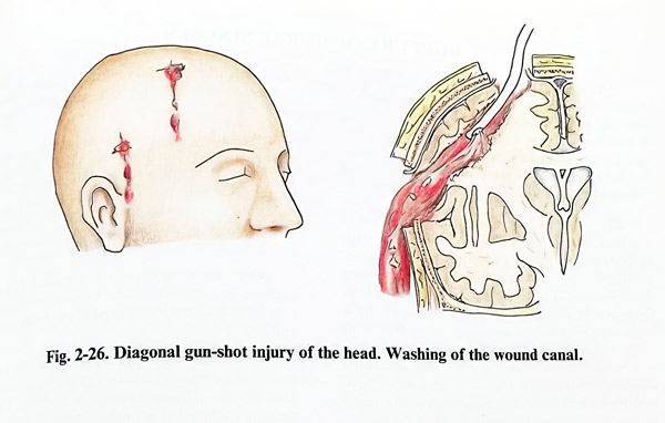
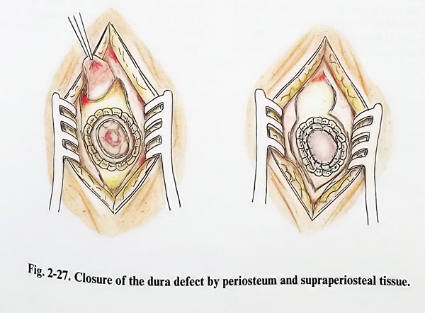
Fixed fragments or foreign bodies seen in the depth after the retraction can be removed using microinstruments and magnification. The saline stream is introduced into the canal for cleaning clots, debris and fragments. A fine, soft, elastic tube with a blunt tip or catheter can be inserted into the canal for flushing. The flushing fluid at the end of the procedure (this step is always repeated several times) must indicate the absence of necrotic and contaminating particles being flushed out of the canal. This will also be an indication of successful haemostasis (Fig. 2-26). Hydrogen peroxide (diluted 1:2 v/v) can be used for flushing and haemostasis. If the distance between entrance and exit wounds is not large, and if it does not traverse ventricles, brain stem, basal ganglia, central and midline structures, the flushing fluid can drain through the exit wound only.Once haemostasis is assured, the surgeon can go on to closure starting with dural plastic repair. The dura is the most important barrier for penetrating secondary infection. The most suitable tissue for plastic repair is periosteum, but fascia can be obtained from the wound or elsewhere in the body for the same purposes (Fig. 2-27). Usually a patch of intact periosteum, slightly exceeding the size of the dural defect, is taken from a part of the scalp distant from the injury wound. It is fixed to the already excised edges of the dural defect by interrupted sutures. Watertightness in closure is always obligatory.
Usually the presence of a certain degree of brain oedema prevents epidural bleeding, so no duro-periosteal sutures are required. The scalp is then closed in the standard manner.