Legacy: "Atlast of Neurosurgery" / L.Karaguiosov, A. Ramadan, K.Karaguiosov / Kiwait/ 1998
Prev < 5. Skull and Brain Malformations
Hydrocephalus is the condition in which the ventricular volume is abnormally large in relation to the volume of the brain. The cause of the hydrocephalus is an important factor in patient management. If the hydrocephalus is due to blockage of the CSF pathways, it will be progressive and will require active management. If the hydrocephalus is secondary to cerebral tissue loss, as can be found in cases of severe head trauma or dementing processes, the hydrocephalus is usually arrested and does not require treatment.
The diagnosis may be obvious in newborns and infants with macrocephaly, irritability, lethargy, vomiting, and poor feeding. Bulging of the anterior fontanelle and scalp vein distension are typical of infantile hydrocephalus. The diagnostic work-up must exclude other pathology, such as subdural haematomas and metabolic disorders as well as more benign conditions such as familial megalencephaly. In older children and adults the manifestations of increased intracranial pressure and impaired consciousness are possible.
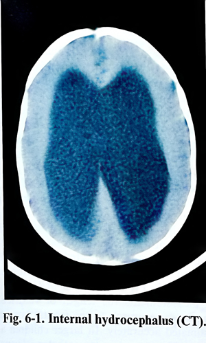
Non-invasive imaging techniques, such as CT, MRI and cranial ultrasonography are essential diagnostic methods to disclose hydrocephalus, to assist in attributing a cause and estimating prognosis and therapeutic requirements. Serial clinical follow-up scanning is often required to detect progression or evolution of the condition (Figs.6-1). Gestational cases of hydrocephalus are now detected by obstetrical ultrasonography. Serial sonography occasionally points to the need for early delivery and treatment of the hydrocephalus after 34-weeks gestation.
After detection of the hydrocephalus, its treatment depends upon the answers to two main questions: is the causing condition curable and is the condition progressive. Removal of an offending neoplasm occasionally provides a permanent cure of the condition. More than half the children with posterior fossa tumours do not require CSF shunting after resection of the tumour.
Hydrocephalus is treated by CSF derivation. It can be intermittent or permanent. Intermittent derivation is obtained by tapping the CSF spaces, but this is reserved for use only when needed on a single occasion or for a short period of time. Communicating hydrocephalus can be tapped in the lumbar CSF compartment (lumbar tap), as can be done in all infants through the open anterior fontanel. In adults derivation for a period of days can be managed externally - by ventricular drainage (ventriculostomy). A longer period requires either the implantation of a reservoir or a shunt system.
From the different types of shunt operations the ventriculoperitoneal shunt is to be preferred, as it has the advantage of easy insertion and revision, relatively benign complications, and the ability of the peritoneal cavity to accept a larger loop of tubing to accommodate axial growth of the patient. In selected patients with noncommunicating hydrocephalus and with competent subarachnoid CSF pathways, as determined by flow studies, third ventriculostomy is once again under evaluation. Newer stereotactic and endoscopic procedures may produce an arrest of the hydrocephalus through the creation of internal fistulae.
Selection of patients and timing of surgery. In congenital hydrocephalus detected at the time of delivery or manifested later, decisions on surgery and timing are usually not a complex problem.
Even in the most extreme cases with a marginal cerebral mantle, social pressure and nursing considerations usually dictate treatment. Extensive experience indicates that early operation produces the best results. The natural history of hydrocephalus points to only a 20% probability of an infant reaching adult life. The variety of handicap includes serious physical, neurological, visual, and intellectual impairment in the majority of the survivors.
Delay of operation may be necessary in patients with active ventriculitis or bloody, proteinaceous CSF. The insertion of temporary ventricular reservoirs for daily withdrawal of CSF often serves to maintain the patient over until definitive shunting can be accomplished. For this reason, sampling of CSF by a single ventricular tap is recommended before surgery. Although shunts may often function satisfactory in the face of extremely high CSF protein, the preferred practice is to delay the procedure until the protein concentration is below 2 g/L.
For patients with normal pressure hydrocephalus the decision for a shunt operation is more complicated. The improvement rates after operation is approximately 65% using the clinical criteria: dementia, gait disturbances, urinary incontinence and large ventricles. The prominence of gait disturbances and periventricular low absorption after CT are important predictors of shunt success. Lumbar puncture is also helpful, but lack of improvement after lumbar puncture does not always exclude a good response to shunting.
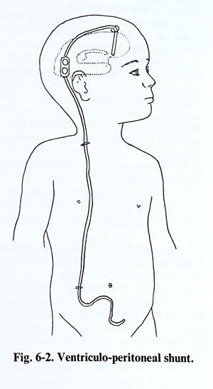
Shunting devices consist of several components, which in spite of variations and technical innovations, always include a ventricular catheter, distal draining catheters (peritoneal or atrial), and a pressure regulator (valve) with a predesigned opening pressure, also compensating orthostatic CSF pressure changes as much as is possible. All the systems are siliconized and once assembled in a 'failsafe' manner, they permit only unidirectional flow of CSF.
Shunt hardware is already sophisticated, and its future development is proceeding in a fast pace that further discussion of current technology is unnecessary. Some general considerations should be kept in mind, however. All the hardware should be seen easily on plain X. rays. Many of the earlier shunting devices were only visible at both ends of the ventricular and peritoneal/atrial catheters, making it impossible to see if the shunt was intact. This is becoming less of a problem as most of manufacturers are producing radioopaque hardware making it possible to see the shunt completely from one end to the other.
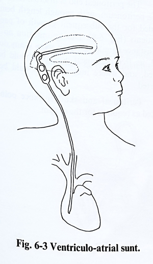
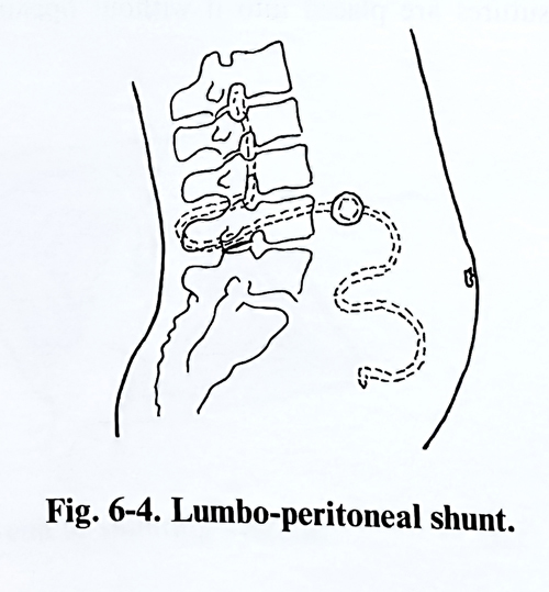
Extracranial shunt operations today include the ventriculoperitoneal (VP), ventriculoatrial (VA), and lumbo-peritoneal (LP) varieties (Figs. 6-2; 6-3; 6-4). Until 20 years ago the VA shunt was preferred for its high degree of success. VA shunts have been largely supplanted by VP shunts, as the latter are technically easier to insert or revise distally and have fewer severe complications. Relative shortening of the shunt can be avoided when enough tubing has been placed in the infant's abdominal cavity to allow for growth into adulthood. On the other hand, VA shunts inserted in infants and children need elective lengthening of the distal end at least once to maintain proper positioning of the tip in the atrium.
It is best to use shunt systems, which are as simple as possible to minimise the chance of malfunction, and it is necessary to have a reservoir in the system. Routine tapping of the reservoir if there is any question as to function or infection of the shunt can avoid unnecessary revision of the system.
TECHNIQUE OF VENTRICULO-PERITONEAL SHUNT
The operation is performed under general anaesthesia in supine position, with the head turned to the opposite side. A pad is placed under the shoulders to raise the neck region and make the angle between the head and chest as flat as possible, and by that making a straight line from the cranial and abdominal entries, so it easier to pass the tubing subcutaneously from the head to the abdomen. That should make the subcutaneous trajectory as straight as possible. After the operative field has been prepared with antiseptic solutions, a small curvilinear incision is marked on the head immediately behind and above the auricle or in the frontal area. An incision of the abdomen 3.5 - 4 cm long is marked on the point of Mc Burney or in the midline just below of xyphoid process. The skin is injected with a 0.25% lidocain with 1:400,000 epinephrine solution to decrease bleeding. Saline is injected along the course of insertion of the catheter.
The small scalp flap on the cranial vault is turned inferiorly, preserving the periosteum and the supraperiosteal tissue intact, leaving only a small part of the skull surface for the burr hole and the dura exposed. In the frontal area the burr hole should be done immediately anterior to the coronal suture along the vertical line, passing through the ipsilateral pupil. The operative wound of the head is covered, and the attention of the surgeon is turned to the abdomen, where a subxiphoid midline incision is made through the abdominal wall. In a stepwise manner, it transects all layers until the peritoneum, and 2 - 4 absorbable sutures are placed into it without opening the membrane to be ready for doing that later.
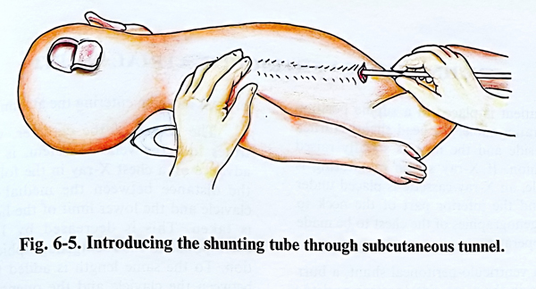
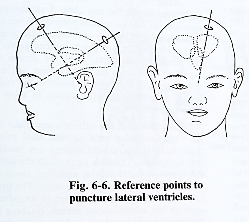
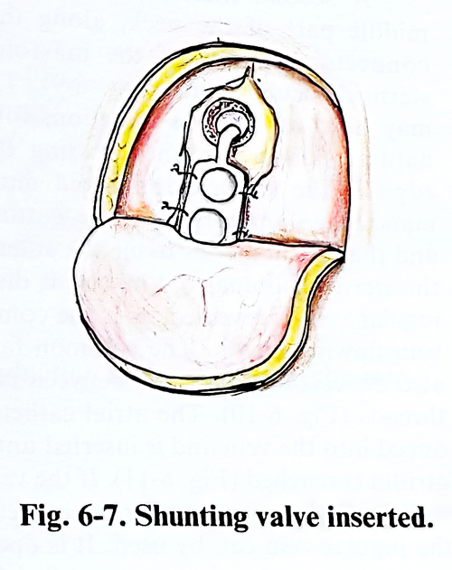
With a special maleable guide is made a tunnel connecting the two operative wounds. If it is not possible to reach the other wound, a small transverse incision must be made on the skin of the neck, just supraclavicular, allowing the proper passage of the tubing over the clavicle which is the biggest obstacle in the course. The shunting tube is passed through the tunnel (Fig. 6-5). After that the dura is opened and the lateral ventricle is punctured. The cannula is directed from the burr hole to the ipsilateral pupil, so that the catheter is introduced in the body of the ventricle and reaches the frontal horn bulb. If the burr hole is in the frontal region, the cannula is directed to the sagittal plane where the biauricular line is supposed to cross it (Figs. 6-6; 6-7). From the CT or MRI scan and skull measurements the length of ventricular tubing needed to reach to the frontal horn can be estimated accurately. In bigger children and adults, a 12 cm ventricular catheter is usually adequate.
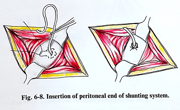
The peritoneal end is inserted after opening the peritoneum, with enough length being provided for the subsequent growth of paediatric patients (Fig. 6-8). During insertion no strong resistance should be felt; if there is any doubt at all, the catheter free peritoneal movement should be clarified, and where indicated it should be reinserted or even confirmed by X-ray image.
At the peritoneal and skull entrance points the elements of the system are fixed as required.
TECHNIQUE OF VENTRICULO-ATRIAL SHUNT
The patient is placed in a supine position on the operating table, the head slightly turned to the left side and the neck is slightly raised with a cushion. If X-ray screen monitoring is not available, an X-ray cassette is placed under the chest and the inferior part of the neck to allow X-ray images of the chest to be taken during the operation.
As in a ventriculo-peritoneal shunt placement, a burr hole is made on the same side (postero-parieto-temporal or frontal area without opening of the dura.
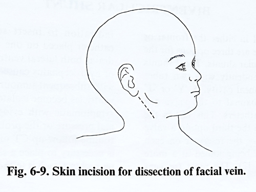
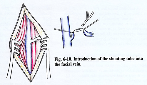
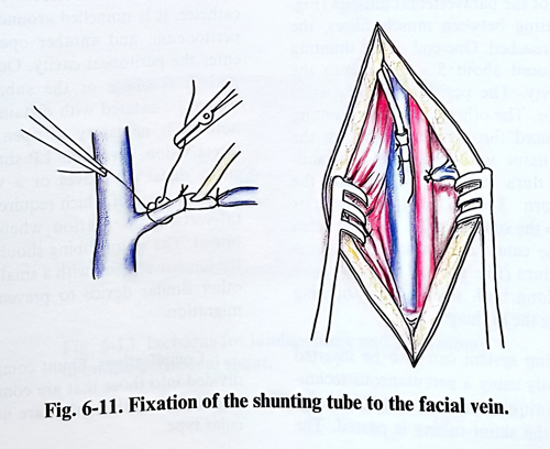
A second incision is then made in the middle part of the neck, along the line that connects the apex of the mastoid with the sternoclavicular joint (Fig. 6-9). The incision may have an oblique direction, following the natural skin creases and crossing the anterior edge of the sterocleidomastoid muscle at the mandibular angle level. After transecting the skin and the subcutaneous tissue, the anterior edge of the sternocleidomastoid muscle is dissected, the jugular vein is identified with the common facial vein inflowing. The common facial vein is also dissected and opened between two holding suture threads (Fig. 6-10). The atrial catheter is introduced into the vein and is inserted until is reached the right atrium (Fig. 6-11). If the common facial vein is not of sufficiently large calibre for the catheter to pass, the jugular vein can by used. It is opened in the same way as the common facial vein and the catheter is fixed to its wall by a purse-string suture so the vein remains patent. The tubing is filled with saline and clamped beforehand to prevent air from entering the right atrium.
The length of the catheter, which penetrates into the venous system, is adjusted in advance on a chest X-ray in the following way: the distance between the medial end of the clavicle and the lower limit of the heart shadow is taken. This is decreased by 10%, which corresponds to the roentgenographic magnification. To that length is added the distance between the clavicle and the vein opening point. The lower end of the catheter usually corresponds to the level between the sixth and seventh thoracic vertebrae.
After the catheter reaches the previously calculated depth, a new chest X-ray is taken to establish its position. When non-radio-opaque catheters are used, contrast medium is injected before the roentgenography. In the adult, the midportion of the atrium is a satisfactory location for the catheter tip; in an infant or child it is best to position the end as low as possible within the atrium to allow for future growth.
A suture fixes the tubing entry place into the common facial vein in its final position so that proper positioning is guaranteed against any inadvertent movement. The common facial vein is next tied around the atrial catheter to secure its position. The proximal part of the atrial catheter is introduced through the subcutaneous tunnel, reaching the previously made burr hole, and the remaing steps of the operation are as described for a VP shunt.
BIVENTRICULAR SHUNT
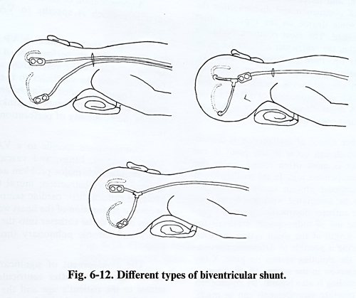
It can be inserted in either the frontal or parietal location. There are three options for the placement of biventricular shunts. The 2 shunts can be treated independently with two tubes tunnelled to the peritoneal cavity. A 'Y' or 'T connector inserted proximal to the valve can connect ventricular catheters. This last is the preferred technique. In the third option a valve can be placed just after the reservoir on each side with the 2 shunts being joined at a more distal location (Fig. 6-12). The most frequent indication to insert second shunt is that a catheter placed on one lateral ventricle initially drains both lateral ventricles satisfactory in case of hydrocephalus caused by a tumour, but then with subsequent tumour growth, the two lateral ventricles become isolated. The patient becomes symptomatic with evidence of raised intracranial pressure or the problem may be detected by routine follow-up CT or MRI. In such a case it is necessary to place a second ventricular catheter and to attach it to the existing shunt.
LUMBO-PERITONEAL SHUNT
A common indication for a LP shunt is in an adult with a communicating hydrocephalus, as in "normal pressure" hydrocephalus. The technique is suitable in patients with small ventricles as well as in cases with benign intracranial hypertension. In the paediatric age, an LP shunt is applied rarely, because communicating hydrocephalus is rarely found in this age group.
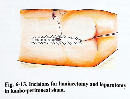
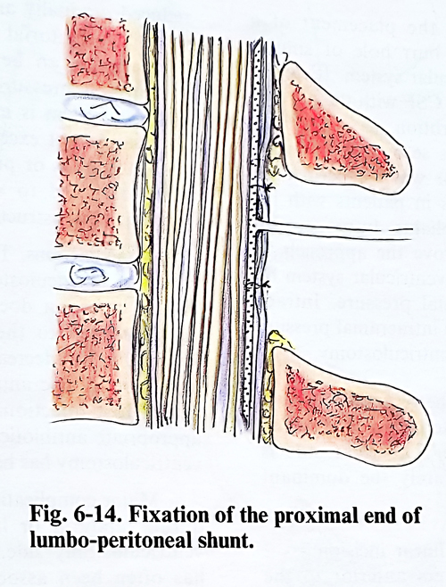
Surgical technique. The patient is placed in a lateral position on the operative table, to make the lumbar spine and lateral abdomen accessible for exploration. A standard laminectomy is performed removing the lamina of L3. The second incision of 5 - 6 cm is made below and parallel to the twelfth rib starting from the lateral border of the paravertebral muscles (Fig. 6-13). Penetrating between muscle fibres, the peritoneum is reached. One end of the shunting tube is introduced about 5 - 10 cm into the peritoneal cavity. The peritoneum is stitched around the tube. The other end of the shunting tube is introduced through the tunnel, in the subcutaneous tissue until it reaches the laminectomy. The dura mater and the arachnoid are opened. The end of the catheter is introduced into the subarachnoid space between the roots of the cauda equina and the tube is fixed to the dura (Fig. 6-14). There are many side holes along the shunting tube, facilitating the drainage.
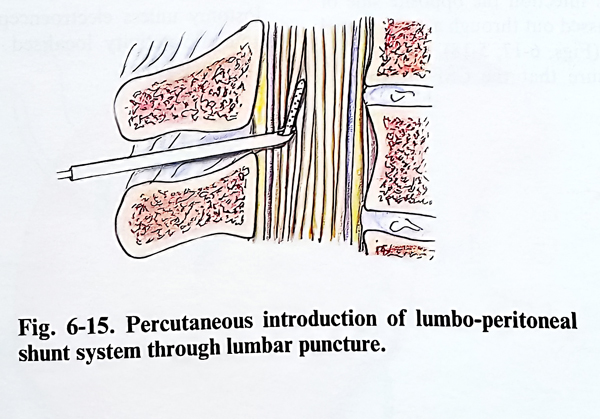
The shunting system can also be inserted more easily and less invasively using a percutaneous technique. This technique employs a 14G needle through which the shunt tubing is passed. The ability of the tubing to pass through the needle should be checked before the shunt placement. A small incision is made in the low midline lumbar area through which the needle is advanced with its tip bevel parallel to the longitudinal fibres of the dura mater to allow easier penetration. When CSF fluid is obtained, the tip is advanced slightly, rotated 90 degrees so that its opening is facing cephalic, and a predetermined length of tubing is then passed through the needle. The position of the catheter tip should be above the conus into the low thoracic region, thereby avoiding later traction of the lumbar roots (Fig. 6-15). The needle is withdrawn over the tubing, with care taken not to pull the catheter back.
When CSF flow is uninterruptedly running from the catheter, it is tunnelled around the flank to the peritoneum, and another opening is made to enter the peritoneal cavity. Occasionally, if the patient is obese or the subarachnoid space cannot be entered with certainty, a small laminotomy is necessary to open the dura under direct vision. Available LP shunt systems have either distal slit valves or a variety of in-line systems, some of which require careful horizontal-vertical orientation when they are positioned. The shunt tubing should be anchored at the lumbar incision with a small silastic collar or other similar device to prevent its subsequent migration.
Complications. Shunt complications can be divided into those that are common to all types of shunts and those that are unique to a particular type.
All shunts carry the risk of obstruction, disconnection and infection. The frequency and location of obstruction and disconnection depend to some degree on the type of shunt hardware used. The most common obstruction place is within the ventricular catheter lumen or around its tip. Paediatric patients most frequently suffer an obstruction of the ventricular catheter by choroid plexus, ependymal, or glial tissue, which can grow into or surround the catheter. A distal slit-valve catheter is the next most frequent site of obstruction.
Another source of malfunction is disconnection, which can occur at any point in the system but is most often found where the different shunt components join. In the paediatric age group, increased protein levels in the CSF appear to be associated with more frequent ventricular catheter obstruction. As noted previously, the use of radio-opaque materials for the entire length of the shunt system is recommendable, making it possible to determine the continuity of the shunt system on plain X-ray films. A reservoir in the system allowing access to CSF sampling is also helpful. By tapping the reservoir, pressure measurements can be made, the system's function can be ascertained, and CSF can be obtained for examination. Each type of shunting system has its own peculiarities which dictate how best to establish its functional adequacy. Most systems contain a pumping mechanism, either in the valve or the reservoir, which should test proper functioning, but the response to pumping and proper functioning of the shunt alone may not correlate absolutely with the overall pressure regulating function.
The management of shunt infection is subject to considerable debate. In principle it requires complete removal of the infected shunt system, with drainage intermittently through a reservoir or continuously by the establishment of an external ventricular drainage system.
A chronic low-grade infection shunt complication specific to VA shunts is nephritis.
Complications specific to VP shunts are ascites; pseudocyst; perforation of viscus; spread of infection or very rarely seeding of a neoplasm from the ventricles to the abdominal cavity. The use of coiled spring tubing inside the peritoneal cavity prevents its kinking but increases the possibility of perforation of abdominal organs.
Complications specific to a VA shunt involve the heart, lungs, and vascular system. They include such major problems as subclavian and vena cava obstruction, mural thrombosis, bacterial endocarditis, cardiac tamponade (secondary to perforation of the heart wall), embolization by the distal catheter into the pulmonary artery and chronic pulmonary thromboembolism.
The development of significant subdural fluid accumulations after ventricular shunting relates to the patient's age and the size of the ventricles; the older the patient, the bigger the risk for this complication, both during and after shunt insertion. Even a minor head injury may produce a significant subdural fluid accumulation if the shunted ventricles are large. The use of a high-pressure valve helps to reduce but does not eliminate this problem. If the subdural collection is asymptomatic and relatively small, treatment is not necessarily indicated, but if a progressive increase in volume is documented with clinical symptoms, it is necessary to drain the subdural fluid. This can be treated with a subdural peritoneal shunt without a valve to provide a pressure gradient across the cortical mantle between the two shunting systems.
VENTRICULOSTOMY
Ventriculostomy is the placement of a drainage catheter via a burr hole or similar technique into the ventricular system. It can be used to temporarily drain CSF with the hope of normalising CSF reabsorbtion, as in patients with acute hydrocephalus secondary to intra-ventrcular haemorrage, or when a permanent shunt is inappropriate, as in patients with infected CSF and hydrocephalus. Intraoperative ventriculostomy can improve the approach to the lesion adjacent to the ventricular system by diminishing the intracranial pressure. Intra-operative and post-operative intracranial pressure can be monitored via the ventriculostomy too.
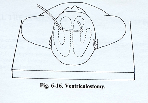
The insertion site of the ventriculostomy is usually the frontal or posterior parietal region as in a shunting technique. The right side is usually chosen, as it is rarely the dominant hemisphere (fig. 6-16).
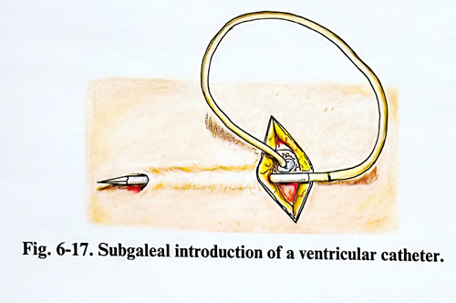
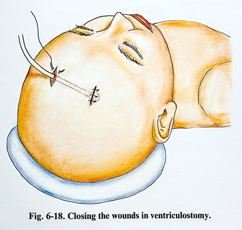
Surgical technique. A linear incision 1 - 2 cm long is made 1 to 2 cm anterior to the coronal suture. The coronal suture gap itself is used as the entry site in the infant. A hand or twist drill may be used to make the opening through the skull. The lateral ventricle is punctured and a plastic catheter is introduced through this puncture. To avoid infection penetration, the opposite end of the catheter is passed out through a small tunnel under the scalp (Figs. 6-17; 5-18). Care should be taken to ensure that the CSF pressure is reduced gradually and not suddenly, since upward transtentorial herniation or a subdural haematoma can be induced by any sudden decrease. A sterile ventricular drainage system is inserted and the maintained CSF pressure should not exceed 100 mm of water. The CSF pulsations in the system or pressure waves are continuously observed to ensure that the drainage system is not obstructed.
Complications. The most common complication of ventriculostomy is CSF infection. The rate of infection does not seem to be directly related to the duration of placement. Tunnelling has decreased the risk of CSF leakage. Prophylactic antibiotics can be used routinely. Most infections are easily treated with appropriate antibiotic therapy, especially if the ventriculostomy has been removed.
Major complications are haematomas: epidural, subdural or intraparenchimal, at the ventriculostomy side. This rare complication has often been associated with a known or unsuspected coagulation disorder.
Seizures originating at the insertion site are a potential risk. As these patients often have one or more reasons for their seizures, it is usually not possible to attribute seizures to the ventriculostomy unless electroencephalography shows epileptic activity localised to the site of the ventriculostomy.