Hydrocephalus is the condition in which the ventricular volume is abnormally large in relation to the volume of the brain. The cause of the hydrocephalus is an important factor in patient management. If the hydrocephalus is due to blockage of the CSF pathways, it will be progressive and will require active management. If the hydrocephalus is secondary to cerebral tissue loss, as can be found in cases of severe head trauma or dementing processes, the hydrocephalus is usually arrested and does not require treatment.
The diagnosis may be obvious in newborns and infants with macrocephaly, irritability, lethargy, vomiting, and poor feeding. Bulging of the anterior fontanelle and scalp vein distension are typical of infantile hydrocephalus. The diagnostic work-up must exclude other pathology, such as subdural haematomas and metabolic disorders as well as more benign conditions such as familial megalencephaly. In older children and adults the manifestations of increased intracranial pressure and impaired consciousness are possible.
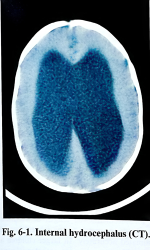
Non-invasive imaging techniques, such as CT, MRI and cranial ultrasonography are essential diagnostic methods to disclose hydrocephalus, to assist in attributing a cause and estimating prognosis and therapeutic requirements. Serial clinical follow-up scanning is often required to detect progression or evolution of the condition (Figs.6-1). Gestational cases of hydrocephalus are now detected by obstetrical ultrasonography. Serial sonography occasionally points to the need for early delivery and treatment of the hydrocephalus after 34-weeks gestation.
After detection of the hydrocephalus, its treatment depends upon the answers to two main questions: is the causing condition curable and is the condition progressive. Removal of an offending neoplasm occasionally provides a permanent cure of the condition. More than half the children with posterior fossa tumours do not require CSF shunting after resection of the tumour.
Hydrocephalus is treated by CSF derivation. It can be intermittent or permanent. Intermittent derivation is obtained by tapping the CSF spaces, but this is reserved for use only when needed on a single occasion or for a short period of time. Communicating hydrocephalus can be tapped in the lumbar CSF compartment (lumbar tap), as can be done in all infants through the open anterior fontanel. In adults derivation for a period of days can be managed externally - by ventricular drainage (ventriculostomy). A longer period requires either the implantation of a reservoir or a shunt system.
From the different types of shunt operations the ventriculoperitoneal shunt is to be preferred, as it has the advantage of easy insertion and revision, relatively benign complications, and the ability of the peritoneal cavity to accept a larger loop of tubing to accommodate axial growth of the patient. In selected patients with noncommunicating hydrocephalus and with competent subarachnoid CSF pathways, as determined by flow studies, third ventriculostomy is once again under evaluation. Newer stereotactic and endoscopic procedures may produce an arrest of the hydrocephalus through the creation of internal fistulae.
Selection of patients and timing of surgery. In congenital hydrocephalus detected at the time of delivery or manifested later, decisions on surgery and timing are usually not a complex problem.
Even in the most extreme cases with a marginal cerebral mantle, social pressure and nursing considerations usually dictate treatment. Extensive experience indicates that early operation produces the best results. The natural history of hydrocephalus points to only a 20% probability of an infant reaching adult life. The variety of handicap includes serious physical, neurological, visual, and intellectual impairment in the majority of the survivors.
Delay of operation may be necessary in patients with active ventriculitis or bloody, proteinaceous CSF. The insertion of temporary ventricular reservoirs for daily withdrawal of CSF often serves to maintain the patient over until definitive shunting can be accomplished. For this reason, sampling of CSF by a single ventricular tap is recommended before surgery. Although shunts may often function satisfactory in the face of extremely high CSF protein, the preferred practice is to delay the procedure until the protein concentration is below 2 g/L.
For patients with normal pressure hydrocephalus the decision for a shunt operation is more complicated. The improvement rates after operation is approximately 65% using the clinical criteria: dementia, gait disturbances, urinary incontinence and large ventricles. The prominence of gait disturbances and periventricular low absorption after CT are important predictors of shunt success. Lumbar puncture is also helpful, but lack of improvement after lumbar puncture does not always exclude a good response to shunting.
Shunting devices consist of several components, which in spite of variations and technical innovations, always include a ventricular catheter, distal draining catheters (peritoneal or atrial), and a pressure regulator (valve) with a predesigned opening pressure, also compensating orthostatic CSF pressure changes as much as is possible. All the systems are siliconized and once assembled in a 'failsafe' manner, they permit only unidirectional flow of CSF.
Shunt hardware is already sophisticated, and its future development is proceeding in a fast pace that further discussion of current technology is unnecessary. Some general considerations should be kept in mind, however. All the hardware should be seen easily on plain X. rays. Many of the earlier shunting devices were only visible at both ends of the ventricular and peritoneal/atrial catheters, making it impossible to see if the shunt was intact. This is becoming less of a problem as most of manufacturers are producing radioopaque hardware making it possible to see the shunt completely from one end to the other.
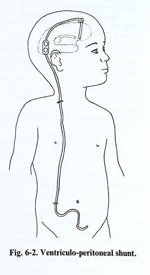
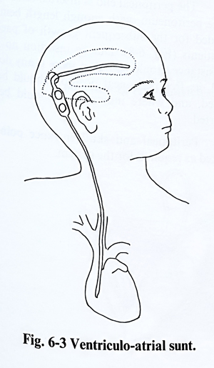
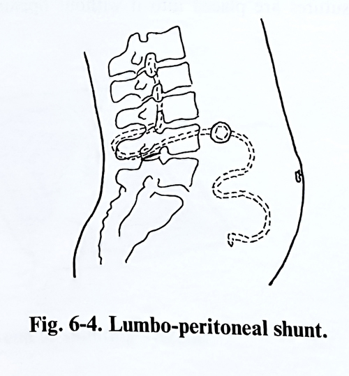
Extracranial shunt operations today include the ventriculoperitoneal (VP), ventriculoatrial (VA), and lumbo-peritoneal (LP) varieties (Figs. 6-2; 6-3; 6-4). Until 20 years ago the VA shunt was preferred for its high degree of success. VA shunts have been largely supplanted by VP shunts, as the latter are technically easier to insert or revise distally and have fewer severe complications. Relative shortening of the shunt can be avoided when enough tubing has been placed in the infant's abdominal cavity to allow for growth into adulthood. On the other hand, VA shunts inserted in infants and children need elective lengthening of the distal end at least once to maintain proper positioning of the tip in the atrium.
It is best to use shunt systems, which are as simple as possible to minimise the chance of malfunction, and it is necessary to have a reservoir in the system. Routine tapping of the reservoir if there is any question as to function or infection of the shunt can avoid unnecessary revision of the system.