TECHNIQUE OF VENTRICULO-ATRIAL SHUNT
The patient is placed in a supine position on the operating table, the head slightly turned to the left side and the neck is slightly raised with a cushion. If X-ray screen monitoring is not available, an X-ray cassette is placed under the chest and the inferior part of the neck to allow X-ray images of the chest to be taken during the operation.
As in a ventriculo-peritoneal shunt placement, a burr hole is made on the same side (postero-parieto-temporal or frontal area without opening of the dura.
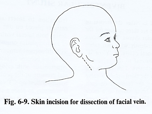
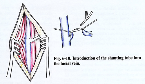
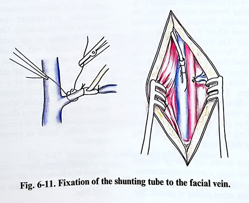
A second incision is then made in the middle part of the neck, along the line that connects the apex of the mastoid with the sternoclavicular joint (Fig. 6-9). The incision may have an oblique direction, following the natural skin creases and crossing the anterior edge of the sterocleidomastoid muscle at the mandibular angle level. After transecting the skin and the subcutaneous tissue, the anterior edge of the sternocleidomastoid muscle is dissected, the jugular vein is identified with the common facial vein inflowing. The common facial vein is also dissected and opened between two holding suture threads (Fig. 6-10). The atrial catheter is introduced into the vein and is inserted until is reached the right atrium (Fig. 6-11). If the common facial vein is not of sufficiently large calibre for the catheter to pass, the jugular vein can by used. It is opened in the same way as the common facial vein and the catheter is fixed to its wall by a purse-string suture so the vein remains patent. The tubing is filled with saline and clamped beforehand to prevent air from entering the right atrium.
The length of the catheter, which penetrates into the venous system, is adjusted in advance on a chest X-ray in the following way: the distance between the medial end of the clavicle and the lower limit of the heart shadow is taken. This is decreased by 10%, which corresponds to the roentgenographic magnification. To that length is added the distance between the clavicle and the vein opening point. The lower end of the catheter usually corresponds to the level between the sixth and seventh thoracic vertebrae.
After the catheter reaches the previously calculated depth, a new chest X-ray is taken to establish its position. When non-radio-opaque catheters are used, contrast medium is injected before the roentgenography. In the adult, the midportion of the atrium is a satisfactory location for the catheter tip; in an infant or child it is best to position the end as low as possible within the atrium to allow for future growth.
A suture fixes the tubing entry place into the common facial vein in its final position so that proper positioning is guaranteed against any inadvertent movement. The common facial vein is next tied around the atrial catheter to secure its position. The proximal part of the atrial catheter is introduced through the subcutaneous tunnel, reaching the previously made burr hole, and the remaing steps of the operation are as described for a VP shunt.