TECHNIQUE OF VENTRICULO-PERITONEAL SHUNT
The operation is performed under general anaesthesia in supine position, with the head turned to the opposite side. A pad is placed under the shoulders to raise the neck region and make the angle between the head and chest as flat as possible, and by that making a straight line from the cranial and abdominal entries, so it easier to pass the tubing subcutaneously from the head to the abdomen. That should make the subcutaneous trajectory as straight as possible. After the operative field has been prepared with antiseptic solutions, a small curvilinear incision is marked on the head immediately behind and above the auricle or in the frontal area. An incision of the abdomen 3.5 - 4 cm long is marked on the point of Mc Burney or in the midline just below of xyphoid process. The skin is injected with a 0.25% lidocain with 1:400,000 epinephrine solution to decrease bleeding. Saline is injected along the course of insertion of the catheter.
The small scalp flap on the cranial vault is turned inferiorly, preserving the periosteum and the supraperiosteal tissue intact, leaving only a small part of the skull surface for the burr hole and the dura exposed. In the frontal area the burr hole should be done immediately anterior to the coronal suture along the vertical line, passing through the ipsilateral pupil. The operative wound of the head is covered, and the attention of the surgeon is turned to the abdomen, where a subxiphoid midline incision is made through the abdominal wall. In a stepwise manner, it transects all layers until the peritoneum, and 2 - 4 absorbable sutures are placed into it without opening the membrane to be ready for doing that later.
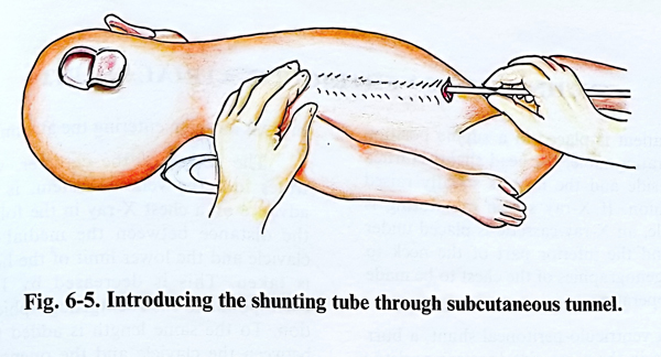
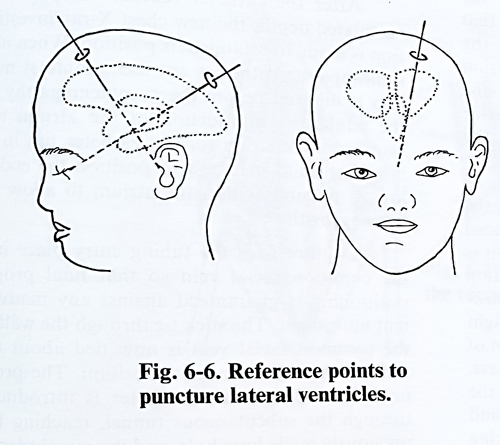
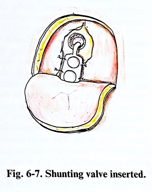
With a special maleable guide is made a tunnel connecting the two operative wounds. If it is not possible to reach the other wound, a small transverse incision must be made on the skin of the neck, just supraclavicular, allowing the proper passage of the tubing over the clavicle which is the biggest obstacle in the course. The shunting tube is passed through the tunnel (Fig. 6-5). After that the dura is opened and the lateral ventricle is punctured. The cannula is directed from the burr hole to the ipsilateral pupil, so that the catheter is introduced in the body of the ventricle and reaches the frontal horn bulb. If the burr hole is in the frontal region, the cannula is directed to the sagittal plane where the biauricular line is supposed to cross it (Figs. 6-6; 6-7). From the CT or MRI scan and skull measurements the length of ventricular tubing needed to reach to the frontal horn can be estimated accurately. In bigger children and adults, a 12 cm ventricular catheter is usually adequate.
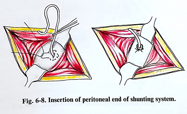
The peritoneal end is inserted after opening the peritoneum, with enough length being provided for the subsequent growth of paediatric patients (Fig. 6-8). During insertion no strong resistance should be felt; if there is any doubt at all, the catheter free peritoneal movement should be clarified, and where indicated it should be reinserted or even confirmed by X-ray image.
At the peritoneal and skull entrance points the elements of the system are fixed as required.