VENTRICULOSTOMY
Ventriculostomy is the placement of a drainage catheter via a burr hole or similar technique into the ventricular system. It can be used to temporarily drain CSF with the hope of normalising CSF reabsorbtion, as in patients with acute hydrocephalus secondary to intra-ventrcular haemorrage, or when a permanent shunt is inappropriate, as in patients with infected CSF and hydrocephalus. Intraoperative ventriculostomy can improve the approach to the lesion adjacent to the ventricular system by diminishing the intracranial pressure. Intra-operative and post-operative intracranial pressure can be monitored via the ventriculostomy too.
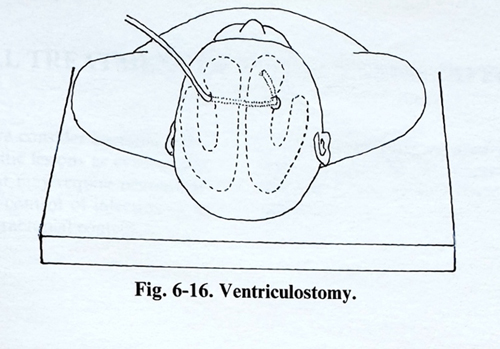
The insertion site of the ventriculostomy is usually the frontal or posterior parietal region as in a shunting technique. The right side is usually chosen, as it is rarely the dominant hemisphere (fig. 6-16).
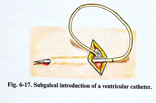
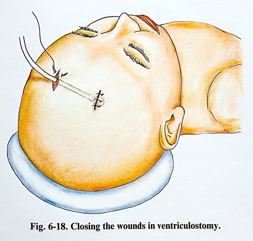
Surgical technique. A linear incision 1 - 2 cm long is made 1 to 2 cm anterior to the coronal suture. The coronal suture gap itself is used as the entry site in the infant. A hand or twist drill may be used to make the opening through the skull. The lateral ventricle is punctured and a plastic catheter is introduced through this puncture. To avoid infection penetration, the opposite end of the catheter is passed out through a small tunnel under the scalp (Figs. 6-17; 5-18). Care should be taken to ensure that the CSF pressure is reduced gradually and not suddenly, since upward transtentorial herniation or a subdural haematoma can be induced by any sudden decrease. A sterile ventricular drainage system is inserted and the maintained CSF pressure should not exceed 100 mm of water. The CSF pulsations in the system or pressure waves are continuously observed to ensure that the drainage system is not obstructed.
Complications. The most common complication of ventriculostomy is CSF infection. The rate of infection does not seem to be directly related to the duration of placement. Tunnelling has decreased the risk of CSF leakage. Prophylactic antibiotics can be used routinely. Most infections are easily treated with appropriate antibiotic therapy, especially if the ventriculostomy has been removed.
Major complications are haematomas: epidural, subdural or intraparenchimal, at the ventriculostomy side. This rare complication has often been associated with a known or unsuspected coagulation disorder.
Seizures originating at the insertion site are a potential risk. As these patients often have one or more reasons for their seizures, it is usually not possible to attribute seizures to the ventriculostomy unless electroencephalography shows epileptic activity localised to the site of the ventriculostomy.